
Bicarbonate Transport in Cystic Fibrosis and Pancreatitis
by
 , and
, and
Dora Angyal
 ,
,
Marcel J. C. Bijvelds
,
Marco J. Bruno
,
Maikel P. Peppelenbosch
 and
and
Hugo R. de Jonge
* Department of Gastroenterology and Hepatology, Erasmus MC University Medical Center Rotterdam, P.O. Box 2040, 3000 CA Rotterdam, The Netherlands
*
Author to whom correspondence should be addressed.
Cells 2022, 11(1), 54; https://doi.org/10.3390/cells11010054
Submission received: 20 November 2021
/
Revised: 20 December 2021
/
Accepted: 21 December 2021
/
Published: 24 December 2021
(This article belongs to the Collection Cystic Fibrosis: Cells, Physiopathology and Emerging Therapies)
Abstract
:CFTR, the cystic fibrosis (CF) gene-encoded epithelial anion channel, has a prominent role in driving chloride, bicarbonate and fluid secretion in the ductal cells of the exocrine pancreas. Whereas severe mutations in CFTR cause fibrosis of the pancreas in utero, CFTR mutants with residual function, or CFTR variants with a normal chloride but defective bicarbonate permeability (CFTRBD), are associated with an enhanced risk of pancreatitis. Recent studies indicate that CFTR function is not only compromised in genetic but also in selected patients with an acquired form of pancreatitis induced by alcohol, bile salts or smoking. In this review, we summarize recent insights into the mechanism and regulation of CFTR-mediated and modulated bicarbonate secretion in the pancreatic duct, including the role of the osmotic stress/chloride sensor WNK1 and the scaffolding protein IRBIT, and current knowledge about the role of CFTR in genetic and acquired forms of pancreatitis. Furthermore, we discuss the perspectives for CFTR modulator therapy in the treatment of exocrine pancreatic insufficiency and pancreatitis and introduce pancreatic organoids as a promising model system to study CFTR function in the human pancreas, its role in the pathology of pancreatitis and its sensitivity to CFTR modulators on a personalized basis.
1. Introduction
Cystic fibrosis (CF) is a potentially fatal multi-organ disease caused by mutation of the cystic fibrosis transmembrane conductance regulator (CFTR) gene. It is estimated that CF currently affects >100,000 people worldwide, and that by 2025, the number of patients will increase by 50% [1,2]. CFTR encodes a phosphorylation-regulated, adenosine triphosphate (ATP)-gated, epithelial anion channel that mediates chloride (Cl−) and bicarbonate (HCO3−) transport across epithelia, principally located in the respiratory, gastrointestinal and male reproductive tracts. The term cystic fibrosis refers to the CF-typical fibrotic lesions in the pancreas, first described in the 1930s [3]. In approximately 85% of CF patients, fibrosis of the pancreas starts in utero, progressing to a complete loss of exocrine pancreas function soon after birth, i.e., exocrine pancreatic insufficiency [4]. The severely reduced release of HCO3− and digestive enzymes into the upper intestinal tract impairs the neutralization of gastric acid and causes malabsorption of nutrients.
Pancreatitis is a complex inflammatory disease of the acinar and ductal epithelia, which probably results from premature activation of digestive enzymes [5]. Currently, there is no specific therapy available, and treatment relies on supportive care. Pancreatitis is one of the three most common causes of gastrointestinal disease-related hospitalizations and is associated with high morbidity and mortality [6]. Acute pancreatitis (AP), recurrent AP (RAP) and chronic pancreatitis (CP) are thought to represent a disease continuum [7]. As opposed to the monogenic disease CF, in pancreatitis, multiple (epi-)genetic, metabolic and environmental factors combine and lead to a complex pathology [8,9]. A substantial body of evidence indicates that the loss of ductal CFTR-dependent fluid and HCO3− secretion may precipitate the development of pancreatitis [10]. This is perhaps most poignantly illustrated by the fact that pancreas-sufficient CF patients, i.e., those who have retained some level of exocrine pancreatic function, are at increased risk of developing (R-)AP and CP [11]. Furthermore, it has been shown that carriers of a CF allele are at increased risk of developing pancreatitis [12].
CFTR-mediated HCO3− secretion not only drives osmotic fluid secretion in ductal structures but is also required for controlling the pH at the epithelial surface, and the proper expansion of secreted mucins. Thus, in CF, a lowering of the luminal pH is thought to result in the accumulation of a hyper-viscid mucus that blocks the ductal structures and promotes microbial colonization and inflammation. In addition, in the pancreas, high levels of HCO3− are thought to maintain secreted digestive enzymes in an inactive state while still located in the ductal tree [13].
In view of the apparent relevance of HCO3− in the pathophysiology of CF and CFTR-related disorders (CFTR-RD) such as pancreatitis, it is perhaps surprising that most research into the function of CFTR has focused on its role as a Cl− channel [14]. Perhaps due to the fact that it is readily formed from and converted to CO2 and H2O, accurate assessment of HCO3− concentrations and transport is challenging, which may explain why CF biomarkers and therapy testing are Cl− biased [15]. This is true for clinical nasal potential difference (NPD) and sweat Cl− tests which are Cl− based, as well as CFTR modulator testing which is based on iodide quenching or membrane potential measurements, while intestinal current measurements (ICMs) and forskolin-induced swelling (FIS) assays measure combined Cl− and HCO3− transport.
This review summarizes our current knowledge of the role of CFTR in pancreatic ductal fluid and HCO3− transport, and the role of CFTR dysfunction in pancreatitis. As is outlined in the next section, CFTR is a critical element of the ion transport machinery in ductal epithelial cells, which, remarkably, is able to accumulate HCO3− in human pancreatic juice to levels of up to 140 mmol/L. Several seminal studies published in the last decade point towards a prominent role of impaired CFTR-mediated HCO3− transport in the pathophysiology of pancreatitis [16,17]. This offers the encouraging prospect that CFTR modulators, originally developed for treatment of CF, may also be of potential benefit for other pancreas-related diseases.
2. Bicarbonate Transport in the Exocrine Pancreas
2.1. CFTR Is Indispensable for the Accumulation of Bicarbonate in Pancreatic Juice
In the exocrine pancreas, acinar cells secrete various digestive enzymes in a small volume of isotonic, NaCl- and H+-rich fluid, after which the ductal epithelium serves to exchange the secreted Cl− for HCO3−, to produce an alkaline fluid (pH 8.0–8.5) [13,18,19]. It is estimated that the human exocrine pancreas secretes up to 1 L of pancreatic juice per day [18,20,21]. Apart from delivering digestive enzymes to the proximal small intestine, making it essential for digestion of (macro-)nutrients, pancreatic juice serves to supply base equivalents for neutralization of the gastric acid entering the small intestine from the stomach. In humans, the HCO3− concentration in pancreatic juice can reach levels of 140 mmol/L [22,23]. Similarly high levels of HCO3− were observed in pancreatic juice of guinea pigs, whereas, in contrast, in mice and rats, HCO3− levels did not exceed 70 mmol/L [24,25,26,27,28].
In humans, the production of pancreatic juice is strongly dependent on postprandial release of the hormone secretin, whereas production is low during fasting. Secretin, through activation of Gα,s-coupled receptors, triggers cyclic AMP (cAMP) production and a consequent protein kinase-mediated phosphorylation and activation of CFTR. In humans, CFTR-mediated, cAMP-dependent ductal fluid secretion is strongly potentiated by parasympathetic, vagal stimulation. Most plausibly, the activation of muscarinic (Gα,q) receptors by acetylcholine triggers K+ efflux through Ca2+-dependent K+ channels, which sustains the negative electrical potential across the luminal plasma membrane driving Cl− and HCO3− efflux through CFTR. In addition to cholinergic neurons, the parasympathetic system comprises neurons that produce vasoactive intestinal polypeptide (VIP), which, as with secretin, activates Gα,s-coupled receptors.
2.2. Molecular Mechanisms of CFTR-Dependent Bicarbonate Transport
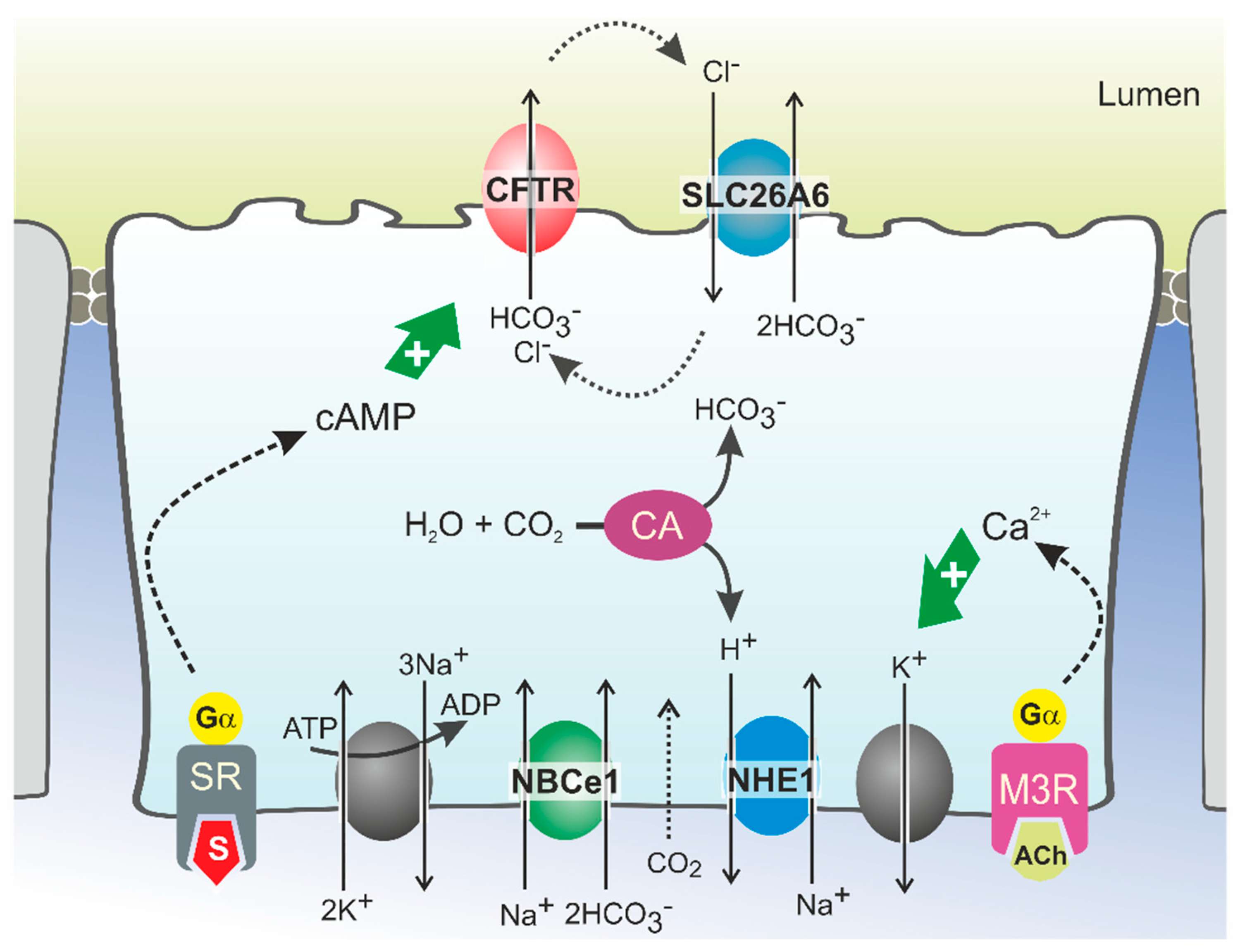
HCO3− secretion involves the coordinated activity of ion transporters located at the basolateral and luminal membranes of pancreatic ductal cells (PDCs) (Figure 1). According to current models, which are discussed in detail elsewhere, uptake of HCO3− from the interstitial space is principally mediated by a Na+/HCO3− cotransporter (NBCe1, SLC4A4) located in the basolateral plasma membrane [13,29]. This transport is driven by the steep electrochemical Na+ gradient across the plasma membrane (generated by Na+,K+-ATPase activity) and allows for intracellular accumulation of HCO3− up to levels well above electrochemical equilibrium. In addition, HCO3− may be produced intracellularly from carbonic anhydrase-catalyzed hydration of CO2, which enters the cells through diffusion (Figure 1). While the protons produced in this reaction are extruded across the basolateral plasma membrane via a Na+/H+ exchange mechanism, HCO3− is secreted across the apical membrane into the ductal lumen via the coordinated activity of the CFTR channel and the Cl−/HCO3− exchangers SLC26A6 and, more tentatively, SLC26A3 [13,30,31,32]. According to this model, which may also be relevant for other HCO3−-secreting epithelia such as the intestinal and biliary epithelia, CFTR serves to facilitate Cl−/HCO3− exchanger-mediated HCO3− secretion by extruding the Cl− ions absorbed through the exchangers [22,30,33,34]. As for CFTR, the activity of Cl−/HCO3− exchangers is stimulated by cAMP and Ca2+ agonists, which implies that their activity is also controlled through neuro-endocrine stimulation of Gα,s- and Gα,q-coupled receptors [35]. CFTR may not only couple functionally to SLC26A6, but also physically, through binding of its regulatory (R) domain to the sulfate transporter anti-sigma (STAS) domain of SLC26A6 [36,37]. This interaction was shown to promote Cl−/HCO3− exchange, even in the absence of CFTR activity [22,36].
Although there is ample evidence for the role of SLC26-type Cl−/HCO3− exchangers in ductal HCO3− secretion, mathematical modeling of the ion motive forces in the ductal epithelium predicted that Cl−/HCO3− exchange mechanisms only significantly contribute to cellular HCO3− extrusion up to luminal HCO3− levels of circa 70 mmol/L [29,38]. Consequently, SLC26-type HCO3− transport mechanisms may serve to secrete HCO3− in the proximal segments of the ductal tree, but to reach the final, high HCO3− levels observed in human pancreatic juice, an alternative transport mechanism must prevail in the distal part. In view of these considerations, it seems plausible that CFTR has a direct role in ductal HCO3− secretion. The CFTR channel itself was shown to directly mediate cellular HCO3− transport in studies on both cell models and epithelial tissues, including pancreatic ducts [39,40,41,42]. It has, however, also been noted that the permeability of the CFTR channel pore for HCO3− is 4–5 times lower than for Cl− [39,40,43,44]. This implies that CFTR can only selectively secrete HCO3− when very low intracellular Cl− and comparatively high HCO3− concentrations are attained. This situation may occur in the distal part of the ductal tree where, upon hormonal stimulation of pancreatic secretion, Cl− levels in PDCs decrease to values as low as 5 mmol/L [29]. In these settings, assuming that, concurrently, intracellular HCO3− is actively accumulated through NBCe1-mediated uptake, it is conceivable that CFTR may function primarily, if not exclusively, as a HCO3− transporter.
To account for the high HCO3− and low Cl− levels found in pancreatic juice, it has been further proposed that the Cl− over HCO3− conductance ratio of CFTR is not static but can be lowered to allow selective HCO3− secretion [45,46,47,48]. This dynamic regulation of CFTR anion conductance is thought to be mediated by members of the WNK (with no lysine) type of protein kinases [13,18,49,50]. WNK kinases (the family consists of four members) regulate an array of ion transporters through activation of downstream protein kinases OSR1 (oxidative stress-responsive kinase 1) and the highly homologous SPAK (STE20/SPS1-related proline/alanine-rich kinase) [51]. The WNK kinases are thought to function as osmotic stress sensors and are activated by a lowering of intracellular Cl− levels. Notably, WNK1 and WNK4 control renal electrolyte transport, and mutations in either cause hypertension as a result of an unchecked tubular Na+ re-absorption. In most cases, WNK kinases regulate the activity of ion transport mechanisms by controlling their localization in the plasma membrane.
At low extracellular Cl− concentrations, WNK1-mediated OSR1 and SPAK activation was shown to markedly increase the HCO3− permeability of CFTR in CFTR-transfected HEK293 cells and in guinea pig PDCs [52]. Interestingly, WNK1 activation depended on CFTR function as the change in HCO3− permeability was not observed in cells expressing non-functional, mutant (F508del) CFTR, suggesting CFTR is required for lowering of the intracellular Cl− concentration. More recently, it was proposed that WNK1 increases the HCO3− permeability of CFTR by a direct interaction that does not require SPAK activation (Figure 2) [53]. WNK kinases are also thought to reduce the surface expression of Cl−/HCO3− exchangers of the SLC26A family [54]. At high luminal HCO3− levels such as those encountered in the distal ductal tree, this silencing of Cl−/HCO3− exchange activity may prevent re-uptake of HCO3− and, consequently, promote its net secretion. Paradoxically, both WNK1 and WNK4 were also shown to reduce CFTR protein levels at the cell surface and suppress CFTR-mediated anion efflux [55]. Moreover, WNK kinases may also reduce NBCe1 activity, suggesting that their activation opposes CFTR-mediated HCO3− secretion [56]. However, the inhibition imposed by the WNK/SPAK pathway on ductal fluid and HCO3− secretion is counteracted by the action of the scaffolding protein inositol-1,4,5-trisphosphate (IP3) receptor-binding protein released with IP3 (IRBIT) [56]. Through its PDZ domain, IRBIT co-localizes at the apical and basolateral plasma membrane with CFTR and NBCe1, respectively, and prevents WNK1/SPAK-mediated endocytosis by recruiting a protein phosphatase (PP1) that counters SPAK-mediated phosphorylation of CFTR and NBCe1 [56,57,58]. In addition to its effect on surface expression, IRBIT may also increase the CFTR open channel probability [59]. Recruitment of IRBIT to these molecular complexes appears to be stimulated through cAMP and Ca2+ signaling, and IRBIT is required for the synergism observed between these signaling pathways [60,61].
3. Bicarbonate Transport in Subjects with CFTR Mutations
The past decade saw great advances in understanding the genetics of pancreatitis, including the role of CFTR mutations. Genes in which mutation confers an increased risk of developing (chronic) pancreatitis are grouped in three categories: (1) the trypsin-dependent pathway including PRSS1, PRSS2, SPINK1, CTRC and CTRB1 [62,63,64]; (2) the misfolding-dependent pathway including CPA1 [65] and CEL [66]; and (3) the ductal pathway which, aside from CFTR, includes CLDN2 and CASR [5]. CFTR mutations are associated with development and earlier age of onset of RAP [67,68,69] and CP [70,71,72,73]. In a large pediatric cohort, 34% of RAP and 23% of CP patients carried CFTR mutations [74]. In a group of RAP patients with a mean age of 23 years, 15% carried CFTR mutations [69], and in a cohort of idiopathic CP patients, this number was nearly 20% [72]. Up to 7% of all CP patients carry CFTR mutations, and depending on the type of CFTR mutation, the risk for developing CP may increase 1.5- to 16-fold [75]. This risk increases 3- to 21-fold when CFTR mutations are combined with a mutation in SPINK1, which encodes the pancreatic secretory trypsin inhibitor [75]. The latter observation underlines the contention that pancreatitis is a multifactorial, complex disorder that develops through an interaction of multiple genetic and/or environmental factors. Consequently, the CFTR alleles that confer an increased risk of developing pancreatitis at the population level only have a small effect on the individual risk [76].
Typical environmental factors that, in conjunction with genetic factors, modify the risk of pancreatitis are alcohol consumption and inhalation of tobacco smoke. As many as 40 to 70% of CP patients drink excessive amounts of alcohol [77,78,79,80,81,82]. Similarly, smoking is an independent risk factor for the onset and recurrence of AP [83], and 60% of CP patients regularly consume tobacco [75]. CFTR mutations are more prevalent in alcohol- and smoking-related CP patients than in the general population, suggesting that partial loss of CFTR function aggravates the effect of such environmental factors [75]. Rosendahl et al. elegantly depicted the range of genetic and environmental interactions in different CFTR-associated diseases [63]. In CF and in hereditary forms of pancreatitis due to mutations in genes other than CFTR (such as PRSS1), genetics are dominant, and environmental factors have little influence on disease onset or severity. Conversely, for CFTR mutations that do not cause typical CF, the interplay with environmental risk factors becomes increasingly important.
To date, more than 2000 mutations in the CFTR gene have been described. These cover a wide spectrum, from apparently functionally silent mutations on one end, to the severe, CF-causing mutations on the opposite end of the scale. Information on the pathophysiological and clinical consequences of these variants has increased markedly over the last decade [1]. The diagnosis of CF requires two CFTR mutations on different alleles which severely impair CFTR channel function. Diagnosis of subjects who carry CFTR gene variants associated with residual protein function is often considerably less straightforward. The terms CF screen-positive, inconclusive diagnosis (CFSPID, European term) and CFTR-related metabolic syndrome (CRMS, North American term) describe patients with elevated immune-reactive trypsinogen who carry no or only one apparently CF-causing allele, which do not fulfil the diagnostic criteria for CF [84,85]. So-called CFTR-related disorders (CFTR-RD) are single-organ diseases with evidence of CFTR dysfunction in the absence of a CF diagnosis [1]. CFTR-RD are commonly associated with the presence of at least one CFTR allele with undefined clinical consequences.
3.1. Impaired Pancreatic Bicarbonate Transport in CF Patients
Impaired pancreatic HCO3− secretion in CF patients was recognized far before the discovery of the disease-causing gene [86]. Follow-up studies indicated that both pancreatic HCO3− and Cl− secretion were strongly reduced in patients and contribute to the CF-typical fluid secretory defect [87,88].
As indicated previously, pancreatic dysfunction in CF is variable and correlates with genotype. In approximately 95% of the CF patients carrying severe CFTR mutations, i.e., resulting in an (truncated) immature protein that is not inserted into the plasma membrane (classes I, II), or in a channel with severely impaired gating (class III), the ion and fluid secretory function of the pancreatic duct is strongly impaired. This leads to atrophy of the ductal and, ultimately, also the acinar structures, culminating in extensive fibrosis and exocrine pancreatic insufficiency. In contrast, CF patients carrying “mild” class IV or V mutations are almost exclusively pancreatic sufficient [89]. Generally, pancreatic-sufficient CF patients also have a milder respiratory phenotype, lower mean sweat Cl− concentrations and a higher life expectancy than pancreatic-insufficient CF patients [90]. The latter group of patients displays overt fibrosis of the exocrine pancreas and a virtually complete loss of HCO3− and enzyme secretion. In the pancreatic-sufficient CF group, enzyme production is apparently adequate, but HCO3− secretion may nevertheless be significantly diminished [91,92]. Pancreatic-sufficient CF patients are at an increased risk of developing pancreatitis, with a median age of onset of 18 years. Pancreatitis in this cohort is often precipitated by fatty meals and alcohol ingestion [93].
3.2. Bicarbonate and Viscid Mucus
It is well established that CFTR-dependent HCO3− secretion is essential for solubilization of mucins, i.e., the polymeric glycoproteins that form the main constituent of mucus, in most CF-relevant epithelia [94,95]. In the respiratory and intestinal tracts, loss of CFTR-mediated HCO3− secretion causes mucins to remain densely packed and attached to the epithelial surface. This leads to the formation of an abnormally viscid and strongly adherent mucus layer [96,97]. In the airways, reduced HCO3− secretion and the concomitant accumulation of viscid mucus are also thought to affect pH regulation at the luminal surface, thought to be crucial for the defense against microbial colonization [98,99]. Furthermore, accumulation of hyper-viscid mucus impedes mucociliary clearance [100]. Thus far, the importance of HCO3− in preventing mucus plugging and in anti-microbial defense in human pancreatic ducts has not been assessed, mainly because of the current lack of representative in vitro models and bona fide in vivo assays. However, ultrastructural and histochemical studies on autopsy pancreatic tissue and in newborn CF pigs and ferrets demonstrated that early acinar plugs consist of zymogen material, not mucus, but that subsequent mucous metaplasia occurs as the obstruction and exocrine atrophy progress [101,102]. Therefore, acinus plugging in CF is primarily due to a defect in ductal fluid secretion, not to accumulation of viscous mucus.
3.3. Bicarbonate Transport in Subjects with Non-CF-Causing CFTR Mutations
CFTR-RD include diseases of the pancreas (i.e., acute recurrent or chronic pancreatitis), the male reproductive tract (congenital bilateral absence of the vas deferens) and the upper respiratory tract (i.e., chronic sinusitis) [103,104]. It has been suggested that, for proper function, these tissues are particularly dependent on CFTR-mediated HCO3− transport, and that the non-CF-causing CFTR mutations associated with these disease syndromes primarily affect the capacity of CFTR to mediate HCO3− transport [105,106]. Whether carrying a CFTR mutation on only one allele can give rise to these phenotypes is still unresolved [105].
In line with the concept that CFTR permeability can be dynamically regulated (Figure 2), it has been proposed that a select group of CFTR mutations specifically reduce the permeability of CFTR to HCO3−. Nine such mutations were identified (CFTR R74Q, R75Q, R117H, R170H, L967S, L997F, D1152H, S1235R and D1270N) in the North American Pancreatitis Study 2 cohort [105]. Patients were screened for CFTR mutations typically not associated with severe CF. These mutations were not only associated with pancreatitis but were additionally associated with rhinosinusitis and male infertility. They were termed bicarbonate-defective CFTR (CFTRBD) because they showed normal Cl− but decreased HCO3− permeability in response to WNK1/SPAK activation [105]. Two of these mutations, R74Q and R75Q, showed a reduced association with WNK1 [53]. However, more recent studies could not confirm that the R75Q mutation is a risk factor for CP, neither in the presence nor absence of a concurrent SPINK1 mutation [107].
4. Acquired CFTR Dysfunction in Pancreatitis
Pancreatitis may not only be linked to CFTR mutations but may also result from environmental factors that reduce (wild-type) CFTR function.
4.1. Alcohol
Early studies showed that alcohol precipitates pain attacks in pancreatic-sufficient CF patients, as well as in patients suffering from hereditary pancreatitis and alcoholic CP [108]. Low concentrations of ethanol (0.3–30 mmol/L) acutely increased secretin-stimulated fluid secretion of guinea pig PDCs [109], while high concentrations (100 mmol/L) decreased ductal fluid and HCO3− secretion by reducing CFTR expression and function [17,109]. Increased sweat Cl− levels suggested that CFTR function was impaired in patients with excessive alcohol consumption [17]. CFTR protein expression was decreased in pancreatic tissues of AP and CP patients obtained from autopsies and surgical resections [17]. Ethanol or its metabolites (palmitoleic acid and palmitoleic acid ethyl ester) were also shown to decrease cellular cAMP and ATP levels in ductal cells, possibly by impairing oxidative phosphorylation [17,110,111]. Accordingly, inhibition of the carboxylester lipase, a key enzyme in the non-oxidative ethanol metabolic pathway, decreased the severity of alcoholic AP [112].
4.2. Bile Acids
Biliary pancreatitis is the leading cause of AP in both children and adults [113]. A proposed mechanism is the reflux of bile into the pancreatic duct, caused by gallstones or sludge within the distal common bile duct. Bile acid exposure causes pancreatic acinar cell injury through a sustained rise in cytosolic Ca2+ and activation of the Ca2+-activated phosphatase calcineurin [114]. Bile acids also exert a dose-dependent effect on PDC functions. A low concentration of chenodeoxycholate (CDC; 0.1 mmol/L) was shown to stimulate apical Cl−/HCO3− exchange activity in guinea pig ducts [115] and CFPAC-1 cells only after CFTR expression [116]. However, CDC administration did not activate the CFTR Cl− channel [115,116]. In contrast, a high CDC concentration (1 mmol/L) inhibited HCO3− secretion in isolated guinea pig pancreatic ducts by causing severe mitochondrial damage [115,117].
4.3. Smoking
Smoking is associated with multiple systemic disorders and leads to acquired CFTR dysfunction in the airways, sweat glands and intestine [118]. The effects of smokers’ plasma on bronchial epithelial cells suggested the involvement of a circulating component of smoke [118]. Acrolein, nicotine, cadmium and manganese have been implicated in CFTR inhibition [118,119,120,121]. While some studies reported a transient activation of CFTR in airway epithelia [122], others showed rapid internalization of CFTR from the plasma membrane [123,124,125]. Importantly, the CFTR potentiator ivacaftor (VX-770) reversed the acute CFTR inhibition caused by cigarette smoke extract exposure in human bronchial epithelial cells [126].
Smoking is also an independent risk factor for the development of chronic pancreatitis [79]. Past and current smokers had lower secretin-stimulated peak HCO3− concentrations in pancreatic fluid, indicating impaired CFTR-mediated pancreatic ductal secretion [127]. Cigarette smoke extract seemed to inhibit CFTR activity and HCO3− secretion in guinea pig pancreatic ducts [119]. Smoking elevated sweat Cl− concentrations in CP patients and decreased CFTR protein expression at the cell surface [128]. Incubation with cigarette smoke extract decreased CFTR expression in CAPAN1 cells and HCO3− secretion in guinea pig pancreatic ducts [128]. Whether this decrease in HCO3− secretion can be reversed by ivacaftor is not known but worthy of investigation.
4.4. Susceptibility to Pancreatitis Inducers
Since not all patients exposed to established environmental risk factors develop pancreatitis (e.g., the majority of heavy alcohol users do not develop pancreatitis), it is probable that additional (genetic) factors play a role. CFTR may be one of those genetic modifiers. Children diagnosed with pancreatitis who were exposed to smoke and had CFTR mutations were admitted to the hospital more often than children without CFTR mutations [129]. In patients with wild-type CFTR, a reserve CFTR activity may compensate for the deleterious effects of alcohol, but in patients carrying CFTR mutations, alcohol consumption may compromise CFTR function sufficiently to trigger symptomatic disease. Non-CF-causing CFTR mutations are also included in experimental pancreatitis models and underline the essential role of CFTR function in PDC (patho-)physiology. In transgenic mice with impaired Cl− transport but significant residual CFTR function (CFTRtm1HGU), the severity of cerulean-induced pancreatitis was increased, mostly via impairment of PDC function and a shift towards a pro-inflammatory phenotype [130].
5. CFTR Modulators
Whereas symptomatic therapy of CF is only able to ameliorate the pathological consequences of CF, CF modulator therapy directly targets the roots of CF, namely, CFTR dysfunction [131,132,133]. Modulators are pharmacological compounds that can be classified on the basis of their different modes of action. So-called potentiators, e.g., ivacaftor (VX-770), restore CFTR channel gating; correctors, e.g., lumacaftor (VX-809), tezacaftor (VX-661) and elexacaftor (VX-445), improve CFTR folding and trafficking to the cell surface, and some such as elexacaftor may also act as co-potentiators [133]; amplifiers, e.g., PTI-CH, co-translationally enhance CFTR biosynthesis by stabilizing CFTR mRNA [134]; and read-through agents for CFTR nonsense mutations, e.g., aminoglycosides [135] and analogs (ELX-02) [136,137], or the recently developed compound SRI-37240, suppress premature termination codons [138]. The spectrum of mutations for which each modulator is clinically approved was reviewed recently [139].
5.1. Modulator Effects in the Pancreas
Thus far, only a few studies have addressed the effectiveness of CFTR modulators in animal models of pancreatitis. In a murine AP model induced by cerulean, pretreatment with tezacaftor and ivacaftor reduced the extent of tissue damage but did not affect other parameters, while in vitro administration increased fluid secretion in pancreatic ducts of AP animals [140]. Furthermore, in a murine autoimmune pancreatitis model, the CFTR corrector C18 rescued CFTR expression and localization in the pancreas [141]. Although these model studies provide valuable insight into the course of CFTR function and expression during pancreatitis, species differences should be considered, not only in epithelial physiology but also in drug sensitivity. For example, ivacaftor and multiple other potentiators have been shown to act on human but not murine F508del-CFTR [142].
Recent studies of ivacaftor with or without lumacaftor contradicted the assertion that exocrine pancreatic insufficiency is irreversible in CF children [143,144,145]. However, while pancreatic function improved, as evidenced by the increased fecal elastase-1 concentration and decreased immunoreactive trypsinogen levels, some studies reported an increase in pancreatitis episodes, resembling the phenotype of pancreatic-sufficient CF patients [143,144]. However, in pancreatic-sufficient CF adults, CFTR modulator therapy reduced the risk and frequency of RAP [146,147]. One study included 1800 CF patients who received CFTR modulators and showed a 67% and 62% reduction in AP hospitalizations in pancreatic-sufficient and pancreatic-insufficient CF patients, respectively [148]. These results suggest that early treatment with CFTR modulators might prevent pancreatic damage, and that CFTR modulators can ameliorate pancreatic complications of CFTR dysfunction. Interestingly, a recent case report documented a patient with RAP and an R117H/7T/F508del CFTR genotype without respiratory symptoms but with elevated sweat Cl−, who was effectively treated with ivacaftor [149]. This suggests a role for CFTR genotype testing in pancreatitis patients to identify those who could benefit from CFTR modulator therapy.
5.2. Modulator Effects on HCO3− Transport
Multiple in vitro and a few in vivo studies have reported various effects of CFTR modulators on HCO3− transport. For example, telemetric measurements of luminal pH in the duodenum of CF patients carrying the G551D gating mutation showed a profound increase in duodenal pH upon ivacaftor treatment, but in this study, the origin of HCO3− (intestinal, biliary or pancreatic duct) was unclear [150]. In another in vivo study, ivacaftor/lumacaftor treatment of CF patients increased renal pendrin (SLC26A4)-mediated HCO3− excretion through correction of F508del-CFTR activity in the β-intercalated cells of the collecting duct [37]. Interestingly, surface liquid alkalization in primary human airway epithelial cell cultures triggered by a triple combination of ivacaftor, elexacaftor and tezacaftor (marketed as Trikafta) was likewise pendrin mediated, but in this cell type, pendrin expression was dependent on pretreatment with the pro-inflammatory cytokines TNFα and IL-17 [151]. In Fischer rat thyroid cells, a common model to study CFTR function, the CFTR corrector lumacaftor was shown to increase the HCO3− permeability of F508del-CFTR, and surprisingly to a greater extent than its Cl− permeability [152]. Similarly, ivacaftor was reported to repair the specific HCO3− conductance defect of the D1152H CFTRBD mutation in monolayers of human nasal epithelial cells [153].
5.3. Modulator Studies in Pancreatic Ductal Organoids
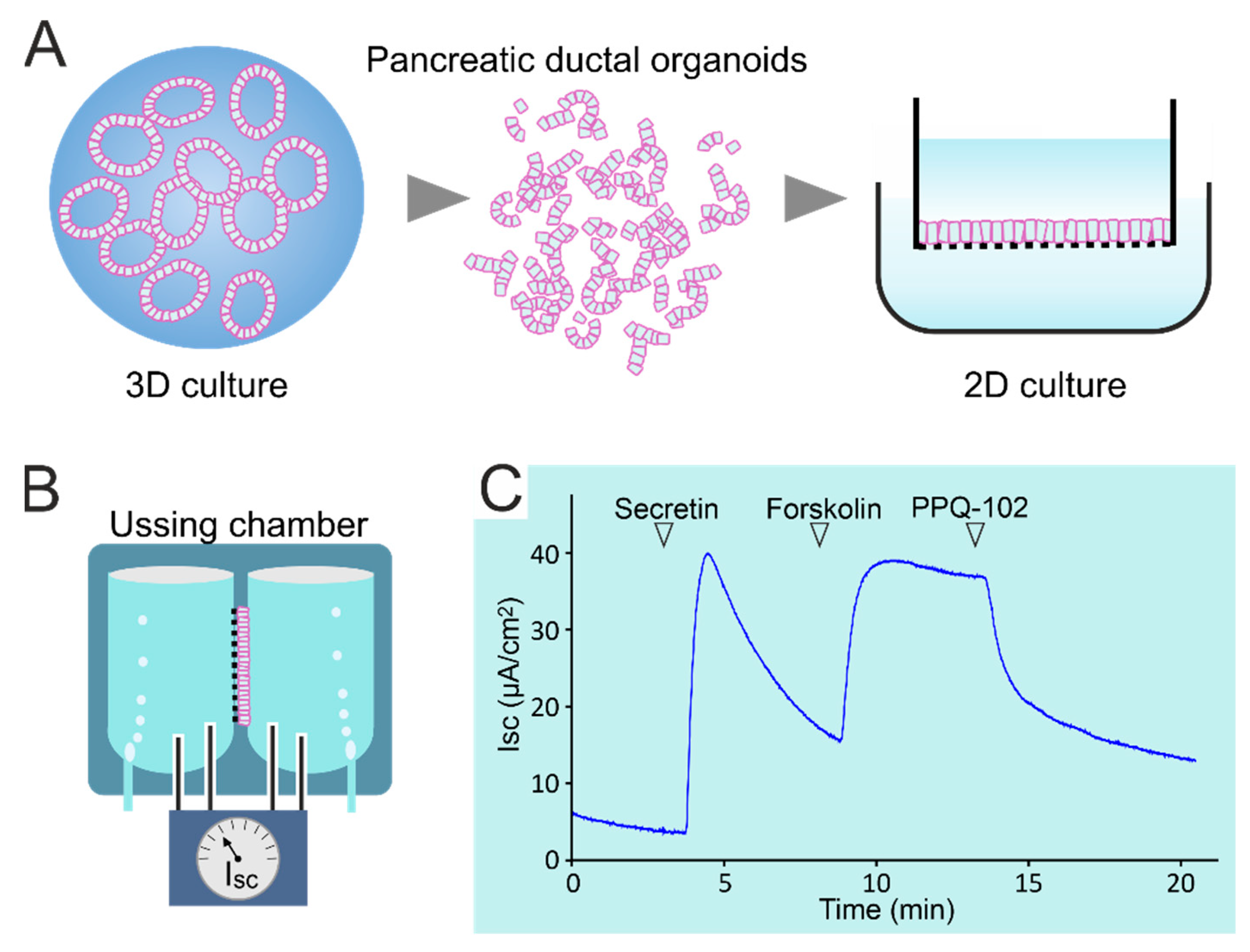
Organoids generated from stem cells in intestinal biopsies and cultured in an extracellular matrix (3D) or as monolayers (2D) on filters or on microfluidic chips have been used as novel tools to study epithelial (patho-)physiology and to predict clinical effects of CFTR modulators in individual patients (personalized medicine) [154,155,156]. Recently, culturing methods have been adapted to allow a similar long-term expansion of mouse and human pancreatic ductal organoids (PDOs) [157,158,159]. Pancreatic organoids can be generated from adult stem cells in pancreatic juice collected during endoscopic ultrasound [160], from microdissected animal- or patient-derived pancreatic ducts [159,161] or, possibly, even from microbiopsies collected during ERCP [162]. PDOs can be cryopreserved, clonally expanded and genetically manipulated and retain the characteristics of the tissue of origin, including gene expression and function of ion channels and transporters [157,161]. They can be grown 2D as polarized monolayers on filters or chips, offering highly suitable models to study Cl− and HCO3− transport pathways and their regulation [159,161] (Figure 3). However, it will be a challenge to reproduce the unique ability of PDCs to accumulate HCO3− up to a 140 mmol/L concentration in luminal baths.
Human PDOs may also facilitate drug development and drug screening for pancreatic disorders such as CF or genetic and acquired forms of AP or CP. One restriction is the poor availability of human biopsies or tissue explants from the pancreatic duct of CF, CFSPID or CFTRBD patients. In this case, creating a human model to study the effect of CFTR mutations and modulators on PDC properties and function would require gene editing of CFTR in non-CF PDOs. Additionally, it would be of great interest to study the ability of CFTR modulators to restore CFTR function in PDCs exposed to alcohol, tobacco smoke or bile salts. In particular, the therapeutic potential of VX-770 in a human PDO model deserves further investigation, considering the strong potentiating effect of this drug on wild-type CFTR channels [163].
6. Concluding Remarks
Whereas complete loss of CFTR function leads to the CF-typical fibrosis of the exocrine pancreas and pancreatic insufficiency, it has become clear in recent years that milder forms of CFTR dysfunction, whether congenital or acquired, are involved in the pathophysiology of pancreatitis. Congruently, recent studies suggest that CFTR modulators originally developed for CF therapy may also be of potential benefit in this context. However, not all patients suffering from pancreatitis carry CFTR mutations, and not all CFTR mutations may be amenable to correction. Therefore, it will be important to identify those CFTR variants that are potentially responsive to drug therapy. PDOs provide a promising model to achieve this objective, as they may be used to select modulators on a personalized basis. Finally, clinical testing of CFTR modulators is indicated to further clarify the role of CFTR dysfunction in the development of AP and CP, and to validate the therapeutic potential of CFTR modulators.
Author Contributions
Conceptualization, H.R.d.J., D.A. and M.J.C.B.; writing—original draft preparation, D.A.; writing—review and editing, H.R.d.J., M.J.C.B., M.J.B. and M.P.P.; visualization, M.J.C.B.; supervision, H.R.d.J.; funding acquisition, H.R.d.J. All authors have read and agreed to the published version of the manuscript.
Funding
This study was funded by the Cystic Fibrosis Foundation (CFF-USA; DEJONG19G0), the Dutch Cystic Fibrosis Foundation (NCFS; HIT-CF2) and the Fondazione Ricerca Fibrosi Cistica (FFC; #3/2016; #9/2020).
Institutional Review Board Statement
Not applicable.
Informed Consent Statement
Not applicable.
Data Availability Statement
Not applicable.
Acknowledgments
The authors wish to thank Wichor Bramer from the Erasmus MC Medical Library for developing and updating the search strategies.
Conflicts of Interest
The authors declare no conflict of interest. The funders had no role in the design of the study; in the collection, analyses or interpretation of data; in the writing of the manuscript, or in the decision to publish the results.
References
- Shteinberg, M.; Haq, I.J.; Polineni, D.; Davies, J.C. Cystic fibrosis. Lancet 2021, 397, 2195–2211. [Google Scholar] [CrossRef]
- Burgel, P.R.; Bellis, G.; Olesen, H.V.; Viviani, L.; Zolin, A.; Blasi, F.; Elborn, J.S. Future trends in cystic fibrosis demography in 34 European countries. Eur. Respir. J. 2015, 46, 133–141. [Google Scholar] [CrossRef] [Green Version]
- Andersen, D.H. Cystic fibrosis of the pancreas and its relation to celiac disease: A clinical and pathologic study. Am. J. Dis. Child. 1938, 56, 344–399. [Google Scholar] [CrossRef]
- Singh, V.K.; Schwarzenberg, S.J. Pancreatic insufficiency in cystic fibrosis. J. Cyst. Fibros. 2017, 16, S70–S78. [Google Scholar] [CrossRef] [Green Version]
- Mayerle, J.; Sendler, M.; Hegyi, E.; Beyer, G.; Lerch, M.M.; Sahin-Tóth, M. Genetics, cell biology, and pathophysiology of pancreatitis. Gastroenterology 2019, 156, 1951–1968.e1951. [Google Scholar] [CrossRef] [PubMed] [Green Version]
- Peery, A.F.; Crockett, S.D.; Murphy, C.C.; Lund, J.L.; Dellon, E.S.; Williams, J.L.; Jensen, E.T.; Shaheen, N.J.; Barritt, A.S.; Lieber, S.R.; et al. Burden and cost of gastrointestinal, liver, and pancreatic diseases in the United States: Update 2018. Gastroenterology 2019, 156, 254–272.e211. [Google Scholar] [CrossRef] [PubMed] [Green Version]
- Petrov, M.S.; Yadav, D. Global epidemiology and holistic prevention of pancreatitis. Nat. Rev. Gastroenterol. Hepatol. 2019, 16, 175–184. [Google Scholar] [CrossRef]
- Whitcomb, D. Framework for interpretation of genetic variations in pancreatitis patients. Front. Physiol. 2012, 3, 440. [Google Scholar] [CrossRef] [Green Version]
- Whitcomb, D.C.; North American Pancreatitis Study Group. Pancreatitis: TIGAR-O version 2 risk/etiology checklist with topic reviews, updates, and use primers. Clin. Transl. Gastroenterol. 2019, 10, e00027. [Google Scholar] [CrossRef]
- Balázs, A.; Hegyi, P. Cystic fibrosis-style changes in the early phase of pancreatitis. Clin. Res. Hepatol. Gastroenterol. 2015, 39, S12–S17. [Google Scholar] [CrossRef] [PubMed]
- Ooi, C.Y.; Dorfman, R.; Cipolli, M.; Gonska, T.; Castellani, C.; Keenan, K.; Freedman, S.D.; Zielenski, J.; Berthiaume, Y.; Corey, M.; et al. Type of CFTR mutation determines risk of pancreatitis in patients with cystic fibrosis. Gastroenterology 2011, 140, 153–161. [Google Scholar] [CrossRef]
- Miller, A.C.; Comellas, A.P.; Hornick, D.B.; Stoltz, D.A.; Cavanaugh, J.E.; Gerke, A.K.; Welsh, M.J.; Zabner, J.; Polgreen, P.M. Cystic fibrosis carriers are at increased risk for a wide range of cystic fibrosis-related conditions. Proc. Natl. Acad. Sci. USA 2020, 117, 1621–1627. [Google Scholar] [CrossRef] [Green Version]
- Lee, M.G.; Ohana, E.; Park, H.W.; Yang, D.; Muallem, S. Molecular mechanism of pancreatic and salivary gland fluid and HCO3-secretion. Physiol. Rev. 2012, 92, 39–74. [Google Scholar] [CrossRef] [Green Version]
- Quinton, P.M. The neglected ion: HCO3-. Nat. Med. 2001, 7, 292–293. [Google Scholar] [CrossRef]
- Hug, M.J.; Clarke, L.L.; Gray, M.A. How to measure CFTR-dependent bicarbonate transport: From single channels to the intact epithelium. Methods Mol. Biol. 2011, 741, 489–509. [Google Scholar]
- Schneider, A.; Larusch, J.; Sun, X.; Aloe, A.; Lamb, J.; Hawes, R.; Cotton, P.; Brand, R.E.; Anderson, M.A.; Money, M.E.; et al. Combined bicarbonate conductance-impairing variants in CFTR and SPINK1 variants are associated with chronic pancreatitis in patients without cystic fibrosis. Gastroenterology 2011, 140, 162–171. [Google Scholar] [CrossRef] [Green Version]
- Maléth, J.; Balázs, A.; Pallagi, P.; Balla, Z.; Kui, B.; Katona, M.; Judák, L.; Németh, I.; Kemény, L.V.; Rakonczay, Z.; et al. Alcohol disrupts levels and function of the cystic fibrosis transmembrane conductance regulator to promote development of pancreatitis. Gastroenterology 2015, 148, 427–439. [Google Scholar] [CrossRef] [PubMed] [Green Version]
- Ishiguro, H.; Yamamoto, A.; Nakakuki, M.; Yi, L.; Ishiguro, M.; Yamaguchi, M.; Kondo, S.; Mochimaru, Y. Physiology and pathophysiology of bicarbonate secretion by pancreatic duct epithelium. Nagoya J. Med. Sci. 2012, 74, 1–18. [Google Scholar] [PubMed]
- Petersen, O.H. Physiology of acinar cell secretion. In The Pancreas: An Integrated Textbook of Basic Science, Medicine and Surgery, 3rd ed.; John Wiley & Sons: Hoboken, NJ, USA, 2018; pp. 41–55. [Google Scholar]
- Gullo, L.; Priori, P.; Pezzilli, R.; Biliotti, G.; Mattioli, G.; Barbara, L. Pancreatic secretory response to ordinary meals: Studies with pure pancreatic juice. Gastroenterology 1988, 94, 428–433. [Google Scholar] [CrossRef]
- Bernacki, E.; Podkowicz, K.; Rublewska, M.; Klepacki, A. Studies on the exocrine function of healthy human pancreas: Pancreatic juice and its certain components. Acta Physiol. Pol. 1976, 27, 337–345. [Google Scholar]
- Lee, M.G.; Wigley, W.C.; Zeng, W.; Noel, L.E.; Marino, C.R.; Thomast, P.J.; Muallem, S. Regulation of Cl-/ HCO3- exchange by cystic fibrosis transmembrane conductance regulator expressed in NIH 3T3 and HEK 293 cells. J. Biol. Chem. 1999, 274, 3414–3421. [Google Scholar] [CrossRef] [Green Version]
- Domschke, S.; Domschke, W.; Rösch, W.; Konturek, S.J.; Wünsch, E.; Demling, L. Bicarbonate and cyclic AMP content of pure human pancreatic juice in response to graded doses of synthetic secretin. Gastroenterology 1976, 70, 533–536. [Google Scholar] [CrossRef]
- Sewell, W.A.; Young, J.A. Secretion of electrolytes by the pancreas of the anaestetized rat. J. Physiol. 1975, 252, 379–396. [Google Scholar] [CrossRef] [PubMed] [Green Version]
- Mangos, J.A.; McSherry, N.R.; Nousia-Arvanitakis, S.; Irwin, K. Secretion and transductal fluxes of ions in exocrine glands of the mouse. Am. J. Physiol. 1973, 225, 18–24. [Google Scholar] [CrossRef] [PubMed]
- Steward, M.C.; Ishiguro, H.; Case, R.M. Mechanisms of bicarbonate secretion in the pancreatic duct. Annu. Rev. Physiol. 2005, 67, 377–409. [Google Scholar] [CrossRef]
- Padfield, P.J.; Garner, A.; Case, R.M. Patterns of pancreatic secretion in the anaesthetised guinea pig following stimulation with secretin, cholecystokinin octapeptide, or bombesin. Pancreas 1989, 4, 204–209. [Google Scholar] [CrossRef]
- Stewart, A.K.; Shmukler, B.E.; Vandorpe, D.H.; Reimold, F.; Heneghan, J.F.; Nakakuki, M.; Akhavein, A.; Ko, S.; Ishiguro, H.; Alper, S.L. SLC26 anion exchangers of guinea pig pancreatic duct: Molecular cloning and functional characterization. Am. J. Physiol. Cell Physiol. 2011, 301, C289–C303. [Google Scholar] [CrossRef] [Green Version]
- Yamaguchi, M.; Steward, M.C.; Smallbone, K.; Sohma, Y.; Yamamoto, A.; Ko, S.B.H.; Kondo, T.; Ishiguro, H. Bicarbonate-rich fluid secretion predicted by a computational model of guinea-pig pancreatic duct epithelium. J. Physiol. 2017, 595, 1947–1972. [Google Scholar] [CrossRef] [PubMed]
- Quinton, P.M. Too much salt, too little soda: Cystic fibrosis. Sheng Li Xue Bao 2007, 59, 397–415. [Google Scholar]
- Fong, P. CFTR–SLC26 transporter interactions in epithelia. Biophys. Rev. 2012, 4, 107–116. [Google Scholar] [CrossRef]
- Lohi, H.; Lamprecht, G.; Markovich, D.; Heil, A.; Kujala, M.; Seidler, U.; Kere, J. Isoforms of SLC26A6 mediate anion transport and have functional PDZ interaction domains. Am. J. Physiol. Cell. Physiol. 2003, 284, C769–C779. [Google Scholar] [CrossRef] [Green Version]
- Novak, I.; Greger, R. Properties of the luminal membrane of isolated perfused rat pancreatic ducts. Pflugers Arch. 1988, 411, 546–553. [Google Scholar] [CrossRef] [PubMed]
- Wine, J.J. Cystic fibrosis: The ‘bicarbonate before chloride’ hypothesis. Curr. Biol. 2001, 11, R463–R466. [Google Scholar] [CrossRef] [Green Version]
- Namkung, W.; Lee, J.A.; Ahn, W.; Han, W.; Kwon, S.W.; Ahn, D.S.; Kim, K.H.; Lee, M.G. Ca2+ activates cystic fibrosis transmembrane conductance regulator- and Cl--dependent HCO3- transport in pancreatic duct cells. J. Biol. Chem. 2003, 278, 200–207. [Google Scholar] [CrossRef] [PubMed] [Green Version]
- Ko, S.B.H.; Zeng, W.; Dorwart, M.R.; Luo, X.; Kim, K.H.; Millen, L.; Goto, H.; Naruse, S.; Soyombo, A.; Thomas, P.J. Gating of CFTR by the STAS domain of SLC26 transporters. Nat. Cell Biol. 2004, 6, 343–350. [Google Scholar] [CrossRef] [PubMed]
- Berg, P.; Svendsen, S.L.; Sorensen, M.V.; Larsen, C.K.; Andersen, J.F.; Jensen-Fangel, S.; Jeppesen, M.; Schreiber, R.; Cabrita, I.; Kunzelmann, K.; et al. Impaired renal HCO3- excretion in cystic fibrosis. J. Am. Soc. Nephrol. 2020, 31, 1711–1727. [Google Scholar] [CrossRef]
- Sohma, Y.; Gray, M.A.; Imai, Y.; Argent, B.E. HCO3- transport in a mathematical model of the pancreatic ductal epithelium. J. Membr. Biol. 2000, 176, 77–100. [Google Scholar] [CrossRef]
- Illek, B.; Yankaskas, J.R.; Machen, T.E. cAMP and genistein stimulate HCO3-conductance through CFTR in human airway epithelia. Am. J. Physiol. 1997, 272, L752–L761. [Google Scholar] [CrossRef]
- Poulsen, J.H.; Fischer, H.; Illek, B.; Machen, T.E. Bicarbonate conductance and pH regulatory capability of cystic fibrosis transmembrane conductance regulator. Proc. Natl. Acad. Sci. USA 1994, 91, 5340–5344. [Google Scholar] [CrossRef] [Green Version]
- Linsdell, P.; Tabcharani, J.A.; Rommens, J.M.; Hou, Y.-X.; Chang, X.-B.; Tsui, L.-C.; Riordan, J.R.; Hanrahan, J.W. Permeability of wild-type and mutant cystic fibrosis transmembrane conductance regulator chloride channels to polyatomic anions. J. Gen. Physiol. 1997, 110, 355–364. [Google Scholar] [CrossRef] [Green Version]
- Ishiguro, H.; Steward, M.C.; Naruse, S.; Ko, S.B.; Goto, H.; Case, R.M.; Kondo, T.; Yamamoto, A. CFTR functions as a bicarbonate channel in pancreatic duct cells. J. Gen. Physiol. 2009, 133, 315–326. [Google Scholar] [CrossRef] [Green Version]
- Ferrera, L.; Baroni, D.; Moran, O. Lumacaftor-rescued F508del-CFTR has a modified bicarbonate permeability. J. Cyst. Fibros. 2019, 18, 602–605. [Google Scholar] [CrossRef]
- Tang, L.; Fatehi, M.; Linsdell, P. Mechanism of direct bicarbonate transport by the CFTR anion channel. J. Cyst. Fibros. 2009, 8, 115–121. [Google Scholar] [CrossRef] [Green Version]
- Reddy, M.M.; Quinton, P.M. Selective activation of cystic fibrosis transmembrane conductance regulator Cl- and HCO3- conductances. J. Pancreas 2001, 2, 212–218. [Google Scholar]
- Reddy, M.M.; Quinton, P.M. Control of dynamic CFTR selectivity by glutamate and ATP in epithelial cells. Nature 2003, 423, 756–760. [Google Scholar] [CrossRef]
- Shcheynikov, N.; Kim, K.H.; Kim, K.M.; Dorwart, M.R.; Ko, S.B.H.; Goto, H.; Naruse, S.; Thomas, P.J.; Muallem, S. Dynamic control of cystic fibrosis transmembrane conductance regulator Cl-/HCO3- selectivity by external Cl-. J Biol. Chem. 2004, 279, 21857–21865. [Google Scholar] [CrossRef] [PubMed] [Green Version]
- Wright, A.M.; Gong, X.; Verdon, B.; Linsdell, P.; Mehta, A.; Riordan, J.R.; Argent, B.E.; Gray, M.A. Novel regulation of cystic fibrosis transmembrane conductance regulator (CFTR) channel gating by external chloride. J. Biol. Chem. 2004, 279, 41658–41663. [Google Scholar] [CrossRef] [Green Version]
- Pallagi, P.; Hegyi, P.; Rakonczay, Z. The physiology and pathophysiology of pancreatic ductal secretion: The background for clinicians. Pancreas 2015, 44, 1211–1233. [Google Scholar] [CrossRef] [PubMed]
- Park, H.W.; Lee, M.G. Transepithelial bicarbonate secretion: Lessons from the pancreas. Cold Spring Harb. Perspect. Med. 2012, 2, a009571. [Google Scholar] [CrossRef] [Green Version]
- Delpire, E.; Gagnon, K.B.E. SPAK and OSR1: STE20 kinases involved in the regulation of ion homoeostasis and volume control in mammalian cells. Biochem. J. 2008, 409, 321–331. [Google Scholar] [CrossRef] [Green Version]
- Park, H.W.; Nam, J.H.; Kim, J.Y.; Namkung, W.; Yoon, J.S.; Lee, J.S.; Kim, K.S.; Venglovecz, V.; Gray, M.A.; Kim, K.H.; et al. Dynamic regulation of CFTR bicarbonate permeability by [Cl-]i and its role in pancreatic bicarbonate secretion. Gastroenterology 2010, 139, 620–631. [Google Scholar] [CrossRef]
- Kim, Y.; Jun, I.; Shin, D.H.; Yoon, J.G.; Piao, H.; Jung, J.; Park, H.W.; Cheng, M.H.; Bahar, I.; Whitcomb, D.C.; et al. Regulation of CFTR bicarbonate channel activity by WNK1: Implications for pancreatitis and CFTR-related disorders. Cell. Mol. Gastroenterol. Hepatol 2020, 9, 79–103. [Google Scholar] [CrossRef] [Green Version]
- Dorwart, M.R.; Shcheynikov, N.; Wang, Y.; Stippec, S.; Muallem, S. SLC26A9 is a Cl− channel regulated by the WNK kinases. J. Physiol. 2007, 584, 333–345. [Google Scholar] [CrossRef]
- Yang, C.-L.; Liu, X.; Paliege, A.; Zhu, X.; Bachmann, S.; Dawson, D.C.; Ellison, D.H. WNK1 and WNK4 modulate CFTR activity. Biochem. Biophys. Res. Commun. 2007, 353, 535–540. [Google Scholar] [CrossRef]
- Yang, D.; Li, Q.; So, I.; Huang, C.L.; Ando, H.; Mizutani, A.; Seki, G.; Mikoshiba, K.; Thomas, P.J.; Muallem, S. IRBIT governs epithelial secretion in mice by antagonizing the WNK/SPAK kinase pathway. J. Clin. Investig. 2011, 121, 956–965. [Google Scholar] [CrossRef] [PubMed] [Green Version]
- Hong, J.H.; Yang, D.; Shcheynikov, N.; Ohana, E.; Shin, D.M.; Muallem, S. Convergence of IRBIT, phosphatidylinositol (4,5) bisphosphate, and WNK/SPAK kinases in regulation of the Na+-HCO3- cotransporters family. Proc. Natl. Acad. Sci. USA 2013, 110, 4105–4110. [Google Scholar] [CrossRef] [Green Version]
- Shirakabe, K.; Priori, G.; Yamada, H.; Ando, H.; Horita, S.; Fujita, T.; Fujimoto, I.; Mizutani, A.; Seki, G.; Mikoshiba, K. IRBIT, an inositol 1,4,5-trisphosphate receptor-binding protein, specifically binds to and activates pancreas-type Na+/HCO3- cotransporter 1 (pNBC1). Proc. Natl. Acad. Sci. USA 2006, 103, 9542–9547. [Google Scholar] [CrossRef] [Green Version]
- Yang, D.; Shcheynikov, N.; Zeng, W.; Ohana, E.; So, I.; Ando, H.; Mizutani, A.; Mikoshiba, K.; Muallem, S. IRBIT coordinates epithelial fluid and HCO3- secretion by stimulating the transporters pNBC1 and CFTR in the murine pancreatic duct. J. Clin. Investig. 2009, 119, 193–202. [Google Scholar] [CrossRef] [Green Version]
- Hong, J.H.; Park, S.; Shcheynikov, N.; Muallem, S. Mechanism and synergism in epithelial fluid and electrolyte secretion. Pflugers Arch. 2014, 466, 1487–1499. [Google Scholar] [CrossRef] [PubMed] [Green Version]
- Park, S.; Shcheynikov, N.; Hong, J.H.; Zheng, C.; Suh, S.H.; Kawaai, K.; Ando, H.; Mizutani, A.; Abe, T.; Kiyonari, H.; et al. Irbit mediates synergy between ca(2+) and cAMP signaling pathways during epithelial transport in mice. Gastroenterology 2013, 145, 232–241. [Google Scholar] [CrossRef] [PubMed] [Green Version]
- Rosendahl, J.; Witt, H.; Szmola, R.; Bhatia, E.; Ozsvári, B.; Landt, O.; Schulz, H.U.; Gress, T.M.; Pfützer, R.; Löhr, M.; et al. Chymotrypsin C (CTRC) variants that diminish activity or secretion are associated with chronic pancreatitis. Nat. Genet. 2008, 40, 78–82. [Google Scholar] [CrossRef] [Green Version]
- Rosendahl, J.; Landt, O.; Bernadova, J.; Kovacs, P.; Teich, N.; Bödeker, H.; Keim, V.; Ruffert, C.; Mössner, J.; Kage, A.; et al. CFTR, SPINK1, CTRC and PRSS1 variants in chronic pancreatitis: Is the role of mutated CFTR overestimated? Gut 2013, 62, 582–592. [Google Scholar] [CrossRef] [PubMed] [Green Version]
- Rosendahl, J.; Kirsten, H.; Hegyi, E.; Kovacs, P.; Weiss, F.U.; Laumen, H.; Lichtner, P.; Ruffert, C.; Chen, J.M.; Masson, E.; et al. Genome-wide association study identifies inversion in the CTRB1-CTRB2 locus to modify risk for alcoholic and non-alcoholic chronic pancreatitis. Gut 2018, 67, 1855–1863. [Google Scholar] [CrossRef] [Green Version]
- Witt, H.; Beer, S.; Rosendahl, J.; Chen, J.M.; Chandak, G.R.; Masamune, A.; Bence, M.; Szmola, R.; Oracz, G.; Macek, M., Jr.; et al. Variants in CPA1 are strongly associated with early onset chronic pancreatitis. Nat. Genet. 2013, 45, 1216–1220. [Google Scholar] [CrossRef] [Green Version]
- Fjeld, K.; Weiss, F.U.; Lasher, D.; Rosendahl, J.; Chen, J.M.; Johansson, B.B.; Kirsten, H.; Ruffert, C.; Masson, E.; Steine, S.J.; et al. A recombined allele of the lipase gene CEL and its pseudogene CELP confers susceptibility to chronic pancreatitis. Nat. Genet. 2015, 47, 518–522. [Google Scholar] [CrossRef] [Green Version]
- Cavestro, G.M.; Zuppardo, R.A.; Bertolini, S.; Sereni, G.; Frulloni, L.; Okolicsanyi, S.; Calzolari, C.; Singh, S.K.; Sianesi, M.; Del Rio, P.; et al. Connections between genetics and clinical data: Role of MCP-1, CFTR, and SPINK-1 in the setting of acute, acute recurrent, and chronic pancreatitis. Am. J. Gastroenterol. 2010, 105, 199–206. [Google Scholar] [CrossRef]
- Sultan, M.; Werlin, S.; Venkatasubramani, N. Genetic prevalence and characteristics in children with recurrent pancreatitis. J. Pediatr. Gastroenterol. Nutr. 2012, 54, 645–650. [Google Scholar] [CrossRef]
- Werlin, S.; Konikoff, F.M.; Halpern, Z.; Barkay, O.; Yerushalmi, B.; Broide, E.; Santo, E.; Shamir, R.; Shaoul, R.; Shteyer, E.; et al. Genetic and electrophysiological characteristics of recurrent acute pancreatitis. J. Pediatr. Gastroenterol. Nutr. 2015, 60, 675–679. [Google Scholar] [CrossRef] [PubMed]
- Sharer, N.; Schwarz, M.; Malone, G.; Howarth, A.; Painter, J.; Super, M.; Braganza, J. Mutations of the cystic fibrosis gene in patients with chronic pancreatitis. N. Engl. J. Med. 1998, 339, 645–652. [Google Scholar] [CrossRef] [PubMed]
- Cohn, J.A.; Noone, P.G.; Jowell, P.S. Idiopathic pancreatitis related to CFTR: Complex inheritance and identification of a modifier gene. J. Investig. Med. 2002, 50, 247S–255S. [Google Scholar]
- Weiss, F.U.; Simon, P.; Bogdanova, N.; Mayerle, J.; Dworniczak, B.; Horst, J.; Lerch, M.M. Complete cystic fibrosis transmembrane conductance regulator gene sequencing in patients with idiopathic chronic pancreatitis and controls. Gut 2005, 54, 1456–1460. [Google Scholar] [CrossRef] [PubMed] [Green Version]
- Cohn, J.A.; Friedman, K.J.; Noone, P.G.; Knowles, M.R.; Silverman, L.M.; Jowell, P.S. Relation between mutations of the cystic fibrosis gene and idiopathic pancreatitis. N. Engl. J. Med. 1998, 339, 653–658. [Google Scholar] [CrossRef]
- Kumar, S.; Ooi, C.Y.; Werlin, S.; Abu-El-Haija, M.; Barth, B.; Bellin, M.D.; Durie, P.R.; Fishman, D.S.; Freedman, S.D.; Gariepy, C.; et al. Risk factors associated with pediatric acute recurrent and chronic pancreatitis: Lessons from INSPPIRE. JAMA Pediatr. 2016, 170, 562–569. [Google Scholar] [CrossRef] [PubMed]
- Beyer, G.; Habtezion, A.; Werner, J.; Lerch, M.M.; Mayerle, J. Chronic pancreatitis. Lancet 2020, 396, 499–512. [Google Scholar] [CrossRef]
- Weiss, F.U.; Skube, M.E.; Lerch, M.M. Chronic pancreatitis: An update on genetic risk factors. Curr. Opin. Gastroenterol. 2018, 34, 322–329. [Google Scholar] [CrossRef]
- Gastard, J.; Joubaud, F.; Farbos, T.; Loussouarn, J.; Marion, J.; Pannier, M.; Renaudet, F.; Valdazo, R.; Gosselin, M. Etiology and course of primary chronic pancreatitis in western France. Digestion 1973, 9, 416–428. [Google Scholar] [CrossRef]
- Yadav, D.; Lowenfels, A.B. The epidemiology of pancreatitis and pancreatic cancer. Gastroenterology 2013, 144, 1252–1261. [Google Scholar] [CrossRef] [Green Version]
- Coté, G.A.; Yadav, D.; Slivka, A.; Hawes, R.H.; Anderson, M.A.; Burton, F.R.; Brand, R.E.; Banks, P.A.; Lewis, M.D.; Disario, J.A. Alcohol and smoking as risk factors in an epidemiology study of patients with chronic pancreatitis. Clin. Gastroenterol. Hepatol. 2011, 9, 266–273. [Google Scholar] [CrossRef] [Green Version]
- Yadav, D.; Hawes, R.H.; Brand, R.E.; Anderson, M.A.; Money, M.E.; Banks, P.A.; Bishop, M.D.; Baillie, J.; Sherman, S.; DiSario, J.; et al. Alcohol consumption, cigarette smoking, and the risk of recurrent acute and chronic pancreatitis. Arch. Intern. Med. 2009, 169, 1035–1045. [Google Scholar] [CrossRef] [Green Version]
- Frulloni, L.; Gabbrielli, A.; Pezzilli, R.; Zerbi, A.; Cavestro, G.M.; Marotta, F.; Falconi, M.; Gaia, E.; Uomo, G.; Maringhini, A.; et al. Chronic pancreatitis: Report from a multicenter Italian survey (PanCroInfAISP) on 893 patients. Dig. Liver Dis. 2009, 41, 311–317. [Google Scholar] [CrossRef]
- Lankisch, P.G.; Assmus, C.; Maisonneuve, P.; Lowenfels, A.B. Epidemiology of pancreatic diseases in Lüneburg County: A study in a defined German population. Pancreatology 2002, 2, 469–477. [Google Scholar] [CrossRef] [PubMed]
- Munigala, S.; Conwell, D.L.; Gelrud, A.; Agarwal, B. Heavy smoking is associated with lower age at first episode of acute pancreatitis and a higher risk of recurrence. Pancreas 2015, 44, 876–881. [Google Scholar] [CrossRef]
- Ren, C.L.; Borowitz, D.S.; Gonska, T.; Howenstine, M.S.; Levy, H.; Massie, J.; Milla, C.; Munck, A.; Southern, K.W. Cystic fibrosis transmembrane conductance regulator-related metabolic syndrome and cystic fibrosis screen positive, inconclusive diagnosis. J. Pediatr. 2017, 181, S45–S51.e41. [Google Scholar] [CrossRef] [PubMed] [Green Version]
- Munck, A.; Mayell, S.J.; Winters, V.; Shawcross, A.; Derichs, N.; Parad, R.; Barben, J.; Southern, K.W. Cystic fibrosis screen positive, inconclusive diagnosis (CFSPID): A new designation and management recommendations for infants with an inconclusive diagnosis following newborn screening. J. Cyst. Fibros. 2015, 14, 706–713. [Google Scholar] [CrossRef] [Green Version]
- Johansen, P.; Anderson, C.; Hadorn, B. Cystic fibrosis of the pancreas: A generalised disturbance of water and electrolyte movement in exocrine tissues. Lancet 1968, 291, 455–460. [Google Scholar] [CrossRef]
- Kopelman, H.; Forstner, G.; Durie, P.; Corey, M. Origins of chloride and bicarbonate secretory defects in the cystic fibrosis pancreas, as suggested by pancreatic function studies on control and CF subjects with preserved pancreatic function. Clin. Investig. Med. 1989, 12, 207–211. [Google Scholar]
- Kopelman, H.; Corey, M.; Gaskin, K.; Durie, P.; Weizman, Z.; Forstner, G. Impaired chloride secretion, as well as bicarbonate secretion, underlies the fluid secretory defect in the cystic fibrosis pancreas. Gastroenterology 1988, 95, 349–355. [Google Scholar] [CrossRef]
- Ahmed, N.; Corey, M.; Forstner, G.; Zielenski, J.; Tsui, L.C.; Ellis, L.; Tullis, E.; Durie, P. Molecular consequences of cystic fibrosis transmembrane regulator (CFTR) gene mutations in the exocrine pancreas. Gut 2003, 52, 1159–1164. [Google Scholar] [CrossRef] [PubMed] [Green Version]
- Taylor, C.J.; Aswani, N. The pancreas in cystic fibrosis. Paediatr. Respir. Rev. 2002, 3, 77–81. [Google Scholar] [CrossRef] [PubMed]
- Choi, J.Y.; Muallem, D.; Kiselyov, K.; Lee, M.G.; Thomas, P.J.; Muallem, S. Aberrant CFTR-dependent HCO3- transport in mutations associated with cystic fibrosis. Nature 2001, 410, 94–97. [Google Scholar] [CrossRef] [Green Version]
- Choi, J.Y.; Lee, M.G.; Ko, S.; Muallem, S. Cl-dependent HCO3- transport by cystic fibrosis transmembrane conductance regulator. J. Pancreas 2001, 2, 243–246. [Google Scholar]
- Shwachman, H.; Lebenthal, E.; Khaw, K.T. Recurrent acute pancreatitis in patients with cystic fibrosis with normal pancreatic enzymes. Pediatrics 1975, 55, 86–95. [Google Scholar] [CrossRef] [PubMed]
- Morrison, C.B.; Markovetz, M.R.; Ehre, C. Mucus, mucins, and cystic fibrosis. Pediatr. Pulmonol. 2019, 54, S84–S96. [Google Scholar] [CrossRef] [Green Version]
- Hansson, G.C. Mucus and mucins in diseases of the intestinal and respiratory tracts. J. Intern. Med. 2019, 285, 479–490. [Google Scholar] [CrossRef] [PubMed] [Green Version]
- Garcia, M.A.S.; Yang, N.; Quinton, P.M. Normal mouse intestinal mucus release requires cystic fibrosis transmembrane regulator–dependent bicarbonate secretion. J. Clin. Investig. 2009, 119, 3497. [Google Scholar] [CrossRef] [Green Version]
- Gustafsson, J.K.; Ermund, A.; Ambort, D.; Johansson, M.E.V.; Nilsson, H.E.; Thorell, K.; Hebert, H.; Sjövall, H.; Hansson, G.C. Bicarbonate and functional CFTR channel are required for proper mucin secretion and link cystic fibrosis with its mucus phenotype. J. Exp. Med. 2012, 209, 1263–1272. [Google Scholar] [CrossRef] [Green Version]
- Chen, J.-H.; Stoltz, D.A.; Karp, P.H.; Ernst, S.E.; Pezzulo, A.A.; Moninger, T.O.; Rector, M.V.; Reznikov, L.R.; Launspach, J.L.; Chaloner, K. Loss of anion transport without increased sodium absorption characterizes newborn porcine cystic fibrosis airway epithelia. Cell 2010, 143, 911–923. [Google Scholar] [CrossRef] [Green Version]
- Kim, D.; Liao, J.; Scales, N.B.; Martini, C.; Luan, X.; Abu-Arish, A.; Robert, R.; Luo, Y.; McKay, G.A.; Nguyen, D.; et al. Large pH oscillations promote host defense against human airways infection. J. Exp. Med. 2021, 218. [Google Scholar] [CrossRef] [PubMed]
- Simonin, J.; Bille, E.; Crambert, G.; Noel, S.; Dreano, E.; Edwards, A.; Hatton, A.; Pranke, I.; Villeret, B.; Cottart, C.-H. Airway surface liquid acidification initiates host defense abnormalities in cystic fibrosis. Sci. Rep. 2019, 9, 1–11. [Google Scholar]
- Tucker, J.A.; Spock, A.; Spicer, S.S.; Shelburne, J.D.; Bradford, W. Inspissation of pancreatic zymogen material in cystic fibrosis. Ultrastruct. Pathol. 2003, 27, 323–335. [Google Scholar] [CrossRef]
- Gibson-Corley, K.N.; Meyerholz, D.K.; Engelhardt, J.F. Pancreatic pathophysiology in cystic fibrosis. J. Pathol. 2016, 238, 311–320. [Google Scholar] [CrossRef] [Green Version]
- Bombieri, C.; Claustres, M.; De Boeck, K.; Derichs, N.; Dodge, J.; Girodon, E.; Sermet, I.; Schwarz, M.; Tzetis, M.; Wilschanski, M.; et al. Recommendations for the classification of diseases as CFTR-related disorders. J. Cyst. Fibros. 2011, 10, S86–S102. [Google Scholar] [CrossRef] [Green Version]
- Steiner, B.; Rosendahl, J.; Witt, H.; Teich, N.; Keim, V.; Schulz, H.-U.; Pfützer, R.; Lühr, M.; Gress, T.M.; Nickel, R.; et al. Common CFTR haplotypes and susceptibility to chronic pancreatitis and congenital bilateral absence of the vas deferens. Hum. Mutat. 2011, 32, 912–920. [Google Scholar] [CrossRef]
- LaRusch, J.; Jung, J.; General, I.J.; Lewis, M.D.; Park, H.W.; Brand, R.E.; Gelrud, A.; Anderson, M.A.; Banks, P.A.; Conwell, D.; et al. Mechanisms of CFTR functional variants that impair regulated bicarbonate permeation and increase risk for pancreatitis but not for cystic fibrosis. PLoS Genet. 2014, 10, e1004376. [Google Scholar] [CrossRef] [Green Version]
- Xu, W.M.; Shi, Q.X.; Chen, W.Y.; Zhou, C.X.; Ni, Y.; Rowlands, D.K.; Liu, G.Y.; Zhu, H.; Ma, Z.G.; Wang, X.F. Cystic fibrosis transmembrane conductance regulator is vital to sperm fertilizing capacity and male fertility. Proc. Natl. Acad. Sci. USA 2007, 104, 9816–9821. [Google Scholar] [CrossRef] [PubMed] [Green Version]
- Martinez, B.; Heller, M.; Gaitch, N.; Hubert, D.; Burgel, P.R.; Levy, P.; Girodon, E.; Bienvenu, T. p.Arg75Gln, a CFTR variant involved in the risk of CFTR-related disorders? J. Hum. Genet. 2014, 59, 206–210. [Google Scholar] [CrossRef]
- Naruse, S.; Kitagawa, M.; Ishiguro, H. Molecular understanding of chronic pancreatitis: A perspective on the future. Mol. Med. Today 1999, 5, 493–499. [Google Scholar] [CrossRef]
- Yamamoto, A.; Ishiguro, H.; Ko, S.B.; Suzuki, A.; Wang, Y.; Hamada, H.; Mizuno, N.; Kitagawa, M.; Hayakawa, T.; Naruse, S. Ethanol induces fluid hypersecretion from guinea-pig pancreatic duct cells. J. Physiol. 2003, 551, 917–926. [Google Scholar] [CrossRef] [PubMed]
- Judák, L.; Hegyi, P.; Rakonczay, Z., Jr.; Maléth, J.; Gray, M.A.; Venglovecz, V. Ethanol and its non-oxidative metabolites profoundly inhibit CFTR function in pancreatic epithelial cells which is prevented by ATP supplementation. Pflugers Arch. 2014, 466, 549–562. [Google Scholar] [CrossRef] [PubMed]
- Pallagi, P.; Madácsy, T.; Varga, Á.; Maléth, J. Intracellular Ca2+ signalling in the pathogenesis of acute pancreatitis: Recent advances and translational perspectives. Int. J. Mol. Sci. 2020, 21, 4005. [Google Scholar] [CrossRef]
- Huang, W.; Booth, D.M.; Cane, M.C.; Chvanov, M.; Javed, M.A.; Elliott, V.L.; Armstrong, J.A.; Dingsdale, H.; Cash, N.; Li, Y.; et al. Fatty acid ethyl ester synthase inhibition ameliorates ethanol-induced Ca2+-dependent mitochondrial dysfunction and acute pancreatitis. Gut 2014, 63, 1313–1324. [Google Scholar] [CrossRef] [Green Version]
- Mederos, M.A.; Reber, H.A.; Girgis, M.D. Acute pancreatitis: A review. JAMA 2021, 325, 382–390. [Google Scholar] [CrossRef] [PubMed]
- Muili, K.A.; Wang, D.; Orabi, A.I.; Sarwar, S.; Luo, Y.; Javed, T.A.; Eisses, J.F.; Mahmood, S.M.; Jin, S.; Singh, V.P. Bile acids induce pancreatic acinar cell injury and pancreatitis by activating calcineurin. J. Biol. Chem. 2013, 288, 570–580. [Google Scholar] [CrossRef] [Green Version]
- Venglovecz, V.; Rakonczay, Z., Jr.; Ozsvári, B.; Takács, T.; Lonovics, J.; Varró, A.; Gray, M.A.; Argent, B.E.; Hegyi, P. Effects of bile acids on pancreatic ductal bicarbonate secretion in guinea pig. Gut 2008, 57, 1102–1112. [Google Scholar] [CrossRef] [PubMed]
- Ignáth, I.; Hegyi, P.; Venglovecz, V.; Székely, C.A.; Carr, G.; Hasegawa, M.; Inoue, M.; Takács, T.; Argent, B.E.; Gray, M.A.; et al. CFTR expression but not Cl- transport is involved in the stimulatory effect of bile acids on apical Cl-/HCO3- exchange activity in human pancreatic duct cells. Pancreas 2009, 38, 921–929. [Google Scholar] [CrossRef]
- Maléth, J.; Venglovecz, V.; Rázga, Z.; Tiszlavicz, L.; Rakonczay, Z., Jr.; Hegyi, P. Non-conjugated chenodeoxycholate induces severe mitochondrial damage and inhibits bicarbonate transport in pancreatic duct cells. Gut 2011, 60, 136–138. [Google Scholar] [CrossRef] [Green Version]
- Raju, S.V.; Jackson, P.L.; Courville, C.A.; McNicholas, C.M.; Sloane, P.A.; Sabbatini, G.; Tidwell, S.; Tang, L.P.; Liu, B.; Fortenberry, J.A.; et al. Cigarette smoke induces systemic defects in cystic fibrosis transmembrane conductance regulator function. Am. J. Respir. Crit. Care Med. 2013, 188, 1321–1330. [Google Scholar] [CrossRef] [PubMed]
- Hegyi, P.; Wilschanski, M.; Muallem, S.; Lukacs, G.L.; Sahin-Toth, M.; Uc, A.; Gray, M.A.; Rakonczay, Z., Jr.; Maleth, J. CFTR: A new horizon in the pathomechanism and treatment of pancreatitis. Rev. Physiol. Biochem. Pharmacol. 2016, 170, 37–66. [Google Scholar]
- Alexander, N.S.; Blount, A.; Zhang, S.; Skinner, D.; Hicks, S.B.; Chestnut, M.; Kebbel, F.A.; Sorscher, E.J.; Woodworth, B.A. Cystic fibrosis transmembrane conductance regulator modulation by the tobacco smoke toxin acrolein. Laryngoscope 2012, 122, 1193–1197. [Google Scholar] [CrossRef] [Green Version]
- Barreto, S.G. How does cigarette smoking cause acute pancreatitis? Pancreatology 2016, 16, 157–163. [Google Scholar] [CrossRef]
- Schnúr, A.; Premchandar, A.; Bagdany, M.; Lukacs, G.L. Phosphorylation-dependent modulation of CFTR macromolecular signalling complex activity by cigarette smoke condensate in airway epithelia. Sci. Rep. 2019, 9, 12706. [Google Scholar] [CrossRef] [PubMed] [Green Version]
- Rasmussen, J.E.; Sheridan, J.T.; Polk, W.; Davies, C.M.; Tarran, R. Cigarette smoke-induced Ca2+ release leads to cystic fibrosis transmembrane conductance regulator (CFTR) dysfunction. J. Biol. Chem. 2014, 289, 7671–7681. [Google Scholar] [CrossRef] [Green Version]
- Marklew, A.J.; Patel, W.; Moore, P.J.; Tan, C.D.; Smith, A.J.; Sassano, M.F.; Gray, M.A.; Tarran, R. Cigarette smoke exposure induces retrograde trafficking of CFTR to the endoplasmic reticulum. Sci. Rep. 2019, 9, 13655. [Google Scholar] [CrossRef] [PubMed] [Green Version]
- Clunes, L.A.; Davies, C.M.; Coakley, R.D.; Aleksandrov, A.A.; Henderson, A.G.; Zeman, K.L.; Worthington, E.N.; Gentzsch, M.; Kreda, S.M.; Cholon, D.; et al. Cigarette smoke exposure induces CFTR internalization and insolubility, leading to airway surface liquid dehydration. FASEB J. 2012, 26, 533–545. [Google Scholar] [CrossRef] [Green Version]
- Raju, S.V.; Lin, V.Y.; Liu, L.; McNicholas, C.M.; Karki, S.; Sloane, P.A.; Tang, L.; Jackson, P.L.; Wang, W.; Wilson, L. The cystic fibrosis transmembrane conductance regulator potentiator ivacaftor augments mucociliary clearance abrogating cystic fibrosis transmembrane conductance regulator inhibition by cigarette smoke. Am. J. Respir. Cell Mol. Biol. 2017, 56, 99–108. [Google Scholar] [CrossRef]
- Kadiyala, V.; Lee, L.S.; Banks, P.A.; Suleiman, S.; Paulo, J.A.; Wang, W.; Rosenblum, J.; Sainani, N.I.; Mortele, K.; Conwell, D.L. Cigarette smoking impairs pancreatic duct cell bicarbonate secretion. J. Pancreas 2013, 14, 31. [Google Scholar]
- Pallagi, P.; Tálas, D.; Madácsy, T.; Venglovecz, V.; Tóth, E.; Darvasi, E.; Balla, Z.; Tóth, K.; Schnúr, A.; Maléth, J.; et al. Investigation of the pathomechanism of smoke-induced pancreatic damage. Pancreatology 2019, 19, S12. [Google Scholar] [CrossRef]
- Ballengee, C.R.; Brooks, P.; Leong, T.; Geem, D.; Freeman, A.J. Effects of second-hand smoke on pancreatitis in children. Pancreas 2019, 48, 706–710. [Google Scholar] [CrossRef]
- Trapp, S.; Aghdassi, A.A.; Glaubitz, J.; Sendler, M.; Weiss, F.U.; Kühn, J.P.; Kromrey, M.L.; Mahajan, U.M.; Pallagi, P.; Rakonczay, Z.; et al. Pancreatitis severity in mice with impaired CFTR function but pancreatic sufficiency is mediated via ductal and inflammatory cells-not acinar cells. J. Cell. Mol. Med. 2021, 25, 4658–4670. [Google Scholar] [CrossRef]
- Keating, D.; Marigowda, G.; Burr, L.; Daines, C.; Mall, M.A.; McKone, E.F.; Ramsey, B.W.; Rowe, S.M.; Sass, L.A.; Tullis, E.; et al. VX-445-tezacaftor-ivacaftor in patients with cystic fibrosis and one or two Phe508del alleles. N. Engl. J. Med. 2018, 379, 1612–1620. [Google Scholar] [CrossRef]
- Capurro, V.; Tomati, V.; Sondo, E.; Renda, M.; Borrelli, A.; Pastorino, C.; Guidone, D.; Venturini, A.; Giraudo, A.; Mandrup Bertozzi, S.; et al. Partial rescue of F508del-CFTR stability and trafficking defects by double corrector treatment. Int. J. Mol. Sci. 2021, 22, 5262. [Google Scholar] [CrossRef]
- Veit, G.; Roldan, A.; Hancock, M.A.; Da Fonte, D.F.; Xu, H.; Hussein, M.; Frenkiel, S.; Matouk, E.; Velkov, T.; Lukacs, G.L. Allosteric folding correction of F508del and rare CFTR mutants by elexacaftor-tezacaftor-ivacaftor (trikafta) combination. JCI Insight 2020, 5, e139983. [Google Scholar] [CrossRef]
- Giuliano, K.A.; Wachi, S.; Drew, L.; Dukovski, D.; Green, O.; Bastos, C.; Cullen, M.D.; Hauck, S.; Tait, B.D.; Munoz, B.; et al. Use of a high-throughput phenotypic screening strategy to identify amplifiers, a novel pharmacological class of small molecules that exhibit functional synergy with potentiators and correctors. SLAS Discov. 2018, 23, 111–121. [Google Scholar] [CrossRef] [PubMed] [Green Version]
- Prokhorova, I.; Altman, R.B.; Djumagulov, M.; Shrestha, J.P.; Urzhumtsev, A.; Ferguson, A.; Chang, C.-W.T.; Yusupov, M.; Blanchard, S.C.; Yusupova, G. Aminoglycoside interactions and impacts on the eukaryotic ribosome. Proc. Natl. Acad. Sci. USA 2017, 114, E10899–E10908. [Google Scholar] [CrossRef] [PubMed] [Green Version]
- Kerem, E. ELX-02: An investigational read-through agent for the treatment of nonsense mutation-related genetic disease. Expert Opin. Investig. Drugs 2020, 29, 1347–1354. [Google Scholar] [CrossRef]
- de Poel, E.; Spelier, S.; Suen, S.W.F.; Kruisselbrink, E.; Graeber, S.Y.; Mall, M.A.; Weersink, E.J.M.; van der Eerden, M.M.; Koppelman, G.H.; van der Ent, C.K.; et al. Functional restoration of CFTR nonsense mutations in intestinal organoids. J. Cyst. Fibros. 2021, in press. [Google Scholar] [CrossRef] [PubMed]
- Sharma, J.; Du, M.; Wong, E.; Mutyam, V.; Li, Y.; Chen, J.; Wangen, J.; Thrasher, K.; Fu, L.; Peng, N.; et al. A small molecule that induces translational readthrough of CFTR nonsense mutations by eRF1 depletion. Nat. Commun. 2021, 12, 4358. [Google Scholar] [CrossRef]
- Bardin, E.; Pastor, A.; Semeraro, M.; Golec, A.; Hayes, K.; Chevalier, B.; Berhal, F.; Prestat, G.; Hinzpeter, A.; Gravier-Pelletier, C.; et al. Modulators of CFTR. Updates on clinical development and future directions. Eur. J. Med. Chem. 2021, 213, 113195. [Google Scholar] [CrossRef] [PubMed]
- Fűr, G.; Bálint, E.R.; Orján, E.M.; Balla, Z.; Kormányos, E.S.; Czira, B.; Szűcs, A.; Kovács, D.P.; Pallagi, P.; Maléth, J.; et al. Mislocalization of CFTR expression in acute pancreatitis and the beneficial effect of VX-661 + VX-770 treatment on disease severity. J. Physiol. 2021, 599, 4955–4971. [Google Scholar] [CrossRef]
- Zeng, M.; Szymczak, M.; Ahuja, M.; Zheng, C.; Yin, H.; Swaim, W.; Chiorini, J.A.; Bridges, R.J.; Muallem, S. Restoration of CFTR activity in ducts rescues acinar cell function and reduces inflammation in pancreatic and salivary glands of mice. Gastroenterology 2017, 153, 1148–1159. [Google Scholar] [CrossRef]
- Bose, S.J.; Bijvelds, M.J.C.; Wang, Y.; Liu, J.; Cai, Z.; Bot, A.G.M.; de Jonge, H.R.; Sheppard, D.N. Differential thermostability and response to cystic fibrosis transmembrane conductance regulator potentiators of human and mouse F508del-CFTR. Am. J. Physiol. Lung Cell. Mol. Physiol. 2019, 317, L71–L86. [Google Scholar] [CrossRef]
- Sellers, Z.M. Pancreatic complications in children with cystic fibrosis. Curr. Opin. Pediatr. 2020, 32, 661–667. [Google Scholar] [CrossRef]
- Gariepy, C.E.; Ooi, C.Y.; Maqbool, A.; Ellery, K.M. Demographics and risk factors for pediatric recurrent acute pancreatitis. Curr. Opin. Gastroenterol. 2021, 37. [Google Scholar] [CrossRef]
- Hoppe, J.E.; Chilvers, M.; Ratjen, F.; McNamara, J.J.; Owen, C.A.; Tian, S.; Zahigian, R.; Cornell, A.G.; McColley, S.A. Long-term safety of lumacaftor–ivacaftor in children aged 2–5 years with cystic fibrosis homozygous for the F508del-CFTR mutation: A multicentre, phase 3, open-label, extension study. Lancet Respir. Med. 2021, 9, 977–988. [Google Scholar] [CrossRef]
- Akshintala, V.S.; Kamal, A.; Faghih, M.; Cutting, G.R.; Cebotaru, L.; West, N.E.; Jennings, M.T.; Dezube, R.; Whitcomb, D.C.; Lechtzin, N.; et al. Cystic fibrosis transmembrane conductance regulator modulators reduce the risk of recurrent acute pancreatitis among adult patients with pancreas sufficient cystic fibrosis. Pancreatology 2019, 19, 1023–1026. [Google Scholar] [CrossRef] [PubMed]
- Carrion, A.; Borowitz, D.S.; Freedman, S.D.; Siracusa, C.M.; Goralski, J.L.; Hadjiliadis, D.; Srinivasan, S.; Stokes, D.C. Reduction of recurrence risk of pancreatitis in cystic fibrosis with ivacaftor: Case series. J. Pediatr. Gastroenterol. Nutr. 2018, 66, 451–454. [Google Scholar] [CrossRef]
- Ramsey, M.L.; Gokun, Y.; Sobotka, L.A.; Wellner, M.R.; Porter, K.; Kirkby, S.E.; Li, S.S.; Papachristou, G.I.; Krishna, S.G.; Stanich, P.P.; et al. Cystic fibrosis transmembrane conductance regulator modulator use is associated with reduced pancreatitis hospitalizations in patients with cystic fibrosis. Am. J. Gastroenterol. 2021, in press. [Google Scholar] [CrossRef] [PubMed]
- Johns, J.D.; Rowe, S.M. The effect of CFTR modulators on a cystic fibrosis patient presenting with recurrent pancreatitis in the absence of respiratory symptoms: A case report. BMC Gastroenterol. 2019, 19, 1–4. [Google Scholar] [CrossRef]
- Gelfond, D.; Heltshe, S.; Ma, C.; Rowe, S.M.; Frederick, C.; Uluer, A.; Sicilian, L.; Konstan, M.; Tullis, E.; Roach, R.N.C. Impact of CFTR modulation on intestinal pH, motility, and clinical outcomes in patients with cystic fibrosis and the G551D mutation. Clin. Transl. Gastroenterol. 2017, 8, e81. [Google Scholar] [CrossRef]
- Rehman, T.; Karp, P.H.; Tan, P.; Goodell, B.J.; Pezzulo, A.A.; Thurman, A.L.; Thornell, I.M.; Durfey, S.L.; Duffey, M.E.; Stoltz, D.A.; et al. Inflammatory cytokines TNF-α and IL-17 enhance the efficacy of cystic fibrosis transmembrane conductance regulator modulators. J. Clin. Investig. 2021, 131, e150398. [Google Scholar] [CrossRef]
- Fiore, M.; Picco, C.; Moran, O. Correctors modify the bicarbonate permeability of F508del-CFTR. Sci. Rep. 2020, 10, 8440. [Google Scholar] [CrossRef]
- Laselva, O.; Moraes, T.J.; He, G.; Bartlett, C.; Szàrics, I.; Ouyang, H.; Gunawardena, T.N.A.; Strug, L.; Bear, C.E.; Gonska, T. The CFTR mutation c.3453G>C (D1152H) confers an anion selectivity defect in primary airway tissue that can be rescued by ivacaftor. J. Pers. Med. 2020, 10, 40. [Google Scholar] [CrossRef]
- Dekkers, J.F.; Berkers, G.; Kruisselbrink, E.; Vonk, A.; de Jonge, H.R.; Janssens, H.M.; Bronsveld, I.; van de Graaf, E.A.; Nieuwenhuis, E.E.; Houwen, R.H.; et al. Characterizing responses to CFTR-modulating drugs using rectal organoids derived from subjects with cystic fibrosis. Sci. Transl. Med. 2016, 8, 344ra384. [Google Scholar] [CrossRef]
- de Winter-de Groot, K.M.; Janssens, H.M.; van Uum, R.T.; Dekkers, J.F.; Berkers, G.; Vonk, A.; Kruisselbrink, E.; Oppelaar, H.; Vries, R.; Clevers, H.; et al. Stratifying infants with cystic fibrosis for disease severity using intestinal organoid swelling as a biomarker of CFTR function. Eur. Respir. J. 2018, 52, 1702529. [Google Scholar] [CrossRef]
- Berkers, G.; van Mourik, P.; Vonk, A.M.; Kruisselbrink, E.; Dekkers, J.F.; de Winter-de Groot, K.M.; Arets, H.G.M.; Marck-van der Wilt, R.E.P.; Dijkema, J.S.; Vanderschuren, M.M.; et al. Rectal organoids enable personalized treatment of cystic fibrosis. Cell Rep. 2019, 26, 1701–1708 e1703. [Google Scholar] [CrossRef] [Green Version]
- Georgakopoulos, N.; Prior, N.; Angres, B.; Mastrogiovanni, G.; Cagan, A.; Harrison, D.; Hindley, C.J.; Arnes-Benito, R.; Liau, S.-S.; Curd, A. Long-term expansion, genomic stability and in vivo safety of adult human pancreas organoids. BMC Dev. Biol. 2020, 20, 1–20. [Google Scholar] [CrossRef] [Green Version]
- Driehuis, E.; Gracanin, A.; Vries, R.G.J.; Clevers, H.; Boj, S.F. Establishment of pancreatic organoids from normal tissue and tumors. STAR Protoc. 2020, 1, 100192. [Google Scholar] [CrossRef] [PubMed]
- Shik Mun, K.; Arora, K.; Huang, Y.; Yang, F.; Yarlagadda, S.; Ramananda, Y.; Abu-El-Haija, M.; Palermo, J.J.; Appakalai, B.N.; Nathan, J.D.; et al. Patient-derived pancreas-on-a-chip to model cystic fibrosis-related disorders. Nat. Commun. 2019, 10, 3124. [Google Scholar] [CrossRef] [Green Version]
- Levink, I.J.M.; Nesteruk, K.; Visser, D.I.; Sieuwerts, A.M.; Fernandes, C.J.C.; Jansen, M.; van Driel, L.; Poley, J.W.; Peppelenbosch, M.P.; Cahen, D.L.; et al. Optimization of pancreatic juice collection: A first step toward biomarker discovery and early detection of pancreatic cancer. Am. J. Gastroenterol. 2020, 115, 2103–2108. [Google Scholar] [CrossRef] [PubMed]
- Molnár, R.; Madácsy, T.; Varga, Á.; Németh, M.; Katona, X.; Görög, M.; Molnár, B.; Fanczal, J.; Rakonczay, Z.; Hegyi, P.; et al. Mouse pancreatic ductal organoid culture as a relevant model to study exocrine pancreatic ion secretion. Lab. Investig. 2020, 100, 84–97. [Google Scholar] [CrossRef] [PubMed] [Green Version]
- Silverman, W.B.; Putnam, P.E.; Orenstein, S.R.; del Rosario, J.F.; Wilson, J.; Kocoshis, S.A. Influence of ERCP on the diagnosis and treatment of pediatric patients with symptomatic pancreatitis and cystic fibrosis: Series of case reports. Gastrointest. Endosc. 1998, 48, 534–536. [Google Scholar] [CrossRef]
- Langron, E.; Prins, S.; Vergani, P. Potentiation of the cystic fibrosis transmembrane conductance regulator by VX-770 involves stabilization of the pre-hydrolytic, O1 state. Br. J. Pharmacol. 2018, 175, 3990–4002. [Google Scholar] [CrossRef] [PubMed]
Figure 1.
Model of HCO3− secretion in pancreatic ducts. HCO3− secretion is controlled by neuro-endocrine input, chiefly by secretin (S) and acetylcholine (ACh), which increase cellular cAMP and Ca2+ levels, respectively, via stimulation of G protein-coupled receptors. This triggers Na+-coupled HCO3− uptake across the basolateral plasma membrane through NBCe1 and luminal HCO3− efflux through CFTR and SLC26A6. HCO3− may also be accumulated intracellularly via the coordinated activity of carbonic anhydrases (CA) and a proton extrusion mechanism, NHE1. Opening of K+ channels sustains the negative membrane potential which drives (CFTR-mediated) anion efflux. Cl− ions absorbed via SLC26A6 may be recycled to the ductal lumen via CFTR. Ultimately, ductal ion and fluid secretion is driven by ion and electrical gradients that are maintained by Na+,K+-ATP-ase, located in the basolateral membrane. SR: secretin receptor; M3R: muscarinic acetylcholine receptor 3.

Figure 2.
Hypothetical model of the role of WNK and IRBIT in ductal HCO3− secretion. (A) Activation of CFTR decreases intracellular Cl−, which relieves the inhibition imposed by Cl− on WNK and stimulates auto-phosphorylation of the protein kinase. Activated WNK binds to CFTR and increases the HCO3− permeability (P) of the channel pore, independent of its protein kinase activity. However, WNK also recruits SPAK, which leads to a phosphorylation-mediated decrease in the cell surface expression of HCO3− transporters, including CFTR. (B) IRBIT is bound to IP3 receptors (IP3R) on cellular Ca2+ stores, e.g., the endoplasmic reticulum (ER). Conceivably, IRBIT is displaced from IP3R upon a phospholipase C (PLC)-mediated increase in IP3 production, after which it translocates to the plasma membrane. Through its PDZ domain, IRBIT anchors to the actin cytoskeleton (not shown) and recruits a protein phosphatase (PP1) to the WNK/SPAK complex. PP1 activity counteracts SPAK-dependent phosphorylation of HCO3− transporters, thus stabilizing their expression at the cell surface. IRBIT does not affect the WNK-dependent but rather the SPAK-independent increase in CFTR HCO3− permeability.

Figure 3.
(A) Pancreatic ducts were isolated from a porcine pancreas by enzyme treatment and suspended in an extracellular matrix. From these ductal structures, spherical organoids are formed within a few days. Cultures are renewed and expanded by mechanical disruption and reseeding of the resulting cell clusters. These can also be used to initiate epithelial monolayers (2D cultures). (B) When cultured on a permeable substrate, monolayers can be used to assess epithelial anion transport in an Ussing chamber, analogous to ICM. (C) Current (Isc) response of a pancreatic organoid-derived monolayer upon stimulation by secretin and the cAMP agonist forskolin, and the effect of the CFTR blocker PPQ-102.

Publisher’s Note: MDPI stays neutral with regard to jurisdictional claims in published maps and institutional affiliations. |
© 2021 by the authors. Licensee MDPI, Basel, Switzerland. This article is an open access article distributed under the terms and conditions of the Creative Commons Attribution (CC BY) license (https://creativecommons.org/licenses/by/4.0/).
Share and Cite
MDPI and ACS Style
Angyal, D.; Bijvelds, M.J.C.; Bruno, M.J.; Peppelenbosch, M.P.; de Jonge, H.R. Bicarbonate Transport in Cystic Fibrosis and Pancreatitis. Cells 2022, 11, 54. https://doi.org/10.3390/cells11010054
AMA Style
Angyal D, Bijvelds MJC, Bruno MJ, Peppelenbosch MP, de Jonge HR. Bicarbonate Transport in Cystic Fibrosis and Pancreatitis. Cells. 2022; 11(1):54. https://doi.org/10.3390/cells11010054
Chicago/Turabian StyleAngyal, Dora, Marcel J. C. Bijvelds, Marco J. Bruno, Maikel P. Peppelenbosch, and Hugo R. de Jonge. 2022. "Bicarbonate Transport in Cystic Fibrosis and Pancreatitis" Cells 11, no. 1: 54. https://doi.org/10.3390/cells11010054
Note that from the first issue of 2016, this journal uses article numbers instead of page numbers. See further details here.