


Genetic background affects susceptibility in nonfatal pneumococcal bronchopneumonia
- 1Discipline of Immunology & Microbiology, School of Biomedical Sciences, Faculty of Health, University of Newcastle, 2Respiratory and Sleep Medicine, Hunter Medical Research Institute, and School of Medical Practice, John Hunter Hospital, Newcastle, and 3Vaccines, Immunity/Infection, Viruses and Asthma Group, Hunter Medical Research Institute, New Lambton, New South Wales, Australia
- P. Hansbro, Bacteriology Research Unit, Vaccines Immunity/Infection Viruses and Asthma, Discipline of Immunology & Microbiology, Level 3, David Maddison Clinical Sciences Building, Royal Newcastle Hospital, Newcastle, New South Wales, 2300, Australia. Fax: 61 249236814. E‐mail: Philip.Hansbro@newcastle.edu.au
Abstract
A nonfatal pneumococcal lung infection model was required to investigate immune responses during recovery, and the interaction of other diseases subsequent to infection. A murine model of nonfatal pneumococcal lung infection was developed and the effect of genetic background on susceptibility was determined in BALB/c and C57BL/6 mice.
Bacteria colonised the lungs and mice developed mild clinical illness with pathophysiology similar to human bronchopneumonia. Recovery was associated with immune cell influx, which cleared bacteria but induced tissue damage characteristic of pneumococcal bronchopneumonia.
After clearance, immune cell populations returned to normal and tissues appeared less inflamed. Although bacterial exposure and clearance were similar, the extent of immune cell influx and tissue damage differed significantly. Larger numbers of neutrophils and lymphocytes entered lung tissue and the affected area was greater in BALB/c compared with C57BL/6 mice.
An inflammatory basis for differences was determined with greater levels of phagocytosis and oxidative burst observed in BALB/c mice. C57BL/6 mice cleared the low inoculum with a reduced immune response; however, C57BL/6 mice are more susceptible to larger inocula, which overwhelms the immune system. These different susceptibilities result from a greater inflammatory response in BALB/c compared with C57BL/6 mice.
- genetic background
- immune response
- infection
- nonfatal
- pneumococcus
- Streptococcus pneumoniae
This work was funded by a Clive & Vera Ramaciotti Research Foundation Grant, the Respiratory SP&T funds, John Hunter Hospital, and the Hunter Medical Research Institute, Newcastle, Australia.
Streptococcus pneumoniae (pneumococcus) is the most common pathogen associated with community-acquired pneumonia 1 and despite antibiotic treatment, has an unacceptably high mortality rate 2–4. Since the rapid emergence of antibiotic resistance 5 there has been a renewed effort directed towards developing protective antipneumococcal vaccines 6 and investigating immune responses to pneumococcal lung infections 7–12. If the major immune responses that are protective against pneumococcal disease can be elucidated, then these responses can be targeted for upregulation by appropriate vaccination and therapeutics to reduce the need for antibiotics.
To test these processes, appropriate animal models of human disease are required. The murine models that are available represent lethal human disease 8, 11, 13–16, with mice developing severe lobar pneumonia with bacteraemia that is generally fatal. There are increasing ethical arguments against the use of lethal models. In addition, these models may not necessarily reflect responses that can successfully clear pneumococcal infection or that occur with the more common, less severe infections that result in bronchopneumonia. There is therefore a need to develop and study pneumococcal infection in recovery models.
Definitive studies in murine recovery models are just beginning. Preliminary data suggest that neutrophil influx and increased tumour necrosis factor (TNF)‐α in bronchoalveolar lavage (BAL) fluid and lung tissue are important in pneumococcal clearance 17, 18. There is increasing evidence that links the outcome of infectious disease to variations in the intensity of inflammatory responses that are determined by host characteristics such as gene polymorphisms 19–21. Thus susceptibility to pneumococcal diseases such as bronchopneumonia may result from host genetic factors, and the genetic background of an individual could play an important role in determining vulnerability. A model of Leishmania major has been used extensively to show that mice with a BALB/c background have a tendency to respond with a greater T‐helper cell (Th) type‐2 response (resulting from interleukin (IL)‐1β, IL‐4, immunoglobulin E production) bias to a given microbial infection. This is in contrast to Th1 (interferon‐γ release) responses in other inbred mouse strains, i.e. C57BL/6 mice 22, 23. Dominant genetic loci have also been implicated in conferring natural resistance, and different strains of mice also show varying susceptibility to pneumococcal infection 18. The impact of genetic variation on recovery from pneumococcal infection is unknown.
The objectives of this study were to develop a nonlethal murine model of pneumococcal lung infection that reflects theimmunological, histological and pathological features of human pneumococcal bronchopneumonia and to compare BALB/c and C57BL/6 mice for differences in immune and inflammatory responses associated with susceptibility to and recovery from pneumococcal lung infection.
Materials and methods
Bacteria
A human clinical isolate (pneumococcus type 3) from an adult with a lower respiratory tract infection was obtained from C. Johnson, John Hunter Hospital, NSW, Australia. A mouse-adapted strain (pneumococcus D39, type 2) was obtained from J. Paton, Adelaide Children's Hospital, South Australia. A well-characterised pneumococcus type‐3 strain (NC012695 (ACTC6303), National Culture Collection, Egham, UK) was provided by J. Kyd, University of Canberra, Australia. Strains were stored at −80°C in tryptone soya broth (Oxoid, Melbourne, Victoria, Australia) with 5% defibrinated horse blood, 0.5% glucose and 20% glycerol. Regression curves were determined to estimate comparable numbers of bacterial cells.
Animals
Female BALB/c and C57BL/6 mice (specific pathogen-free (SPF), 6–8 weeks of age) were obtained from the University of Newcastle and maintained in SPF housing. All procedures were approved by the University of Newcastle Animal Care and Ethics Committee.
Infection
Bacteria were inoculated on Tryptone Soy Agar plates supplemented with 5% blood and 0.5% glucose and incubated for 16 h (37°C, 5% CO2). The following day, colonies were harvested in sterile phosphate-buffered saline (PBS; 2 mL). Viable numbers were estimated from regression curves and were confirmed by plating serial dilutions and calculating results as colony-forming units (cfu). Mice were infected intratracheally after surgical exposure of the trachea 24 at inoculum doses of 2×10⁶ or 2×10⁷ cfu in 50 µL PBS or 2×10⁴ or 2×10⁵ cfu in 30 µL PBS. For the comparison of bacterial strains, three mice for each inoculum concentration were infected and monitored for 72 h, however, several of these mice became sufficiently ill to require euthanasia prior to the end of the experiment (table 1⇓). For the characterisation of the model, groups of eight mice were infected and were sacrificed at 0, 4, 12, 48 and 72 h after inoculation. Four mice were used to recover BAL fluid and four were used for collection of lung tissue. Animals were sacrificed by sodium pentobarbitone (Provet, Keysborough, Victoria, Australia) overdose. Similar results were obtained using a nonsurgical method of intratracheal inoculation 15.
Responses of BALB/c mice infected with different strains and inocula of Streptococcus pneumoniae
Monitoring of symptoms
Animals were monitored every hour for 10 h following inoculation, then every 4 h thereafter. Clinical abnormalities were noted as + or ++ depending on severity. Symptoms included shallow breathing, increased breathing effort, hunched posture, rough coat, shaking, inactivity and isolation. Animals were sacrificed if a score of ++ was noted for more than one abnormality.
Serum collection and bacterial recovery
Animals were sacrificed, blood was collected and bacterial recovery from serum was determined as cfu per total volume of serum collected 25.
Pathogenesis studies
Animals were sacrificed and lungs were lavaged (2×1 mL PBS). Alternatively, whole lungs were removed aseptically and homogenised in 1 mL of PBS using a Heidolph tissue homogeniser. Bacterial recovery from lavage fluid or homogenates was determined from serial 10‐fold dilutions.
Inflammatory cells
Cytospins were prepared and stained with haematoxylin and eosin (DiffQuick®; Provet) 25. Two slides per sample were examined by light microscopy and immune cells were identified and quantitated in duplicate as the number of cells per 100 cells. The change in the numbers of the different immune cells compared with numbers at 0 h was also determined. The differences for each type of immune cell were then combined to give the overall change compared with 0 h. This provided a measure of total immune cell changes during the time course of the infection.
Histology
Lungs were removed, fixed in 10% buffered formalin, sectioned and stained with haematoxylin and eosin. Lung inflammation and damage was assessed as previously described 8, 26. The percentage of inflamed lung tissue was assessed by light microscopy on low power (10×). Inflamed areas were examined on high power (100×) and lung damage scores were determined according to the Davis system 8. This included evaluation of influx of inflammatory cells into lung tissue, haemorrhage, oedema and tissue injury. The overall histological score was calculated as the percentage of lung involvement multiplied by the score obtained from the Davis system 26. Representative photographs were taken of lung tissue.
Phagocytosis and oxidative burst assays of bronchoalveolar macrophages and peripheral phagocytic cells
Pneumococci (type 3, NC012695) were labelled with carboxy-fluorescein diacetate, succinimidyl ester (CFSE) and opsonised by incubation with 50% pooled serum (BALB/c or C57BL/6) 27. Peripheral blood cells and bronchoalveolar macrophages were isolated from uninfected BALB/c or C57BL/6 mice 27.
Phagocytosis and generation of reactive oxygen intermediates against CFSE-labelled bacteria were determined in whole heparinised blood or lung lavage by flow cytometry 27 using a fluorescence-activated cell sorter, Calibur (Becton Dickinson, San Jose, CA, USA) flow cytometer. Controls included blood, blood with pneumococci, blood with dihydroethidium (Sigma, Castle Hill, New South Wales, Australia), and results were read from samples of blood with pneumococci and dihydroethidium.
Statistical analysis
Statistical analysis was performed using the Mann-Whitney test for the comparison of two groups and the Kruskal-Wallis test for comparison of three or more groups. A p‐value of <0.05 was considered statistically significant.
Results
Comparison of pneumococcal strains for symptoms and invasion following infection
The results of intratracheal inoculation of BALB/c mice with different inocula of three pneumococcal strains are shown in table 1⇑. Mice inoculated with the human clinical isolate did not become infected and remained asymptomatic. All mice infected with the type‐2 strain D39 developed bacteraemia and were sacrificed prior to the end of the experiment. Mice infected with the higher doses of the type‐3 strain NC012695 were sacrificed prior to the end of the experiment. Animals infected with 2×105 cfu·30 µL−1 became inactive (after 4–6 h); however, this effect lasted only a few hours. The mice recovered and behaved normally after 24 h. None of the mice infected with 2×104 cfu·30 µL−1 showed any clinical signs of infection. The NC012695 strain was used in subsequent studies.
Characterisation of the model and determination of the effect of genetic background on bacterial clearance and tissue damage
Signs of infection
All BALB/c and C57BL/6 mice infected with 2×10⁵ cfu in 30 µL of strain NC012695 showed signs of inactivity 4–6 h after inoculation compared with noninfected mice but the animals recovered within 24 h.
Bacterial clearance
Bacterial numbers in BAL fluid decreased from initial inoculum levels to zero after 48 h (fig. 1⇓) and at 12 h there were significantly (p<0.025) lower numbers of bacteria in the BAL fluid of C57BL/6 at 6 h compared with BALB/c mice. Over the first 6 h, the number of bacteria recovered from lung tissue increased as bacterial growth occurred and was greater in BALB/c compared with C57BL/6 mice, though not significantly so. Bacterial numbers then fell after 12 h, and by 48 h there were no measurable pneumococci in lung tissue from either mouse strain. No animals showed measurable bacteria in serum at any time point. Bacteria were cleared between 24–48 h (results not shown).

Detection of bacteria in bronchoalveolar lavage fluid (○) and lung tissue (▪) following intratracheal inoculation of a) BALB/c and b) C57BL/6 mice with 2×10⁵ colony-forming units (cfu) Streptococcus pneumoniae NC012695 in 30 µL sterile phosphate-buffered saline. Values are expressed as mean±sem cfu per pair of lungs. n=4 for each time point. *: p<0.05 at 48 and 72 h postinoculation compared with initial inoculum, and at 48 h compared with 12 h; #: p<0.05 between BALB/c and C57BL/6 at 12 h postinoculation.
Inflammatory cell influx
After infection, neutrophils and lymphocytes were recruited into the lung tissue but returned to normal after resolution in both mouse strains (fig. 2⇓ and table 2⇓). Throughout the time course, neutrophil influx correlated with changes in pneumococcal numbers (table 2⇓, figs 1 and 2⇑⇓). The proportion of neutrophils was significantly increased at 4 h, peaked at 12 h, remained significantly elevated at 48 h and decreased at 72 h. At 4 h, there was a significant increase in lymphocytes, which then decreased significantly at 12 h and then returned to pre-infection levels at72 h. Overall changes in numbers of all immune cells weredetected within 4 h, reached maximal levels by 12 h and gradually returned to original levels over the next 60 h (fig. 2⇓ and table 2⇓). BAL fluid from BALB/c mice contained significantly more neutrophils at 4, 12 and 48 h than C57BL/6 mice (table 2⇓).

Immune cell influx into bronchoalveolar lavage fluid of a) BALB/c and b) C57BL/6 mice following intratracheal inoculation with 2×10⁵ colony-forming units Streptococcus pneumoniae NC012695 in 30 µL sterile phosphate-buffered saline. Mean±sem macrophage (○), lymphocyte (□) and neutrophil (▵) cell numbers per 100 cells are shown. ▪: the total deviation of immune cell numbers from levels at 0 h. n=4 for each time point. For significant differences see table 2⇓.
The types and numbers of immune cells in lung tissue of BALB/c and C57BL/6 in response to pneumococcal bronchopneumonia
Histology
After inoculation (12–24 h), there was evidence of awidespread parenchymal inflammatory response with multifocal perivascular and peribronchial aggregates of neutrophils and lymphocytes but little if any alveolar exudation or pneumonic consolidation (results not shown). This pattern was consistent with a bronchopneumonia (lobular pneumonia), although there was no evidence of suppuration. Inflammatory cell influx became visible at 4 h, reached maximum levels by 12 h in BALB/c and 48 h in C57BL/6 mice and remained elevated until 72 h. Lung haemorrhage was indicated by the presence of erythrocytes and was only seen in the alveolar spaces of lung tissue of BALB/c mice. This was mild and occurred only at 4 h (fig. 3⇓ and table 3⇓).
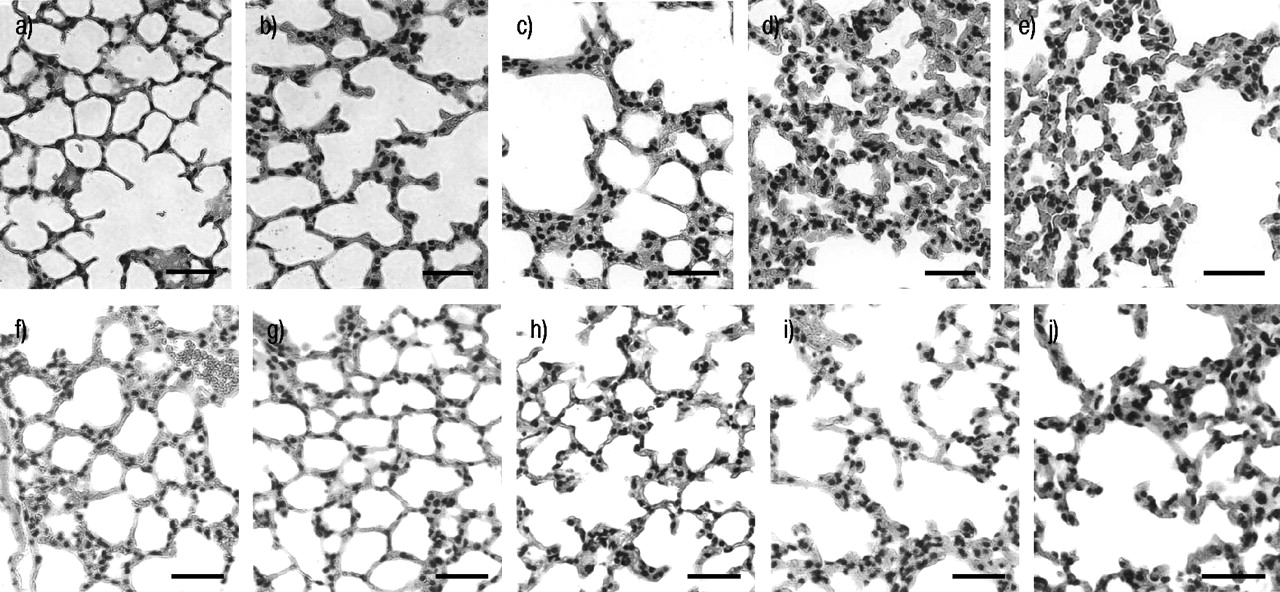
Photographs of histopathology changes in the lungs of BALB/c (a–e) and C57BL/6 (f–j) mice following inoculation with 2×10⁵ colony-forming units Streptococcus pneumoniae NC012695 in 30 µL of sterile phosphate-buffered saline. Lung sections were stained with haematoxylin and eosin at 0 (a and f), 4 (b and g), 12 (c and h), 48 (d and i) and 72 (e and j) h after inoculation. Scale bars=12.5 µm.
Histopathological scores relating tissue damage to pneumococcal lung infection in BALB/c and C57BL/6 mice
Lung tissue swelling was apparent by 4 h and was still significant at 72 h in both strains. Tissue injury was evaluated by assessing the extent of modification of alveolar structure and disorganisation of lung tissue. Tissue injury became apparent at 4 h in BALB/c but not until 12 h in C57BL/6, and remained present until 72 h in both strains. The percentage of lung tissue involved in inflammation and remodelling was lower at all time points in C57BL/6 compared with BALB/c mice, significantly so after 12 (p<0.025) and 48 (p<0.05) h. In both strains the percentage of tissue involvement increased significantly compared with control levels to 5% at 4 h, 20–30% at 12 h and 33–40% at 48 h, but decreased to 30% by 72 h as the lung tissue began to recover (table 3⇑).
Summary
A summary of the results of bacterial infection and clearance, circulating immune cell influx and histological changes is shown in figure 4⇓. Histological changes follow the influx of immune cells in response to bacterial infection and clearance. The infection was completely cleared and immune cell distribution and histological scores were returning to preinfection levels by 72 h.

Summary of the characterisation of the recovery model of mouse pneumococcal pneumonia (n=4). Bacterial recovery from lung tissue (▪) and histopathological changes (○) with time in the lungs of a) BALB/c and b) C57BL/6 mice infected with 2×10⁵ colony-forming units of Streptococcus pneumoniae strain NC12695 in 30 µL sterile phosphate-buffered saline are shown. Percentage changes in relative numbers of immune cells in BAL fluid over the same time period are also shown for c) BALB/c and d) C57BL/6 mice.
Functional differences
Significantly greater phagocytic activity in bronchoalveolar macrophages and peripheral phagocytes, and oxidative burst of bronchoalveolar macrophages were observed in BALB/c compared with C57BL/6 mice (p<0.05; fig. 5⇓).

Phagocytosis of Streptococcus pneumoniae and the production of reactive oxygen intermediates in a) bronchoalveolar macrophages and b) whole heparinised blood from naive female BALB/c (□) and C57BL/6 (└) mice (n=9–16). *: p<0.05.
Discussion
This study reports a reproducible recovery model of pneumococcal lung infection in mice that has similarities to human disease, and compares the susceptibility of mice with different genetic backgrounds. The pneumococcus type‐3 strain NC012695 at an inoculum concentration of 2×10⁵ cfu in 30 µL induced mild clinical disease, with no bacteraemia, from which mice recovered.
Bacterial numbers in lung tissue increased over the first 4 h as bacteria initially adhered to and colonised lung tissue and grew, indicating that infection was occurring, but were then cleared by induced immune responses at 48 h. Inflammatory responses were characterised by a neutrophil influx into BAL and lung tissue that peaked by 12 h, and then fell as bacterial numbers decreased. Histopathological examination of lung tissue showed that lung damage increased after infection to 48 h. At this time, immune responses had cleared all viable bacterial cells and the numbers of inflammatory cells had decreased correspondingly. These results demonstrate that a recovery model of pneumococcal lung infection has been established in which mice initially become infected, the bacteria are cleared by induced immune responses and mice recover from the infection.
The effects of genetic background in this model were investigated and it was found that BALB/c had a more intense neutrophil influx and more severe lung damage than C57BL/6 mice. These results indicate a quantitative difference in immune response between the two strains.
The nonfatal model described has a number of similarities to human disease. Infected animals recover from the clinical illness and detectable bacteraemia occurs in only a small proportion of cases. The gross pathology of the model was consistent with bronchopneumonia 28. Although lobar pneumonia observed in other mouse models is the usually recognised pattern of disease in young adults, atypical forms are increasingly recognised. Pneumococcal bronchopneumonias are more common in the very young, the elderly and immunocompromised. The time course of histological changes is also similar, where the early response involves tissue oedema and the presence of erythrocytes in alveolar spaces, followed by a neutrophil influx, which are gradually replaced by macrophages (fig. 2⇑). During recovery from disease in humans, neutrophils progressively decline and are replaced by macrophages and finally infiltrating lymphocytes 29, 30.
This nonfatal model differs from lethal models in the quantitative bacterial clearance profile and the extent of tissue injury, especially lung haemorrhage. In lethal models, bacterial numbers decline over the first 16 h but then increase to massive levels (>108 cfu·g−1) after 96 h and remain at these levels until death 8, 11. The present model showed complete clearance of bacteria by 48 h. Tissue damage is different in lethal models where erythrocyte extravasation increases over the whole time course up to 96 h and is so abundant after 72–96 h that erythrocytes become visible in the BAL fluid 8. During recovery, lung haemorrhage was only noted at 4 h in BALB/c mice, was mild and correlated with the clinical signs and symptoms of mild pneumococcal lung infection. The inflammatory cell profile was qualitatively similar in lethal and recovery models. Relative macrophage numbers decrease and neutrophil numbers increase by 12 h reaching maximal levels at 24 h. In BAL, polymorphonuclear neutrophils (PMN) numbers remain elevated, whereas in lung tissue PMN numbers then decline as they are replaced by monocytes and lymphocytes between 72–96 h 8, 10, 11.
It is still unknown, however, whether the precise immune responses that are induced in the recovery model and lead to bacterial clearance, are the same as those in lethal models. The roles of particular immune factors in the recovery model are currently being elucidated. In murine models, cytokine responses that have been implicated in and correlate with the clearance of pneumococcal cells from the lung and recovery from infection include TNF‐α, IL‐1β, IL‐6 and IL‐10 and granulocyte/macrophage colony-stimulating factor 7, 17, 18, 31. Studies of genetic susceptibility in humans has only recently commenced but IL‐10 polymorphisms have been implicated in the increased risk of pneumococcal septic shock 32. These immune responses are under genetic control, and in this study the effects of genetic background on recovery from and inflammatory response to pneumococcal infection were examined. Importantly, these results show that although bacterial inoculation, clearance and lung exposure were similar between the two mouse strains, the extent of immune cell influx and tissue damage was significantly different. This demonstrates that the inflammatory changes are mouse strain-dependent rather than resulting from pneumococcal dose or toxicity.
BALB/c mice exhibited clinical symptoms for longer, suffered significantly increased immune cell influx, more immune damage in the form of lung haemorrhage and a larger extent of lung tissue damage than C57BL/6 mice. Significantly increased phagocytic activity and oxidative burst in bronchoalveolar macrophages and oxidative burst of peripheral phagocytes for BALB/c compared with C57BL/6 mice were also observed. Others have also shown significant functional differences in the bronchoalveolar macrophages from BALB/c and C57BL/6 mice 33. Bronchoalveolar macrophages from C57BL/6 mice had a higher capacity for phagocytosis, show a higher level of expression of macrophage receptor with collagenous structure and increased generation of nitric oxide compared with those from BALB/c mice 33. However, in these studies unopsonised particles and not pneumococci were used, which suggests that the two mouse strains respond differently to different inflammatory stimuli.
The present observations show that upon infection, reduced immune and inflammatory responses were elicited in C57BL/6 compared with BALB/c mice, and the immune, inflammatory and cellular basis for the differences are provided. These results expand on the observations of Gingles et al. 18 who showed that BALB/c mice were capable of surviving infection with 1×10⁶ cfu of a mouse-adapted strain D39, whereas ∼60% of C57BL/6 mice died at or before 72 h. At lower inocula levels (this study and those normally encountered in nature), significantly smaller immune cell and local inflammatory responses are induced in C57BL/6 than in BALB/c mice. These responses are sufficient to clear infection with minimal damage to lung tissue resulting from host immune responses. However, when mice are exposed to larger more virulent inocula, the immune responses of C57BL/6 mice are not capable of clearing the bacteria, which overrun the immune system and lead to bacteraemia and death. Conversely, the magnified immune responses in BALB/c mice are capable of clearing stronger inocula, which enable recovery.
Other factors that may affect the levels of immune responses include that intratracheal infection leads to a different type of infection than intranasal 34, or that different strains of mice have diverse susceptibilities to different pneumococcal strains with different virulence factors. The present results and those of Gingles et al. 18 suggest that mice with a Th2 background are less susceptible to pneumococcal infection than mice with a Th1 background, although the observation that bacteria are cleared within the 48 h excludes the involvement of adaptive T‐cells in recovery. The mechanisms are unclear but may involve the interaction of underlying immune responses of mice with a predominantly Th2 background with innate immune responses such as toll-like receptors and polarised dendritic cells. Pneumococcal pneumolysin diversity 35, differences in surfactant protein A activity 36 or as yet unidentified factors may also play a role in differential susceptibility. This model would be useful in the examination of innate immune function in different genetic environments and the identification of important differences offers the possibility of further dissecting immune responses associated with recovery from pneumococcal infection, which may be manipulated to therapeutic advantage.
A recovery model of pneumococcal lung infection in mice has been established that is representative of human bronchopneumonia. This model has been used to show that in C57BL/6 mice, significantly reduced immune and inflammatory responses are elicited to lower inoculum doses of pneumococcal strain NC012695 than in BALB/c mice. This results in less inflammatory-associated damage but responses in C57BL/6 mice are overwhelmed by higher inocula of a more virulent pneumococcal strain 18. Differences are small but reproducible and may represent the difference between mild infection and recovery, or the onset of severe disease in response to some inocula or immune status of the individual. This model is more ethically acceptable than lethal models and can be used to study bronchopneumonia, for antibiotic, vaccine and therapeutic trials, pathogenesis and immune response studies. Importantly this model is suitable for use in studies of diseases that may occur in association with or subsequent to pneumococcal lung infection and recovery such as chronic obstructive pulmonary disease and asthma.
Acknowledgments
The authors would like to thank P. Foster and C. Blackwell for their assistance in the preparation of this manuscript and R. Kumar for aid with histological interpretation.
- Received July 14, 2003.
- Accepted September 16, 2003.
- © ERS Journals Ltd