


Figures
- FIGURE 1
B-cells are a prominent feature in bleomycin (Blm)-induced pulmonary fibrosis in mice. a) Immunohistochemical analysis of B220 staining 28 days post-oropharyngeal Blm treatment reveals an increase in B220+ cells (iv, arrows, and viii) at sites of fibrosis (Martius scarlet blue (MSB) staining, iii, vii collagen blue) compared to saline-treated controls (MSB i, v) and B220-stained (ii, vii). Images i–iv represent low magnification; scale bar=200 µm. Images v–iv represent high magnification; scale bar=100 µm. Images are representative of three mice in each group. b) Flow cytometry did not show a significant increase in circulating or lung resident CD45.2+ CD19+ B-cells in Blm-treated mice 7 days post-treatment (n≥4). c) No significant difference in circulating CD45.2+ CD19+ B-cells was detected in isolated peripheral blood mononuclear cells (PBMCs) 28 days following Blm treatment. There was no significant difference in the number of CD19+ cells detected in dissociated lung tissue post-Blm treatment. Statistical analysis was performed using an unpaired t-test (n≥10).
- FIGURE 2
Anti-CD20-mediated B-cell depletion does not protect mice against oropharyngeal bleomycin (Blm)-induced pulmonary fibrosis. a) Anti-CD20 therapy 7 days prior to and 7 days following Blm successfully reduced circulating CD19+ cells as measured by flow cytometry in mice on day 7 and 28 post Blm treatment (n≥4). b) Immunohistochemical analysis of B220 expression on spleen tissue sections demonstrated a marked decrease in B220+ cell numbers in anti-CD20-treated mice compared to IgG2a-treated control mice. Scale bars=100 µm (25 µm higher power). c) Quantitative flow cytometry analysis showed that anti-CD20 treatment significantly reduced Blm-induced CD19+ cell numbers in the lung on day 7 and day 28 (n≥4). d) Masson's trichrome staining showed no overt difference in the extent of collagen deposition (blue) in mice treated with Blm and anti-CD20 compared to Blm only and mice treated with Blm and IgG2a. Scale bars=200 µm. e–g) Quantitative analysis of fibrosis and collagen deposition was performed using micro computed tomography (μCT) and high-performance liquid chromatography (HPLC) analysis of hydroxyproline, but there was no significant difference in mice treated with Blm and anti-CD20 compared with Blm and vehicle control. e) Represents a preventative model where anti-CD20 was administered 7 days prior to, and 7 days after Blm treatment. μCT analysis was performed ex vivo and the lung density was used as a representation of fibrosis. There was no difference in the amount of Blm-induced fibrosis in IgG2a or αCD20 treated mice (n≥7). f) HPLC analysis did not show any difference in the amount of Blm-induced lung collagen in in IgG2a- or αCD20-treated mice, confirming that αCD20 had no effect on lung fibrosis (n≥6). g) The data represent a therapeutic model where anti-CD20 was administered on days 10 and 19 post-Blm treatment. Similarly, there was no detectable change in the amount of collagen in the lungs of Blm and anti-CD20 treated mice compared to Blm alone or Blm- and IgG2a-treated mice (HPLC n≥5 and μCT n≥6). All images are representative of at least three mice per group. *: p≤0.05, ##: p≤0.005, ###: p≤0.0005, ****: p≤0.0001.
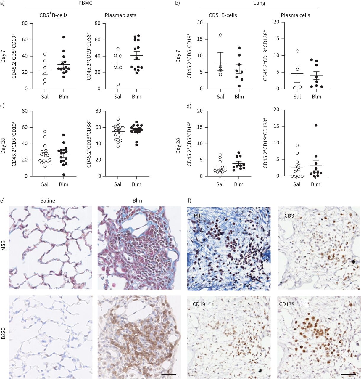
- FIGURE 3
Characterisation of specific B-cell subsets in the peripheral circulation and lung of mice following oropharyngeal bleomycin (Blm)-induced pulmonary fibrosis. a) Blm treatment had no effect on the number of CD5+CD19+ B-cells or CD19+CD38+CD138+ plasma cells detected in peripheral blood mononuclear cells (PBMCs) on day 7 post-Blm treatment (n≥6) or b) CD5+CD19+ B-cells or CD19+CD138+ PCs in the lung 7 days post-Blm treatment (n≥4). c, d) No significant difference was observed in the frequency of CD19+CD138+ plasma cells in the circulation or the lung at day 28 post-Blm treatment (n≥6). No significant increase in CD5+CD19+ cells was observed in the lung 28 days post-Blm treatment (n≥6). Statistical analysis was performed using an unpaired t-test. e) Mouse lung tissue from saline-treated or Blm-treated mice (day 28) was stained with Martius scarlet blue (MSB; collagen blue) or with anti-B220 antibody. B220+ cell aggregates were detected in the lungs of Blm-treated mice (brown 3,3′-diaminobenzidine staining). f) Serial sections of Blm-treated lung stained with Masson's trichrome (MT) and immune markers. CD3+ lymphocytes, CD19+ B-cells and CD138+ plasma cells were detected at sites of fibrosis. All images are representative of at least three mice per group. Scale bars=50 µm.
- FIGURE 4
B-cell composition of the lungs of mice following anti-CD20 and bleomycin (Blm) treatment. a) Mouse lung tissue from Blm-exposed mice treated with anti-CD20 or isotype control (day 28) was stained with Masson's trichrome (collagen blue). Immunohistochemistry shows a marked reduction in B220+ B-cells in 28-day treated mice and retention of CD5+ expressing cells compared with mice treated with Blm and IgG2a. All images are representative of at least three mice per group. Scale bars=50 µm. b) Flow cytometry confirms depletion of CD19+ cells in the circulation, but revealed no increase in circulating CD5+ B-cells, but a significant increase in CD5+ T-cells following anti-CD20 depletion (n≥4). c) However, anti-CD20 treatment did not deplete the plasma cell population in the lung 28 days following Blm (n≥5). ns: nonsignificant. Statistical analysis was performed using one-way ANOVA. ***: p≤0.001.
- FIGURE 5
Plasma cell depletion reduces bleomycin (Blm)-induced lung fibrosis. Mice were given either a vehicle control or bortezomib (Btz) at day −7 and twice weekly until 28 days after intranasal Blm (2 mg·kg−1) treatment (day 0) and the volume of fibrosis was quantified by micro-computed tomography analysis. a) Blm induced significant lung fibrosis compared to saline, which was reduced following bortezomib treatment (n≥4). b) Immunofluorescence staining of lung tissue from Blm+vehicle or bortezomib-treated mice was performed to detect plasma cells within the lungs. The nuclei are stained blue, CD19+ cells are stained red and CD138+ cells are stained green. The plasma cells are positive for both CD19 and CD138. Some epithelial cell staining is seen in the bortezomib-treated group. The figure shows a representative image from each group (n≥4). Scale bars=60 μm. DAPI: 4′,6-diamidino-2-phenylindole. *: p<0.05. A lower-power image showing an extensive area of fibrosis is shown in supplementary figure S3.
- FIGURE 6
Analysis of B-cell populations in the lung and peripheral blood of idiopathic pulmonary fibrosis (IPF) patients and aged matched healthy controls (CTR). a) Martius scarlet blue (MSB) stained and anti-CD20 stained sections of IPF lung tissue demonstrate collagen deposition (blue) and an accumulation of CD20+ B-cells within regions of fibrosis (brown). In addition, CD5+ and CD138+ plasma cells were abundant in IPF lung tissue. All images are representative of at least three mice per group. Scale bars=50 µm. b) Quantitation of the frequency of plasmablasts and memory B-cells in white blood cells of healthy controls (n=27) and IPF patients (n=51) as determined by flow cytometry. Each symbol represents the value from one individual. c) Representative dot plot and contour plots of CD19+ CD20+ B-cells from an IPF patient who was bled on two separate occasions 18 months apart. CD19+ versus CD20+ staining identifies mature B-cells (top), CD20+CD27+ cells identifies memory B-cells (middle) and CD20+ CD38+ identifies plasmablasts (PBs) (bottom), n=4. d) Frequency of CD19+CD20+ CD27+ CD38+ plasmablasts in the peripheral blood of four different individuals bled on two occasions 18 months apart. PCs: plasma cells.


Supplementary Materials
Supplementary Material
Please note: supplementary material is not edited by the Editorial Office, and is uploaded as it has been supplied by the author.
Supplementary material ERJ-01469-2021.Supplement
Supplementary table S1 ERJ-01469-2021.Table
Supplementary figure S1. Gating strategy for mouse PBMCs and mouse lung cell flow cytometry analysis. Mouse PBMCs and dissociated lung cells were stained with a cocktail of fluorescently labelled monoclonal antibodies (mAbs) and analysed by flow cytometry. The leukocyte population was determined by gating CD45.2+ cells. CD45 is a general leukocyte cell surface marker. Leukocytes were subsequently stratified into specific immune cell subsets. a) PCs were identified by selecting CD19+ CD3− cells and then selecting CD38+ and CD138+ cells. b) B regulatory cells were identified by selecting CD5+ and then CD19+. ERJ-01469-2021.Figure_S1
Supplementary figure S2. Gating strategy for human white blood cell analysis using flow cytometry. Representative flow cytometry plots were shown for white blood cells from one control patient that was washed and stained with a cocktail of fluorescently labelled mAbs to distinguish T and B cell subsets. Lymphocytes were gated on FSC, and SSC properties and viable cells were gated and analysed for CD3+ T cells and CD19+ B cells. Mature B cells were identified as CD19+ CD20+ cells. These cells were then analysed for the presence of CD20+ CD27+ memory B cells (Bmem) and the Bmem cells were further analysed for the presence of CD20+ CD38+ plasmablasts (PBs). ERJ-01469-2021.Figure_S2
Supplementary figure S3. a) Low magnification image of a representative lung section from Blm + vehicle treated mice stained with H&E. The image shows areas of normal tissue and dense fibrosis, and multiple mononuclear cell aggregates are present within areas of fibrosis. Inset is enlarged in b) with a mononuclear cell aggregate (arrow). b) High magnification image of mononuclear cell aggregate contains CD19+CD138+ PCs (yellow). The isotype control shows no positive CD19 or CD138 staining. Scale bars 100 μm. ERJ-01469-2021.Figure_S3
Supplementary Material
This one-page PDF can be shared freely online.
Shareable PDF ERJ-01469-2021.Shareable


