


1 INTRODUCTION
Asthma and chronic obstructive pulmonary disease (COPD, also called chronic airflow limitation (CAL)) are the most frequent diagnoses in patients with intrathoracic airways obstruction [1]. Often these patients show a spontaneous variability in the degree of airways obstruction, which can be documented by serial lung function measurements. Large variability in the degree of airways obstruction is indicative of an increased susceptibility of the patient to environmental stimuli that cause acute airway narrowing. Knowledge of the potential severity of these episodes of acute airways obstruction is of clinical interest. Therefore, several quantitative measures of the response of the airways to bronchoconstrictors in vivo have been advocated over the past two decades. The objective of the present guidelines is to address the methodological issues of the various available techniques, and to provide up-to-date international guidelines on standardization. The present recommendations might not represent the potentially best methodologies. However, they do represent the currently validated techniques, by which interchangeable results can be obtained among laboratories.
Variable airways obstruction can be mimicked in the laboratory by challenge tests with bronchoconstrictive stimuli (fig. 1) [2]. This enables one to measure the degree of the so-called «airway responsiveness» of the subject to a particular agent. Since the bronchoconstrictive response varies from one stimulus to another, one needs to specify the challenging agent. Therefore, the term «nonspecific» airway responsiveness should be abandoned.

Dose-response curves to inhaled methacholine using the dosimeter method in 3 subjects. Airway hyperresponsiveness in asthma is characterised by a leftward shift of the curve (hypersensitivity), a steeper slope (hyperreactivity), and an increase in maximal response (excessive airway narrowing). Modified from de Pee et al. [243] with permission.
Airway hyperresponsiveness refers to an exaggerated response to the bronchoconstrictor. This is reflected by an increased sensitivity to the stimulus, which is usually accompanied by an excessive severity of the induced obstructive response [3]. The term «hyperresponsiveness» is recommended as a general description of the phenomenon. «Hypersensitivity» and «hyperreactivity» specifically refer to a leftward shift and an increase in slope, respectively, of the dose-response curve obtained during a challenge test (fig. 1) (see § 4.1) [2, 3]. Because most investigators assume that the bronchi are the major component in these responses, the terms «airway hyperresponsiveness» and «bronchial hyperresponsiveness» are used interchangeably.
The mechanisms underlying airway hyperresponsiveness have not been fully clarified [4]. Both genetic predisposition (associated with atopy) and environmental factors (e.g. virus infections) could be involved in its pathogensis [5]. Airway hyperresponsiveness seems to be a composite physiological disorder, determined by a heterogeneous mechanism in asthma [6] as well as in COPD [7]. It appears to be associated with inflammatory disorders in the airways in both disease entities. In asthma, the mucosal inflammation comprises epithelial desquamation, thickening of the sub-epithelial reticular layer, microvascular congestion, plasma exudate and oedema, smooth muscle hyperplasia and hypertrophy, and (sub)mucosal infiltration with mast cells and activated lymphocytes and eosinophils [8, 9]. In COPD, the inflammatory disorders differ between the various subtypes of the disease: chronic bronchitis, peripheral airways disease or emphysema [9, 10]. (Activated) T-lymphocytes and macrophages seem to be the predominant infiltrating cells in COPD, without concomitant sub-basement membrane thickening [11, 12]. Several of the above inflammatory abnormalities are correlated with the results of inhalation challenge tests [3–8]. Therefore, the degree of airway hyperresponsiveness may indirectly reflect the severity of the disease process in the airways in asthma and COPD. This has been extensively reviewed elsewhere [13].
Inhalation challenge procedures are currently applied in research studies as well as in clinical practice. Both circumstances need descriptions of specific measurement conditions, such as the selection of subjects, the choice of the stimulus, the delivery of the stimulus, the method of measurement of bronchoconstriction, and technical or medical precautions. In addition, it has become evident that challenge tests with each of the various bronchoconstrictor stimuli require distinct laboratory protocols [14]. Therefore, the protocols for the most commonly used challenges with pharmacological agents (histamine, methacholine), physical stimuli (non-isotonic aerosols, cold/dry air, exercise), and sensitizing agents (allergens, occupational sensitizers) will be separately addressed. Finally, attention will be paid to the analysis of the data and the interpretation of the results in the clinical setting and in research studies. It needs to be emphasized that, despite a broad consensus on most of these methodological issues, there are still a number of unsolved dilemma's regarding the standardization of inhalation challenge tests. These are addressed at the end of this report. Since the measurement of airway responsiveness in infants and children is an issue by itself, the present document is focused on adults with only incidental reference to children.
2 MEASUREMENT CONDITIONS
2.1 Indications in clinical diagnosis
Inhalation challenge tests are being used in research studies as well as in clinical practice. The indications for performing inhalation challenge tests in research depend on the hypothesis and objective of the study. The tests are particularly important in follow-up studies in patients with asthma, for which guidelines have been provided [15].
Challenge tests can potentially play a role in daily practice in the clinical diagnosis of patients with variable airways obstruction (§ 4.5) [16, 17]. From clinical and epidemiological studies it appears that airway responsiveness measurements supply valuable information about airways disease, in addition to that from symptoms, spirometry and diurnal peak flow variation [18, 19]. The tests document the potential of variable airways obstruction, and may be useful in those patients in whom this can not be recorded in another way. Does this imply that the tests are useful in diagnostic procedures? Regarding the diagnosis of asthma, the usefulness of pharmacological challenges in clinical practice differs from that in the epidemiological setting [20]. It strongly depends on the prevalence of asthma in the population under investigation [21, 22]. The pharmacological tests are particularly suitable for the exclusion of asthma in the clinic [20, 21, 23, 24], because of the high sensitivity ( = number of asthmatic subjects with a positive test per total number of asthmatic subjects) and high negative predictive value ( = number of non-asthmatic subjects with a negative test per total number of subjects with a negative test). However, the tests are less useful to confirm the diagnosis, particularly in epidemiological studies [20, 21, 23, 24], due to their moderate specificity ( = number of non-asthmatic subjects with a negative test per total number of non-asthmatic subjects) and relatively low positive predictive value ( = number of asthmatic subjects with a positive test per total number of subjects with a positive test). Careful selection of multiple cut-off values can optimize the positive and negative predictive values of the test, even though this introduces a considerable intermediate «grey» area of inconclusive test results [24]. Therefore, the indication of challenge tests in diagnostic procedures of asthma seems to be limited to those patients with typical symptoms without otherwise documented variable airways obstruction [21, 22]. Since airway hyperresponsiveness is also associated with COPD, a positive test result can not be used in the differential diagnosis with asthma [18]. Therefore, in the presence of airways obstruction the tests are hardly indicated. So far there are no challenging agents that allow a clear distinction between asthma and COPD [25].
Serial measurements of airway responsiveness seem to be useful during clinical follow-up, in order to monitor any aggravation of responsiveness following exposure to sensitizing agents, and to document improvement after therapeutic interventions (§ 4.5) [20, 26]. Even though within-subject changes in airway responsiveness to histamine do not consistently reflect the clinical expression of asthma [27], the measurements may indirectly provide additional information above FEV1 about changes in the inflammatory state of the airways and the likelihood of obstruction if an appropriate stimulus is encountered [28]. In addition, the degree of airway hyperresponsiveness reflects the need for medication [16]. However, it needs to be emphasized that the current international consensus on asthma therapy does not recommend hyperresponsiveness to be used as a guide for the level of treatment [17].
Finally, it can be postulated that the level of airway responsiveness has a prognostic value in asthma [26]. Although there is some controversy on this in longitudinal epidemiological studies [29, 30], airway responsiveness appears to be of prognostic significance in prospective clinical studies in children [31] as well as in adults [32]. If these findings can be confirmed in other studies, the prognostic value might become one of the major indications of challenge tests in research and clinical practice.
2.2 Contra-indications
Challenge tests should always be done at the discretion of a physician. There are no data in support of any strict contra-indication of doing an inhalation challenge tests. The current standardized procedures have been shown to be safe during numerous clinical studies (see § 2.4). Nevertheless, the following absolute contra-indications are recommended:
a severe airways obstruction at baseline (FEV1<1.2 l in adults),
b recent myocardial infarction (<3 months),
c recent cerebral vascular accident (<3 months),
d known arterial aneurysmata,
e inability to understand the procedures and the implications of a challenge test.
Relative contra-indications are the following:
a spirometry-induced airways obstruction,
b moderate to severe airways obstruction (e.g. FEV1 < predicted value minus 3·SD of the predicted value: predicted FEV1 minus 1.5 l in males and predicted FEV1 minus 1.2 l in females [33],
c recent upper respiratory tract infection (<2 wks),
d during exacerbations of asthma,
e hypertension,
f pregnancy,
g epilepsia requiring drug treatment.
2.3 Subject characteristics
In addition to the contra-indications mentioned above (§ 2.2), several patient characteristics need attention prior to inhalation challenge testing. Document any noticeable recent relevant allergen or sensitizer exposure. Record all drug therapy, including the last dose and the time it was taken. Oral and inhaled bronchodilators (β-adrenergic agents, ipratropium bromide, theophyllines) should be withheld for their duration of action. This also holds for antihistamines (except for methacholine challenge), that should be stopped 4 days prior to the test (6 wks for asteizole if taken daily for at least one wk [34]). Sodium cromoglycate, nedocromil, and corticosteroids may have acute and long-term effects on airway responsiveness [35]. Even though there is a small acute effect of e.g. steroids on histamine responsiveness [36], these so-called anti-inflammatory drugs are usually not withheld prior to histamine or methacholine challenge. Sodium cromoglycate and nedocromil have an acute protective effect against most non-pharmacological challenges [35, 37], whilst inhaled steroids particularly inhibit the late asthmatic response following allergens or occupational sensitizers [38]. Therefore, these drugs are usually withheld before non-pharmacological challenges. Finally, sodium cromoglycate, nedocromil, and inhaled steroids do have long-term attenuating effects on airway responsiveness which need to be taken into account when interpreting the results [35, 37].
Besides medication usage, baseline lung function, the response to diluent, and the degree of atopy (allergen) are relevant for selecting the starting dose of the challenge (see below). The effect of prior smoking or usage of caffeine-containing beverages on the challenge tests is probably small but is still controversial. Before the challenge, the technician needs to explain and demonstrate the test to the patient.
2.4 Precautions
The precautions that are required when carrying out inhalation challenge tests vary between the different types of stimuli. In general, the safety of standardized histamine and methacholine tests is recognized all over the world. Therefore, the safety requirements for these tests are relatively simple. In contrast, tests with sensitizing agents such as allergens or occupational sensitizers, need extensive precautions and patient-monitoring including the night and day following the experiment. As the experience with the physical tests (exercise, cold air, hypo- and hypertonic aerosols) and with any other pharmacological tests than histamine or methacholine is still relatively limited, the safety requirements for these tests should also be stringent. In case of any doubt, similar precautions as those for allergen challenges should be taken (see § 2.4.3). The precautions for challenge tests include: laboratory materials, personnel training, and written safety protocols.
2.4.1 Histamine and methacholine
Challenge tests with histamine and methacholine, performed in a carefully standardized manner, are safe. Therefore, the precautions during these tests are those that are recommended in general in any clinical lung function laboratory. This includes oxygen and bronchodilators [39]. Personnel should be trained in the management of acute severe asthma [17, 39].
If the test is performed in the context of epidemiological surveys, it can be carried out even if a physician is not present provided that:
1 a standardized protocol is used and the starting dose should be low;
2 baseline FEV1 is ≥ predicted value minus 3·SD of the predicted value [33] (§ 2.2) and ≥2 l;
3 no significant bronchoconstriction occurs after the nebulisation of diluent;
4 a trained and experienced technician performs the test;
5 oxygen and a β2-adrenergical agent can be readily used;
6 the responsible physician or a community physician informed of the protocol and qualified to manage bronchoconstriction can rapidly (within 10 min) see the subject in case of emergency.
If the test is performed in the hospital, a doctor experienced in challenge tests should be present in the hospital and readily available if needed, as criteria no. 2 and 3 listed above might not be fulfilled.
Under all circumstances, the patient must not be left unattended at any time. The patient should be instructed to discontinue inhalation if symptoms become troublesome. At the end of the test the patient should only leave the testing area after his/her bronchoconstriction has adequately improved, either spontaneously or by inhaled bronchodilator (towards FEV1 values >90% of baseline) [40]. The patient should also receive proper instructions in case of relapse of bronchoconstriction during the first 24 h after the test.
2.4.2 Allergens or occupational sensitizers
If challenges with allergens or occupational sensitizers are carried out, in addition to the above (§ 2.4.1), cardiopulmonary resuscitation equipment must be available in the room, together with ready to use oxygen, inhaled and intravenous bronchodilators, intravenous anti-histamines, intravenous steroids and adrenaline (see § 3.5 and § 3.6) [17, 39]. The inhaled doses must be carefully standardized according to the present recommendations (§ 3.5.2–3.5.4, § 3.6.2–3.6.4). The tests can only be performed by a specifically trained technician, and a doctor, experienced in this type of challenge and in acute severe asthma, must be present in the room during the challenge. After the challenge, the doctor should be at close call in the laboratory. The patient is monitored in the laboratory for at least 7 h, and lung function (e.g. PEF) should be measured repetitively during the first 24 h. Severe airways obstruction should be treated adequately [17, 39].
2.4.3 Other inhalation challenges
The very stringent precautions identified in § 2.4.2 are also required for many other inhalation challenges, particularly those tests that have not been fully standardized. This includes tests with any other pharmacological or sensitizing agents. With regard to the physical challenges, there is general consensus that standardized exercise tests are safe (see § 3.4). However, the experience with other physical tests is still relatively limited. Therefore, it is recommended that the safety equipment should be similar as that for allergen challenge, and that a doctor, experienced in this type of challenge, is readily available. If a late response can be expected, lung function should be monitored as after allergen challenge (§ 2.4.2). Alternatively, if bronchoconstriction is adequately improved (see § 2.4.1) the observation period in the laboratory following these challenges may be shorter than for allergens, according to the recommendations for histamine and methacholine.
2.5 Choice of the bronchoconstrictor
Acute airways obstruction can arise from smooth muscle contraction either with or without inflammatory changes in the airway wall. These inflammatory changes include: hyperaemia, plasma exudate, oedema, or hypersecretion, which by themselves may not cause serious airway narrowing, but in combination with smooth muscle contraction will lead to severe, acute obstruction [41, 42]. These bronchoconstrictor mechanisms are involved to a various extent during different types of challenge tests [14, 35, 43]. Some bronchoconstrictors act directly and predominantly on airway smooth muscle itself (e.g. methacholine, histamine), whereas other stimuli depend on the involvement of cellular or neurogenic mechanisms, indirectly leading to smooth muscle contraction and possibly to inflammatory changes in the airway wall (e.g. non-isotonic aerosols, cold/dry air, exercise) [43]. The inflammatory mechanisms predominate after challenge with sensitizing agents, particularly during late asthmatic reactions (e.g. allergens, occupational sensitizers) [38]. In addition, these challenges by themselves can also cause temporary increase in airway responsiveness to other, non-sensitizing stimuli [14, 35]. Therefore, the results of the various challenge tests are only weakly correlated and thereby not interchangeable, each implicitly providing different information. The discordance between the tests may also arise from the distinct ways of expressing the response (e.g. obtained from dose-response or time-response curves) [44]. This warrants further pathophysiological and clinical studies, particularly during follow-up of therapeutical interventions [44].
The choice of the bronchoconstrictor stimulus to be used depends on pathophysiological, methodological, and clinical criteria [14, 35]. Pharmacological challenges with histamine or methacholine have best been standardized and validated in patients with asthma or COPD. It can be argued that physical challenges are better at mimicking naturally encountered bronchoconstrictor stimuli, thereby having more impact for clinical problems. However, these tests have less stringently been standardized. They also have a couple of drawbacks, such as the relatively small range of the doses that can be administered, and the still limited experience with these tests in clinical epidemiology. Challenge tests with sensitizing agents are hardly ever needed in clinical practice (except in the case of agents encountered in the workplace) (§ 3.5.3 and 4.5), but they are extremely useful in pathophysiological studies. In research studies the choice of the challenge depends on the pathophysiological pathway under investigation (§ 3.7).
2.6 Lung function measurements
Airways obstruction can be documented in a number of ways [33]. Two types of measurements need to be distinguished: those preceded by a deep inspiration to total lung capacity (FVC, FEV1, peak expiratory flow, and maximal expiratory flow-volume curves), and those without a deep inspiration (airways resistance or conductance, and partial expiratory flow-volume curves) [2]. Since a deep inspiration can either cause transient bronchodilatation or bronchoconstriction [33, 45], this distinction is highly relevant during challenge procedures, particularly in research.
Although the various methods of lung function assessment highlight different aspects of lung mechanics, their behaviour during challenge tests in clinical practice is very similar [46]. Among the measurements including a deep inspiration, the FEV1 is first choice. There is no clear benefit from using other measurements obtained from the maximal expiratory flow-volume curve [2]. The measurement of FEV1 is well standardized, as is extensively discussed elsewhere in this issue [33]. Even though the recording of FEV1 implicitly affects the degree of obstruction by the preceding deep inspiration, its use in serial measurements of airway responsiveness leads to the most reproducible results [46]. The forced vital capacity (FVC) may provide additional information to FEV1 during challenge tests, particularly on airway closure, which may predict the maximal response to bronchoconstrictors [47]. However, repetitive FVC measurements are exhausting for the patient and, therefore, they are not recommended for routine use.
Lung function tests without a deep inspiration, such as specific airways conductance [48] or partial flow-volume curves [49], are more sensitive to small changes in bronchoconstriction than FEV1 [46]. This makes them more suitable for research studies in normal subjects, in whom the response to bronchoconstrictors is limited [50]. However, the reproducibility of airway responsiveness measurements with these methods is substantially less than with FEV1 [46], so that the latter is recommended in clinical practice and epidemiological studies.
2.7 Symptoms
Even though bronchoconstriction can best be documented by lung function assessment, symptoms of breathlessness during the challenge may provide additional information that is clinically or pathophysiologically relevant. The best validated method for measuring the breathlessness that is perceived during exertion is the Borg category scale [51], which has also been applied to histamine and allergen challenge testing [52, 53]. A promising alternative for pharmacological challenge testing might be the visual analogue scale (VAS) for breathlessness [54]. However, further validation of these techniques during various challenge tests will be required. Therefore, at this stage they cannot be recommended as outcome variables of airway responsiveness measurements.
3 LABORATORY PROTOCOLS
3.1 Pharmacological agents
3.1.1 Background
Pharmacological challenges with aerosolized solutions of carbachol [55] or histamine [56, 57] were introduced on both sides of the Atlantic about half a century ago. The procedures were further developed by De Vries et al. [58] and Orehek [59], and subsequent worldwide application of histamine and methacholine inhalation challenge tests was based on the work of Hargreave et al. [60]. Currently, these pharmacological challenges are the first choice for airway responsiveness measurements in clinical practice as well as in research (§ 2).
Histamine is one of the major inflammatory mediators involved in asthma, producing airways obstruction by smooth muscle contraction, and to some extent by increased microvascular permeability and/or stimulation of (non)cholinergic activity [61]. Carbachol and methacholine are synthetic muscarinic agonists that are more stable than acetylcholine itself and not degradable by cholinesterase [59]. Even though carbachol challenges have been used in asthma [62], most current experience exists for methacholine [60]. The solubility of methacholine allows administration of higher doses than with histamine, without side effects [50]. This may be particularly useful in epidemiological studies. Remarkably, histamine and methacholine provide concordant results (comparable PC or PD values) although they are not fully interchangeable (§ 3.1.8).
3.1.2 Solutions
Histamine acid phosphate (histamine di-phosphate)
Standardized solutions of histamine are usually made from histamine di-phosphate powder (HDP, molecular weight: 307) and phosphate-buffered saline (PBS). Phosphatebuffered saline is used as the diluent because, at higher histamine concentrations, unbuffered solutions become sufficiently acid to alter the response of the airways [63]. PBS and histamine di-phosphate solutions need to be prepared in a carefully standardized manner, particularly paying attention to the molecular water content of the various salts (e.g. HDP.1H2O). Recently, detailed recommendations on this have been supplied (table 1) [64].
To make a solution of 32 mg·mL−1 histamine: weigh 32.00 g HDP (or 33.88 g HDP.1H2O) and add 1000 mL of sterile PBS. Filter through a 0.22 μm filter, put into a sterile vial and autoclave. Histamine di-phosphate does not dissolve easily in phosphate-buffered saline and the higher concentrations may precipitate out when stored at 4°C. These solutions should be shaken well before use.
Methacholine (acetyl-β-methyl choline chloride)
The pH of methacholine solutions is stable and a buffered diluent is not needed. Normal saline should be used as the diluent because solutions made up with phosphate buffered saline have shown chemical instability over a period of three months [65]. Methacholine powder is highly hygroscopic; it must be stored in a dry container in a freezer and handled very carefully to ensure accurate dry concentrations [66]. At higher concentrations, methacholine becomes more viscous such that by 256 mg·mL−1, nebulizer output for a given flow is significantly reduced and adjustments need to be made. If methacholine chloride is not available, methacholine bromide can be used instead. Both methacholine salts have been shown to have equal biological potency, at least when expressed on a molar-base (1 mol methacholine chloride = 195.4 g, 1 mol methacholine bromide = 239.9 g) [67].
To prepare a solution of 100 mg·mL−1 methacholine, weigh out 5 g methacholine powder and dissolve in 45 mL normal saline. Filter through a 0.22 μm filter and put into a sterile vial.
Storage of test solutions
Histamine and methacholine solutions should be stored in the dark at 4°C. At this temperature, both are stable for at least three months [65, 68–71]. However, bacterial contamination enhances degradation rapidly [70]. Therefore, single use ampoules might be preferable. Since temperature affects nebulizer output, solutions should be allowed to equilibrate to room temperature (approximately 30 min) before use [72].
3.1.3 Aerosol generation
Airway responsiveness is defined as the response of the airways to a provoking agent. It is essential for a reliable assessment of responsiveness that both the dose of the provoking agent and the response are measured accurately. Unless both are carefully standardized, results are unreliable and cannot be related to previously established reference values [60]. It is of overriding importance that the aerosol generation for one type of challenge is consistent between and within subjects, so that the same dose is delivered in an identical way on different occasions.
The current standardization of the dose refers to the amount of provocative agent administered to the mouth. The factors that determine the deposition of aerosols in the airways are [73]: the number and size of the droplets delivered to the mouth, air temperature and relative humidity, airways geometry and breathing pattern. Due to oropharyngeal deposition the actual dose that enters the lungs can only be estimated.
The doses of aerosol for the three most commonly used histamine/methacholine test procedures, described below, are generated by jet nebulizers. In this section we examine variables that need to be standardized to ensure that the dose is accurate [74]. These methods are theoretically suitable for adults as well as for children. However, one is reminded that with the tidal breathing and dosimeter method children are inhaling the same dose as adults [75]. There is evidence that for similar doses, the response to histamine and methacholine may be greater in children that in adults [75]. Not only should this be taken into consideration when selecting starting doses for children, but also when interpreting the results. To date it is unclear as to whether the dose in children should be size-corrected [75].
Nebulizer output
As the driving pressure and the flow rate of compressed air to a nebulizer increases, the aerosol output increases and the resultant increased dose provokes greater airway narrowing. Therefore, all nebulizers must be calibrated to operate at a known output. The calibration needs to be performed under exactly the same conditions as those under which the system is used during a challenge test.
First, any extra port or vent of the nebulizer must be closed (except for in the «Yan-method» where the vent is open; see § 3.1.6). The driving pressure of the compressed air upstream of the flowmeter should be about 344 kPa (50 p.s.i.) for the tidal breathing method and about 138 kPa (20 p.s.i.) for the dosimeter method. Then the nebulizer should be adequately filled with liquid, preferably 3 mL, or less if an adequate small reservoir is used [76]. The simplest calibration method is to measure weight loss from the nebulizer at various airflows as indicated by a pressure compensated flowmeter [74]. Weight loss (y-axis) is plotted against airflow (x-axis), and the correct airflow is chosen by linear interpolation at the desired weight loss. For the bolus methods (dosimeter and Yan method) the nebulizer weight loss per actuation is used to calculate the cumulative dose for estimation of the PD20 (see § 3.1.5 and 3.1.6). For the tidal breathing method, the results are expressed in terms of concentration (PC20), and, therefore, nebulizers are adjusted to give a standardized output (§ 3.1.4). The actual output is regularly checked at the calibrated value of airflow. This is adequate for most clinical and research purposes. However, weighing makes no allowance for evaporation of water during nebulization. By using more sophisticated equipment it appears that solute output is not fully proportional to weight loss [74, 77]. Therefore, some research studies require more precise methods of measuring nebulizer output.
Nebulizer output varies considerably between different brands of nebulizers and between specimens of the same brand [74]. However, provided there is adequate cleaning, nebulizer output is highly reproducible for individual nebulizers, even after long-term heavy use [78]. Nevertheless, regular checking of nebulizer output is recommended.
Particle size
The majority of jet nebulizers generate heterodisperse droplets with a mass median aerodynamic diameter (MMAD) of 1 to 4 μm [74]. Variation within this range has little effect on the response [79]. It is wise to check the particle size of new nebulizers as there are occasional rogues, but further calibration is rarely needed. Particle size should be expressed in MMAD with the accompanying GSD (geometric standard deviation), and not in mean size or size range.
Apparatus between nebulizer and mouth
Aerosols evaporate, impact or deposit in apparatus placed between the nebulizer and mouth. In general, the distance between the nebulizer and the mouth should be kept to a minimum. Modifications of the face mask or the mouthpiece, tubing, and valve box may seriously affect the dose delivered at the mouth (fig. 2) [80].
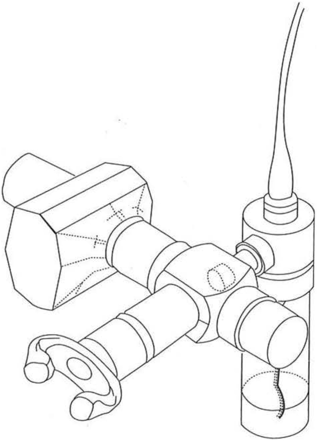
Experimental set-up of a jet nebulizer connected to the central chamber of an in- and expiratory three-way valve box with an expiratory aerosol filter. The subject is connected to the mouthpiece. See tidal breathing method (§ 3.1.4). Modified from Juniper et al. [84], with permission.
Temperature and evaporation
As compressed air passes through jet nebulizers, significant evaporation occurs and test solutions become colder and more concentrated. After 2 min of nebulization, the concentration increases by 10% [81]. Nebulizer output also decreases with cooling (to about 10°C) [72]. To standardize for both these effects, test solutions should be discarded after use and the patients should not be allowed to clasp the nebulizer vial in a warm hand.
3.1.4 Tidal breathing method [82–84]
Aerosols of histamine/methacholine are generated by a validated jet nebulizer with MMAD between 1 and 4 μm, and calibrated to give an output as estimated by weight loss of 0.13 mL·min−1. Aerosols are delivered either through a face mask, held loosely against the face, or through a Hans Rudolph valve box with a mouthpiece [85]. The latter system allows absorption of the expirate in a low resistance aerosol filter attached to the expiratory port of the valve box (fig. 2). Each aerosol is inhaled by quiet tidal breathing at spontaneous frequency through the mouth for 2 min, using a nose clip. The first aerosol inhaled is the diluent and this is followed at 5 min intervals by doubling concentrations of histamine or methacholine from 0.03–32 mg·mL−1 (or higher concentrations if necessary in research). The FEV1 is measured before the test and at 30 and 90 s after each inhalation, the lowest technically satisfactory [33] recording will be used in the analysis. The lowest value reflects maximal bronchoconstriction at a certain dose. It has been chosen in order to take into account slight variations in the time course of bronchoconstriction induced by the agents [86], and because deep inspirations may remove bronchoconstriction [45]. The test is stopped when the FEV1 has fallen by 20% or more from baseline. The results are expressed as the concentration of methacholine/histamine causing a 20% fall in FEV1 (PC20) (§ 3.1.7 and 3.1.8). The reproducibility of the test is described in § 4.3.
To shorten the test procedure, low concentrations may be omitted in some patients. However, it should be emphasized that this can only be recommended when the administrator of the test has extensive experience. The starting concentration is calculated from the baseline FEV1, the response to diluent, and current medication usage [84, 87] (§ 2.3) in the following way:
1 FEV1/VC>80% and FEV1>70% predicted and FEV1 falls <10% after the diluent inhalation and the patient's symptoms are well controlled, use starting concentrations between 0.125 and 2.0 mg·mL−1, depending on the medication being taken:
Medication Starting concentration
Inhaled or ingested corticosteroids 0.125 mg·mL−1
Daily bronchodilators 0.25 mg·mL−1
Occasional bronchodilators (< once/day) 1.0 mg·ms−1
No medication 2.0 mg·mL−1
2 FEV1/VC<80% or FEV1<70% predicted and FEV1 falls <10% after the diluent inhalation and the patient's symptoms are well-controlled, use starting concentrations between 0.03 and 0.125 mg·mL−1, depending on the used medication:
Medication Starting concentration
Inhaled or ingested corticosteroids 0.03 mg·mL−1
Other or no medication 0.125 mg·mL−1
3 If a patient's FEV1 falls by 10% or more after the diluent inhalation, or if asthma symptoms do not appear to be well controlled, do not omit any concentrations - start all patients at 0.03 mg·mL−1.
If, after the first concentration of histamine or methacholine, there has been no significant fall in the FEV1 (less than 5% from best baseline) and there is no clinical evidence of any bronchoconstriction (chest tightness, cough or wheezing), the next dose may be omitted. Again, this can only be done if the technician is highly experienced. For example: if, after 0.03 mg·mL−1, there are no symptoms and the fall in FEV1 is less than 5%, the next concentration may be 0.125 mg·mL−1; if this produces no significant change in FEV1 and there are still no symptoms, then 0.5 mg·mL−1 may be given. As soon as there is any evidence of symptoms or the FEV1 is falling: do not omit any further concentrations. Even after a fall of 5% in the FEV1, the next concentration sometimes gives quite a precipitous fall. If concentrations are omitted, it is important to stress before every 2-min inhalation that the subjects should remove the face mask/mouthpiece as soon as they experience any breathing or chest discomfort.
The test may also be shortened by reducing the nebulization time from 2 min to 0.5 min [58]. This worsens the reproducibility of the test results [88], even though a recent study did report similar reproducibility for the PC20 obtained by 0.5 min and 2 min inhalation [89]. Furthermore, the ratio of the PC20 between the two methods has been reported to be 3.1, in stead of the expected value of 4 [89]. Therefore, at present the 2 min tidal breathing method is recommended.
3.1.5 Dosimeter method [90–93]
Aerosols of methacholine or histamine are generated by a jet nebulizer. A dosimeter is an electrical valve system, which enables one to administer aerosol during inspiration only. A flow sensor in the expiratory port triggers a solenoid which exposes the nebulizer to compressed air at 138 kPa (20 p.s.i.) for about 0.6 s, to give a calibrated output per puff of 9.0 μl [92]. The bolus of aerosol is inhaled through the mouth from the outlet of the nebulizer during an inspiratory capacity breath over 5 s, without breath holding at total lung capacity (TLC) [92]. This is done 5 times per concentration (total output 45 μl) without delay. The first aerosol is diluent followed by doubling concentrations of histamine or methacholine from 0.03 to 32 mg·mL−1 (or larger if necessary).
The doses are given at 5 min intervals. FEV1 is usually measured 30 and 90 s following each inhalation as for the tidal breathing method. The test is stopped when FEV1 has fallen by 20% or more. The tests may be shortened according to the recommendations in § 3.1.4. Results should not be calculated from cumulative breath units (1 unit = 1 inhalation of aerosol from a 1.0 mg·mL−1 solution) [90], because the non-standardized nebulizer outputs in the literature (ranging between 1.0 μl [94], 2.0 μl [95], 7.1 μl [96], 8.9 μl [91] and 9.0 μl [92] per puff) make between-centre comparisons in terms of breath units very unreliable. It is recommended to express the results in cumulative nebulized mol histamine or methacholine to cause a 20% fall in FEV1 (PD20) (§ 3.1.7 and 3.1.8). Alternatively, the results can be presented as PC20, which has been reported to be similar between tidal breathing and dosimeter method when the present standardization is accomplished [92]. The reproducibility of the method is described in § 4.3.
3.1.6 Yan method [94]
This is a hand-operated method, delivering aerosols during inspiration only. Aerosols are generated by five calibrated DeVilbiss 40 glass, handheld, nebulizers (stoppers removed) (2.2–3.8 μl per squeeze). Saline and histamine or methacholine solutions of 3.15, 6.25, 25 and 50 mg·mL−1 are placed in each nebulizer. After assessment of baseline FEV1, the saline nebulizer is placed between the teeth and the patient exhales to FRC. At the beginning of an inspiratory capacity manoeuvre to TLC lasting 1–2 s, the operator gives the nebulizer bulb one firm squeeze. The breath is held at TLC for 3 s, whereafter the subject exhales outside the nebulizer. This is repeated twice for saline but the number of breaths for each concentration of histamine varies according to the dose being administered, which ranges from 0.03 to 7.8 μmol histamine di-phosphate or from 0.05 to 12.3 μmol methacholine chloride (cumulative), or greater if needed [94]. FEV1 is measured 60 s after each dose and the test is stopped when the FEV1 has fallen by 20% or more from post-saline. Results are expressed as the cumulative dose (in μmol) causing a 20% fall in FEV1 (PD20). The reproducibility of PD20 is described in § 4.3.
3.1.7 Calculation of the response
The airway narrowing response can be calculated as % fall in FEV1 in two ways: taking either the post-diluent or baseline value as a reference.
Lowest post-diluent FEV1
With this method one calculates the response of the airways to either histamine or methacholine alone. The lowest, technically satisfactory [33] FEV1 measured at 30 or 90 s after inhalation of diluent is taken as the pretest value. The % fall in FEV1 in response to histamine/methacholine is 100 ×
Mean baseline FEV1
With this method one calculates the overall response of the airways to the diluent + methacholine or histamine. The mean of 3 baseline measurements of FEV1 (within 5% of the largest) is taken as the pretest value. The % fall in FEV1 in response to diluent + histamine/methacholine is
3.1.8 Expression of the response
Calculation of PC20 or PD20
PC20 is used for the tidal breathing method, whereas PD20 is the preferred index for the dosimeter and Yan method. Other outcome variables are discussed in § 4.1.
figure 3 shows the calculation of the PC20 in mg·mL−1 for the tidal breathing method. The method is exactly the same (including the log transformation) for the dosimeter and the 'Yan' method (PD20 μmol). Plot the % fall in FEV1 against the concentration or dose of methacholine/histamine on a log scale. The PC20 or PD20 is obtained by linear interpolation between the last two points, according to the formula [84]: where:

Example of the calculation of the PC20 (provocative concentration at 20% fall in FEV1 from baseline) from a concentration-response curve to inhaled histamine or methacholine in one subject. The PC20 is obtained by linear interpolation between the adjacent data-points. Modified with permission from reference [84].
C1 = second last concentration (<20% FEV1 fall),
C2 = last concentration (>20% FEV1 fall),
R1 = % fall FEV1 after C1,
R2 = % fall FEV1 after C2.
Extrapolation of dose-response curves should be avoided, although it may be appropriate over a dose-range not exceeding one doubling dose [97].
At the recommended nebulizer output a normal PC20 for histamine and methacholine in adults is ≥8 mg·mL−1 (with a «grey zone» between 4 – 16 mg·mL−1) [60] and a normal PD20 is ≥7.8 μmol [94, 98]. These are arbitrary cut-off points (§ 2.1). It means that at these levels of airway responsiveness variable airways obstruction is unlikely at this point. The agreement between the tidal breathing, dosimeter, and Yan method is good [92, 94, 99], although numerical results do not always correspond due to differences in the reported units. In asthma the PC20 is similar for histamine and methacholine [83, 87], whereas the PD20 has been reported to be slightly lower for histamine as compared to methacholine in asthmatic children [75, 98] (§ 3.1.3) as well as in adults with COPD [100].
If two challenge tests are carried out within hours of each other, the bronchoconstrictive response to the second test may be less than to the first test, particularly when high doses are being applied [101, 102]. To minimize this tachyphylactic effect, histamine tests should be done at least 6 h apart. Methacholine tests should preferably be separated by >24 h, even though the methacholine tachyphylaxis, as observed in normals [102], could not be confirmed in asthmatics [103].
3.2 Hypo- and hypertonic aerosols
3.2.1 Background
Already in 1968 De Vries et al. [58] and later, in 1980, Allegra and Bianco [104] reported that the inhalation of an aerosol of distilled water could induce an increase in specific airways resistance (sRaw) in patients with asthma. Thereafter, Schoeffel et al. [105] reported that hypertonic as well as hypotonic aerosols of saline could cause a reduction in FEV1. Over the next 10 years a number of investigators used these aerosols for the assessment of bronchial hyperresponsiveness, and the challenge procedure is now widely used [106–109].
It is now generally accepted that non-isotonic aerosols induce airway narrowing indirectly by causing the release of endogenous mediators which cause bronchial smooth muscle contraction and airway oedema. The release of mediators is thought to be directly caused by the change in osmolarity [110]. Histamine is likely to be an important mediator released because specific antihistamines are very effective in inhibiting the airway response to both hyper- and hypotonic aerosols [109, 111, 112]. However, it is likely that other mediators such as the leukotrienes and prostaglandins are also involved. Further, there may be a neural component involving release of sensory neuropeptides or a parasympathetic reflex [113].
Sodium cromoglycate [114], nedocromil sodium [115] and furosemide [116, 117], drugs which are thought to affect mast cell release of mediators and/or the noncholinergic neural activity, are very effective at inhibiting airway responses to non-isotonic aerosols. The anticholinergic agents have been shown to be inconsistent in their effect and changes in baseline FEV1 due to their action as bronchodilators make the findings difficult to interpret [114, 118, 119]. The sensitivity to hypertonic saline is reduced with daily administration of corticosteroids given as aerosols [120]. This usually occurs after 400–2000 μg has been taken daily for 2–8 weeks.
The responsiveness to 4.5% saline has been shown in asthmatics to correlate with numbers of mast cells obtained by bronchial biopsy [121]. The airways response to distilled water in patients with transplanted lungs has also been shown to correlate with airway inflammation [122].
There now seems to be sufficient evidence to suggest that the severity of the airway response to these aerosols reflects the involvement of inflammatory cells and their mediators in the airways [106]. Thus, in addition to demonstrating the capacity of the airways to narrow in response to the endogenous release of inflammatory mediators, challenges with these aerosols, when followed over time, may be useful in evaluating both the acute and the chronic effect of medications used in the treatment of airway inflammation.
Tests with non-isotonic aerosols are generally considered to be safe. However, there is one report of acute, fatal bronchoconstriction in response to a standardized distilled water challenge (Fabbri, personal communication). Therefore, extensive precautions must be taken (§ 2.4).
3.2.2 Solutions
The most commonly used solutions are distilled water and 4.5% saline. Some laboratories use 3.6% saline, and a concentration of 2.7% saline is usually sufficient to induce a reduction in FEV1 in very sensitive patients. Dose-response curves are obtained by increasing the time of exposure to the single saline concentration. Alternatively, a dose-response curve can be obtained by doubling the concentration (0.9, 1.8, 3.6, 7.2, 14.4%) [112]. This is less practical for routine purposes and should be regarded as a research procedure.
A concentration of 4.5% saline is recommended in preference to the other solutions for several reasons. The test is shorter when this concentration of saline is used compared with lower concentrations, and 80% of clinically recognised asthmatics have a 20% fall in FEV1 after 15 mL has been nebulized or less. A person who responds to 4.5% saline usually also has exercise-induced asthma. The osmolarity is slightly above sea water, and the test is also used for screening SCUBA divers.
3.2.3 Aerosol generation
Ultrasonic nebulizers are recommended for generation of non-isotonic aerosols because they produce aerosols which are more dense than conventional jet nebulizers. The mass-median aerodynamic droplet size is usually between 2 and 10 μm and this is reduced to less than 5 μm when the breathing circuit is attached. It is recommended that a large two-way valve (Hans Rudolph 2700) be used and that the tubing attaching this to the nebulizer should have a smooth internal bore and be of constant length and diameter.
It is important to choose a nebulizer that has a reproducible output of at least 1.2 mL·min−1 with the breathing circuit attached. Further, it is necessary to have a nebulizer with a bowl or canister which can be easily detached for weighing. A volume capacity of 100–200 mL for the bowl is recommended. The temperature of the solution increases with time, but this is minimised with these relatively large volumes, and the small increases in temperature appear to have little or no effect on the response. Preferably use the same volume fill for each subject and the same setting for the nebulizer output. The output of the nebulizer and the total dose of aerosol delivered to each subject can be measured by weighing the bowl and tubing, but not the valve, before and after challenge. The valve is not weighed because of the production of saliva during the test.
For any one individual, the output of the nebulizer during the challenge is linearly related to time. Thus, to obtain the dose delivered for each exposure, it is only necessary to know the output. The output is expressed in mL per min. This can be obtained simply by dividing the total dose delivered during the challenge, measured by a change in weight, over the total time of exposure to the aerosol. The dose of aerosol, in mL, delivered for each interval can be calculated from the time of exposure. For clinical purposes 1 g of 4.5% NaCl is considered as 1 mL and a correction (multiply by 1.03) is not made for specific gravity.
3.2.4 Protocol
At present it is recommended that the dose of aerosol be increased by increasing the length of each challenge interval. Although the nebulizer output or saline concentration could be increased to achieve a higher dose, this may cause cough and be distressing to the patient (see § 3.2.2). In order to prevent any problems with electrical charge in some ultrasonic nebulizers, 0.03% saline can be used instead of distilled water in case of hypotonic challenge. For hypertonic challenge 4.5% saline is recommended.
Measurements of FEV1 or specific airways resistance (sRaw) [2, 48] are made in duplicate or triplicate before and between 60 and 90 s after each exposure to the aerosol. The exposure times are doubled as follows: 30 s, 1 min, 2, 4 and 8 min. If the fall in FEV1 is greater than 10% of baseline the previous exposure time is repeated rather than increased. The challenge is stopped after 15 mL is nebulized or when there is a 20% reduction in FEV1 or a doubling (100% increase) in sRaw.
Although a 20% fall in FEV1 is generally accepted as abnormal, on the basis of the findings in healthy non-asthmatic subjects, a value of 15% or more should probably be regarded as abnormal [123–126]. A broncho-dilator is always administered by aerosol at the end of each challenge.
3.2.5 Expression of the response
The dose-response curve is constructed by plotting the change in FEV1, expressed as a percentage of the baseline value or the predicted value, against the cumulative dose of aerosol delivered, expressed in mL (fig. 4). The dose is calibrated according to § 3.2.3. About 15% of the measured weight loss will be deposited in the intrapul-monary airways. A value for PD15, and PD20 can be obtained by linear interpolation. Within the asthmatic population the values for PD20 are log normally distributed so the PD20 values are log transformed before a statistical analysis is carried out.

Cumulative dose of water or 4.5% saline used to provoke a fall in FEV1, expressed as a % of the pre-challenge level or as a % of predicted FEV1. The cumulative dose is measured by weighing the nebulizer and tubing, but not the valve, before and after challenge. The cumulative time is recorded. The output of the nebulizer is calculated in mL per min. The dose for each exposure can be calculated from the time.
To assess changes in sensitivity after an intervention the values for PD20 are compared using log transformation. For an individual the fold difference in PD20 after an intervention can also be calculated after log transformation [120]. Because baseline lung function can be altered by treatment, or change spontaneously over time, a reactivity index (slope of the dose-response curve) can be useful. This is defined as the change in FEV1, expressed as a percentage of the predicted value, per unit dose of aerosol [120]. To compare the responses after an intervention the index is compared over the same values of percent predicted FEV1. This index can also be expressed as a fold difference. For subjects who do not reach a 15 or 20% fall in FEV1, the maximum % fall in FEV1 and the maximum dose of aerosol delivered should be reported. A person is considered to have a severe response if the PD20 is <2 mL, a moderate response if it is 2.1–6.0 mL, and a mild response if the PD20 is 6–20 mL [123].
3.3 Cold/dry air inhalation
3.3.1 Background
Airway responsiveness can be measured by the inhalation of stimuli which cause bronchoconstriction through the release of mediators from cells within the airways, as well as by the inhalation of these bronchoconstrictor mediators. One such method is by isocapnic hyperventilation of cold and/or dry air. Although the original description of hyperventilation induced bronchoconstriction was made in 1946 [127], renewed interest in this method for inducing bronchoconstriction occurred because of the recognition that cooling and/or drying of the airways is the mechanism of exercise-induced bronchoconstriction. Airway narrowing can result from cooling and subsequently rewarming of the airway mucosa, as well as from local hyperosmolarity due to drying (reviewed in § 3.4). The degree of airway hyperresponsiveness to isocapnic hyperventilation of cold dry air is moderately correlated with the degree of airway hyperresponsiveness to inhaled methacholine [128] and histamine [129] in asthmatic subjects.
Isocapnic hyperventilation of cold/dry air uses a naturally occurring stimulus to provoke bronchoconstriction rather than a chemical stimulus. Whether this implies a better safety profile still needs to be confirmed. Isocapnic hyperventilation can be administered in a dose-response fashion, and, for this reason may in some instances have advantages over exercise for measuring airway responsiveness. When giving doubling doses, the shape of the dose-response curve obtained in asthmatics is similar to other bronchoconstrictor stimuli: a lower threshold, increased slope and absence of a plateau response, when compared to non-asthmatics [130].
3.3.2 Production and conditioning of air
The most convenient source of air is to use dry compressed air, which is cooled by being passed over a cooling coil, through which methanol cooled to −35 °C is passed. This results in inspired air cooled to −12 to −15 °C. In many laboratories, however, just dry air at room temperature is used, because this is adequate to provoke bronchoconstriction in most asthmatics, and avoids the complexities of having to cool the air. This technique leads to similar results as those with cold dry air, even though in animals studies differences between these methods have been observed [131]. Subjects inhale the cold and/or dry air through a valve. A Hans Rudolph valve can be used for this purpose, which can be divided into an inspiratory and expiratory port, if inspired and expired air temperature is being measured. If this method is used, end-tidal CO2 needs to be continuously measured in the expired air, using a capnograph, and CO2 added to the inspired air to keep the subject eucapnic.
3.3.3 Protocol
After measurement of baseline spirometry, the subject breathes at increasing minute volumes, beginning at 7.5 l·min−1, increasing to 15, 30, 60 l·min−1 and maximum minute ventilation, each period of ventilation lasting 3 min. The inspired volumes can be achieved by targeting, using a device which gives a visual display of the inspired volume. The rate of breathing is dictated either by the technician or by a metronome. The tidal volumes and rates which can be used to achieve the desired minute ventilation are: 0.75 l at 10 breaths/min; 1.5 l at 10 breaths/min; 2.0 l at 15 breaths/min. After each step the patient breathes room air, and the FEV1 is measured at 30 s, 90 s, then at 3 min and 5 min and every 2 min until the lowest technically satisfactory value [33] is reached. The challenge is stopped once a 20% fall in FEV1 occurs. A major limitation with the method, however, is that the maximal challenge that can be given is limited to the maximal voluntary ventilation that can be achieved by the subject being studied. On the other hand, achieving maximal ventilation may be an advantage of this technique over exercise. Although this method produces a cumulative bronchoconstrictor effect, the maximal bronchoconstriction obtained is not significantly greater than if maximal minute ventilation alone is used as the stimulus [132]. A simplified method, in which CO2 is added in a fixed amount to the inspired air (FI.CO2 = 0.049), prevents hypocapnia over a wide range of minute ventilations from 40 to 105 l·min−1 [133]. This eliminates the need to measure end-tidal CO2. However, this FI.CO2 may cause hypercapnia at lower minute ventilations used in the challenge procedures (7.5, 15, 30, l·min−1). Therefore, with this technique the ventilation loads of 30%, 60% and 100% predicted maximal voluntary ventilation are used to overcome the possibility of hypercapnia. If patients with severely increased airway hyperresponsiveness are being tested, who may develop significant bronchoconstriction at these lower minute ventilations, it is necessary to monitor end-tidal CO2.
An even more simplified technique has been used in paediatrics: a single-step 4 min isocapnic voluntary hyperventilation at 75% of the maximal voluntary ventilation (MVV, as determined from pre-challenge FEV1) of cold dry air (−10 °C) [134]. Since in adults there are no comparative studies of this test with the multiple-step protocol, at this stage, the dose-response approach is recommended.
3.3.4 Expression of the response
The response is measured as the change in FEV1 from the baseline values. A dose-response curve is obtained by plotting the incremental increase in minute ventilation (V′E) against the change in FEV1, (usually 10% or 20% fall), and the response expressed as the PV′E,10 or PV′E,20 (the provocative minute ventilation causing a 10% or 20% fall in FEV1, respectively). If cold dry air is being used as the stimulus, some investigators have calculated the respiratory heat exchange (RHE) [135]. This requires the measurement of the inspired and expired air temperature, using rapidly responding thermocouples, and water content. If dry air is used, then the water content is 0 mg·l−1. The expired air is assumed to be fully saturated, and therefore the water content can be obtained from standard graphs of air temperature and humidity. A dose-response curve is obtained by plotting the incremental increase in RHE (kcal·min−1) against the change in FEV1, and the results expressed as the RHE giving the predetermined fall in FEV1. However, in most instances, when just dry air is used as the stimulus, reporting minute ventilation is sufficient and the respiratory heat exchange does not need to be calculated.
A fall in FEV1 of at least 10% is generally required to indicate a positive result to the challenge. The magnitude of this response is greater than 2 standard deviations above the response seen in normal subjects [135, 136].
3.4 Exercise
3.4.1 Background
It is now 30 years since Jones [137] described the effects of exercise on the airways resistance in children with asthma. This exercise-induced bronchoconstriction is usually referred to as exercise-induced asthma (EIA), even though it is generally accepted that exercise is not an inducer, but an inciter of asthma. By the early 1970's, Godfrey and his colleagues had identified many of the factors which determined the severity of the airway response [138]. These included the type of exercise, its intensity and duration, and the interval since exercise had last provoked an attack of asthma. By the late 70's it was recognised that the most important determinant was the level of ventilation which was reached and sustained during exercise. It was noted that exercise in itself was not necessary to provoke an attack of asthma, and hyperventilation alone could achieve the same response [139]. At the same time the effects of temperature and water content of the inspired air were being investigated. It was discovered that exercise-induced asthma could be completely inhibited when air at body temperature and water vapour was inhaled during exercise [140]. Thus it was concluded that the stimulus for EIA had to be the loss of either heat or water or both from the airways during exercise.
Initially it was considered that cooling of the airways was the stimulus [141] but later studies demonstrated that water loss was more important than heat loss. From these studies it was proposed that transient hyperosmolarity of the airway surface liquid as a result of evaporative water loss was the stimulus to EIA [142]. It was also demonstrated in vitro that an increase in osmolarity provided an ideal environment for the release of mast cell mediators [143]. These mediators have the potential to cause the airways to narrow by contraction of bronchial smooth muscle. A more recent proposal has suggested that vascular engorgement and oedema of the bronchial circulation is more important than bronchial smooth muscle contraction in causing the airways to narrow [144, 145]. At present there is no direct evidence to support either of these two hypotheses, and further studies are required to clarify this important issue [146].
Exercise provokes an attack of asthma in 70 to 80% of the population with clinically recognized asthma. In clinical situations exercise tests are not very sensitive, but are highly specific for the diagnosis of asthma, and are particularly useful in children [147–149]. It may not be very practical in older patients. In field studies, exercise tests are less specific for the diagnosis of asthma [150], showing a prevalence of positive responses between 7 and 16% [150, 151]. EIA may occur at any age and is equally common in adults and children. The signs and symptoms of EIA do not differ from other forms of acute asthma except that they are of short duration. In addition to increasing airways resistance exercise can induce a transient hyperinflation and arterial hypoxaemia. The majority of persons recover spontaneously from an attack of EIA within 30 min while a minority require oxygen and bronchodilators given by nebulization. In 50% of persons EIA is followed by a refractory period of about 1 hour [152]. The occurrence of a late asthmatic response, several hours after exercise is rare and their association with exercise itself is still a controversial issue [153, 154].
EIA is prevented in most asthmatic patients by premedication with sodium cromoglycate, nedocromil sodium or a β-adrenoceptor agonist [152, 155–158]. In some cases a combination of both types of medication is required. In difficult cases the dose of these agents is doubled and ipratropium bromide added.
3.4.2 Experimental set-up
In the laboratory exercise should be carried out on either a bicycle ergometer or a motor-driven treadmill. There are definite advantages in using a bicycle in that the head is stable and it is easier to administer dry gas and collect expired air. Further, the work intensity during cycling, but not running, is independent of body weight, making it easier to predict the workload required to achieve the target ventilation.
The ventilation in litres per minute should be measured at least during the last 4 min of exercise. The time of exercise should be 6 to 8 min.
Heart rate should be monitored continuously and in the case of persons over 40 yr of age an electrocardiogram should be taken throughout exercise and for 5 min after its completion.
Changes in airways resistance are best measured indirectly by documenting changes in FEV1 or PEF. Although PEF is easier to measure, FEV1 is recommended as it reflects a larger proportion of the flow-volume curve.
It is strongly recommended that air inspired during exercise has a water content as low as possible and less than 10 mg per litre (relative humidity less than 50% between 20–25°C). The best way to achieve this is to have the subject inspire compressed medical air at room temperature via a demand valve and regulator. Although some laboratories generate air of subfreezing temperatures there is no need to have expensive equipment to condition the inspired air. Providing the inspired air is dry it is only necessary to increase the time of the exercise to increase the severity of the airway response.
Medication and baseline lung function alter the response to exercise and need to be taken into consideration when planning the protocol. Measurements of FEV1 should be made on arrival in the laboratory and the stability and reproducibility of the value established. The best values should preferably not vary by more than 10%; the absolute value should be within 80% of the subject's usual value and preferably better than 75% of their predicted value. The subject should have withheld the medications according to recommendations in § 2.3.
3.4.3 Protocol
The highest of the FEV1 measurements taken immediately before exercise is recorded and used in the calculations. The work intensity is selected for the subject to achieve between 40% to 60% of their predicted maximum voluntary ventilation (which can be taken as FEV1 predicted·35) during the last 4 min of exercise. The oxygen consumption to achieve this ventilation can be estimated from the equation relating these measurements in children and in adults [156]. The work load to achieve this oxygen consumption is then determined from the equation. For the first minute the work load is set to 60% of the target load. This is increased to 75% in the second min, 90% in the third min and 100% in the 4th minute. When the subject has achieved the target level of ventilation or reached the ventilatory or working capacity the work load is sustained for 4 min. Minor adjustments may need to be made to this work load but this protocol is good both for general laboratory use and for research [157, 158]. A pilot exercise study may be needed, since it seems unnecessary to have responses above 30% fall in FEV1 if repeated tests are required.
To select a work load for running on a treadmill it is necessary to know the weight of the subject. It is usual to aim for a speed and slope on the treadmill which will induce 30–45 mL of oxygen consumption per kilogram. Running at 5–9 km·h−1 (3–5 mph) up a 10% incline is usually sufficient work for most subjects. Once the target ventilation is achieved it should be sustained for 4 min if possible.
The subject must have the nose clipped to ensure mouth breathing. The measurements of FEV1 are made in duplicate after exercise at 1, 3, 5, 7, 10 and 15 min and the highest value at each time is recorded [156]. A bronchodilator is always given by aerosol upon completion of the protocol.
3.4.4 Expression of the response
The % fall index is the most widely used term to express the severity of EIA (fig. 5). It is calculated by subtracting the lowest value of the measurement of FEV1 recorded after exercise and expressing it as a percentage of the value recorded immediately before exercise. A fall in FEV1 of 10% or more of the pre-exercise value should be considered abnormal when tests are being performed in the laboratory. However, in the field a fall of 15% or more is used [155]. If measurements are made during exercise (this is now unusual) the % rise index can be calculated. This is calculated by subtracting the value recorded immediately before exercise from the highest value measured during exercise and expressing it as a percentage of the value measured before exercise. The exercise lability index is the sum of the % fall and the % rise and is an excellent index for characterising airway responsiveness to exercise (fig. 5) [138].

Changes in peak expiratory flow (PEF) and forced expiratory volume in one second (FEV1) recorded during and after exercise in an asthmatic person who has some airflow limitation at rest. The highest, lowest, and pre-exercise values are used to calculate the % rise and % fall. Modified from Anderson [152], with permission.
It is recommended that in addition to the % fall index, the pre-exercise value for FEV1 and the lowest value after exercise be reported in % predicted. In this way the clinical relevance of response to therapy is more easily ascertained [159].
The ventilation required to induce the observed % fall in FEV1 is also reported and is useful for assessing the effect of therapy. Reporting values for heat and water loss has little meaning in clinical practice. The recording of a potential late response is not recommended, because the interpretation of a fall in lung function at about 3–8 h after exercise is still unclear [153, 154].
3.5 Allergen inhalation test
3.5.1 Background
The airway response to allergen and chemical sensitizers (§ 3.6) is more complex than that to the bronchoconstrictive triggers covered above in § 3.1 to 3.4. The early asthmatic response (EAR) is an episode of airflow obstruction, predominantly airway smooth muscle contraction, maximal between 10 and 20 min after inhalation and resolving within 90 to 120 min [160]. The late asthmatic response (LAR) is an episode of airflow obstruction, probably caused by both airway smooth muscle contraction and inflammation, occurring between 3 and ≥8 h after inhalation [38, 160]. Allergen-induced increase in airway responsiveness to histamine or methacholine ΔPC20), likely an indirect measure of allergen-induced inflammation, occurs between 3 h [161] and several days [162] after allergen exposure, chiefly in subjects manifesting late asthmatic responses. Controlled standardized allergen inhalation tests have a limited, if any, role clinically. They may be helpful in confirming allergic asthmatic responses in exceptional cases where immunotherapy is contemplated. However, they are of great value in research, both for investigating the pathophysiology of asthma [161–163], and studying new, particularly prophylactic, pharmacological agents [164, 165]. The allergen inhalation test protocol outlined below takes into account the three aspects noted above: EAR, LAR, and change in PC20 [160, 162–165]. Standardization is directed primarily towards within-subject reproducibility, which is particularly relevant to research studies. Under all circumstances extensive precautions are needed for safety requirements (§ 2.4).
3.5.2 Solutions and dose range
Solutions for allergen inhalation are made from the best available commercial aqueous allergen standardized extracts. These must be carefully standardized according to recent recommendations [166]. The stock solutions should preferably have a concentration of 10,000 biological units (BU) or 10 histamine equivalent prick test (HEP) per mL (1000 BU·mL−1 = 1 HEP). In weight/volume (w/v) this ranges approximately from 1:10–1:20 w/v (pollen and animal danders) to 1:50–1:100 w/v (house-dust mite). Stock solutions are diluted 1:8 with sterile buffered carbol isotonic saline containing 0.5% phenol. Thereafter, serial doubling dilutions (1:16, 1:32,… ≥1:1024) are made. The weakest allergen solution used for allergen inhalation is determined by the skin test endpoint (§ 3.5.4) in the individual undergoing challenge [167], and may be as dilute as 1:65,536. The most concentrated solution used for inhalations is usually the 1:8 dilution; this represents 1250 BU·mL−1 or 1.25 HEP (1:80 w/v dilution for pollen extracts to 1:800 w/v dilution for house-dust mite extract). Stability of particularly the weaker allergen concentrations is important, and is assured by freshly mixing allergen dilutions before each inhalation challenge.
3.5.3 Aerosol generation
Aerosols are generated with a jet nebulizer (driving pressure up to 344 kPa or 50 p.s.i.) calibrated with airflow between 4 and 9 l·min−1, to give a mass loss of 0.13 g·min−1 (approximately equivalent to 0.13 mL·min−1). A closed system should be used to prevent inadvertent allergen exposure and sensitization of laboratory personnel. The nebulizer is connected to a Hans-Rudolph box and valve system, and to the patient by a mouthpiece. Two ventilator breathing circuit filters are placed in series on the expiratory line to trap the uninspired nebulizate, and the exhalate.
3.5.4 Protocol
A control day is important to ensure stability of FEV1 (within 10% of baseline) over the 8 to 10 h of the study. On the control day, diluent is inhaled for 2 min at 10 min intervals on three occasions. FEV1 is measured initially in triplicate and in duplicate 10 min after each inhalation, and following the last inhalation, every 10 min for the first hour, at 90 min and 2 hours, and then hourly up to 7 h after challenge. On the control day, duplicate prick skin tests with the doubling dilutions of allergen for inhalation are done in order to determine the skin test endpoint, i.e. the weakest concentration producing a wheal of 2 mm or 2 mm larger than control. A histamine or methacholine PC20 is measured at 7 h. The severity of the early asthmatic response (PC20,allergen) can be predicted from the skin test endpoint and the histamine or methacholine PC20 [167]. This is highly relevant for safety purposes. It is being done with the following formula: where SS = the skin test endpoint.
On the allergen challenge day, allergen inhalations are commenced starting two to four concentrations below the predicted PC20,allergen. Doubling concentrations of allergen are inhaled for 2 min at 10 min intervals until the FEV1 measured at 10 min has fallen by ≥15%, or until the top concentration has been reached. If the FEV1 falls by <10%, the next concentration is inhaled. If the FEV1 falls between 10–15%, the next concentration should be inhaled for 1.5 min rather than 2 min. Only if the fall in FEV1 is still <15% should the concentration be inhaled for 2 min. This is an effort to minimize the maximal fall in FEV1 after inhaled allergen. Since the EAR progresses for ≥20 min, even the slightest of symptoms occurring during allergen inhalation is an indication for immediate discontinuation of inhalation. The FEV1 is monitored for at least 7 h as on the control day. It should be emphasized that this monitoring is always required, because even in the absence of an EAR a LAR can not be excluded. The patient then receives written instructions about the medical care during the first 24 h after the challenge (§ 2.4). In exceptional cases, only the EAR is of interest. Then the patient should be given a β-agonist either or not combined with inhaled steroids following the EAR. After full recovery from the EAR the patient is allowed home with adequate instructions on measurements of PEF and medical care during the first 24 h.
Allergen-induced increase in airway responsiveness can be addressed by measuring histamine or methacholine PC20 at 3 h (between the early and late response), or the next day at 24 to 32 h after inhalation. Repeat measurements of PC20 may be made over several days if indicated.
Repeat allergen inhalation tests, generally under experimental conditions, are done with the same dose of allergen throughout, since the cumulative allergen dosage determines the response of the subjects [168]. Both for safety and consistency in timing of pre-allergen medications, one should strive to have three inhalations up to and including the final concentration of allergen delivered for each test. Repeat tests are done a ≥7 day intervals when the baseline FEV1 is stable (within ±10%), the histamine or methacholine PC20 measured before the test is stable (within ± one doubling dilution), and there has been no natural allergen exposure or respiratory tract infection for ≥4 wk [164, 167]. In contrast to repeat histamine/methacholine challenges under experimental conditions, e.g. in situations where the early response is being inhibited by an agent such as a β2-agonist, one should not aim to achieve a fall in FEV1 of 20% during the EAR because of the possibility of severe prolonged late responses. It also needs emphasis that the so-called anti-inflammatory drugs have profound effects on early and late asthmatic responses [164]. Continuous personal attendance by a physician is required during the challenge, and for the duration of the early asthmatic response. Complete cardiopulmonary resuscitation equipment must be readily available including particularly parenteral adrenaline (drawn up and ready to use) and inhaled β-agonists (see § 2.4). These tests should only be carried out in a hospital setting.
3.5.5 Expression of response
The best FEV1 at each time is retained for analysis. A typical example of time-response curves is illustrated in figure 6. The EAR is defined as the maximum per cent reduction in FEV1 (% fall from baseline) occurring in the first hour after challenge. The magnitude of the LAR is defined as the maximum per cent reduction in FEV1 occurring between 3 and 7 h after challenge. An alternative way to express the EAR is the PC20,allergen [167]. In addition, some investigators express the EAR and LAR as the area under the time-response curve [169]: the advantage of this type of analysis is the inclusion of all data-points on the time-response curve. Allergen-induced increase in airway responsiveness is generally defined either as the difference in log PC20 or as the pre/post PC20 ratio using logarithmic transformation for statistical analyses.

Mean (± SEM between subjects) time-response curves of FEV1 (squares, upper panel) and of expiratory flow obtained from partial flow-volume curves (V′40p) (triangles, lower panel) after an allergen challenge with inhaled house-dust mite extract in 10 atopic asthmatic subjects on 2 days (open and closed symbols) separated by a 2 weeks interval. When using the present standardization, the reproducibility of the early and late asthmatic response appears to be good. Modified from Bel et al. [249], with permission.
3.6 Occupational sensitizers
3.6.1 Background
Definition of occupational asthma
Occupational asthma is caused by a specific «sensitizing» agent present at the workplace [170–172]. Asthma of occupational origin may also be caused by an acute exposure to strong respiratory irritant gases at work, a syndrome named «reactive airway dysfunction syndrome» [173–175]. An extensive list of these agents causing occupational asthma has been published in recent review articles [e.g. 170–172].
Diagnosis of occupational asthma
A correct collection of patient's history is the most important step for the diagnosis of occupational asthma [170–172]. Repeated measurements of peak expiratory flow performed at home and at work over several weeks, may provide objective confirmation of a suggestive history of occupational asthma [176, 177]. Although skin tests and specific IgE, when available, are not diagnostic for disease, they may be useful to diagnose sensitization to high molecular weight occupational sensitizers.
In subjects with occupational asthma the degree of airway responsiveness to methacholine or histamine is usually increased [170–172] and remains increased after cessation of exposure [178, 179], but it may also be normal, particularly when the subject is examined after a period of non-exposure [180, 181].
The specific bronchial provocation tests are presently considered as the «gold standard» for the confirmation of occupational asthma [170–172, 182–184], even though false positive or false negative challenges can occur (§ 3.6.5). Extensive safety requirements are needed (see § 2.4 and § 3.6.5).
3.6.2 Agents
Specific bronchial provocation tests with occupational agents
In subjects with reproducible measurements of lung function on a control day, usually 2 or more inhalation challenges are performed on different days, starting with exposure to the diluent or a control product (e.g. filtered air, PBS, chemical diluent, lactose), followed by exposure to increasing concentrations of the occupational agent. Specific bronchial provocation tests with occupational agents may cause early, late, or dual asthmatic and atypical reactions [182–184].
In some cases it is not possible or ethical to perform specific bronchial provocation testing, either because they are risky due to poor baseline lung function, or the general physical conditions of the subject, or because of poor reproducibility of lung function measurements, or simply because some subjects refuse to perform such tests. In these cases other diagnostic approaches must be undertaken to establish the diagnosis. Combined monitoring of lung function parameters, such as PEF at the worksite and of histamine or methacholine airway hyperresponsiveness may provide alternative approaches in these cases [176, 177, 185]; these approaches require further study before they are accepted as definitive and do not actually represent true alternatives to specific bronchial provocation testing [184].
While specific bronchial provocation tests have been used increasingly in clinical practice, some of these tests performed with suspected causative agents for which there has been little previous experience are still considered research procedures, and more validation of their sensitivity, specificity, and reproducibility may be needed before they can be recommended for general clinical use.
Sensitivity, specificity and reproducibility have been reasonably established for bronchial provocation tests with only few agents, such as toluene diisocyanate (TDI) or soluble agents. The specificity of TDI challenge has been demonstrated by the fact that non-exposed control subjects with asthma with documented airway hyperresponsiveness are nonreactive to TDI challenge at 15 to 20 ppb [185]. The reproducibility of the TDI challenge procedure has been demonstrated to be reasonable [186]. Therefore, as an example, most details on the methodology discussed below refer to TDI-challenge.
3.6.3 Dose delivery
Nature of the agent
Agents soluble in water or safe diluent, dust, or vapours can be used for inhalation challenge testing with occupational agents. One should always ensure that the correct chemical is used. All calibration and monitoring devices should be regularly checked.
Water soluble high molecular weight allergens
The methodology to be used with these agents is the same as the one being used for bronchial provocation tests with common allergens.
Agents in powder or dust form
The method of tipping powder from one tray onto another, used in the past, should not be used anymore, because it does not allow for monitoring of the concentration of particles in the air. Indeed, as for all occupational agents, the levels of dust to which the subjects are exposed should never exceed the short-term exposure limit (TLV-STEL) set by the American Conference for Governmental Industrial Hygienists (ACGH) [187]. A recently developed apparatus makes exposure to steady and low concentrations of particles possible and permits monitoring of the diameter of the particles (fig. 7) [188–190]. This consists of a particle generator, an exposure chamber with elimination system, and photometer- and cascade-impactor monitoring devices, all being automated using closed-loop feedback, regulated by computer programme [189, 190].
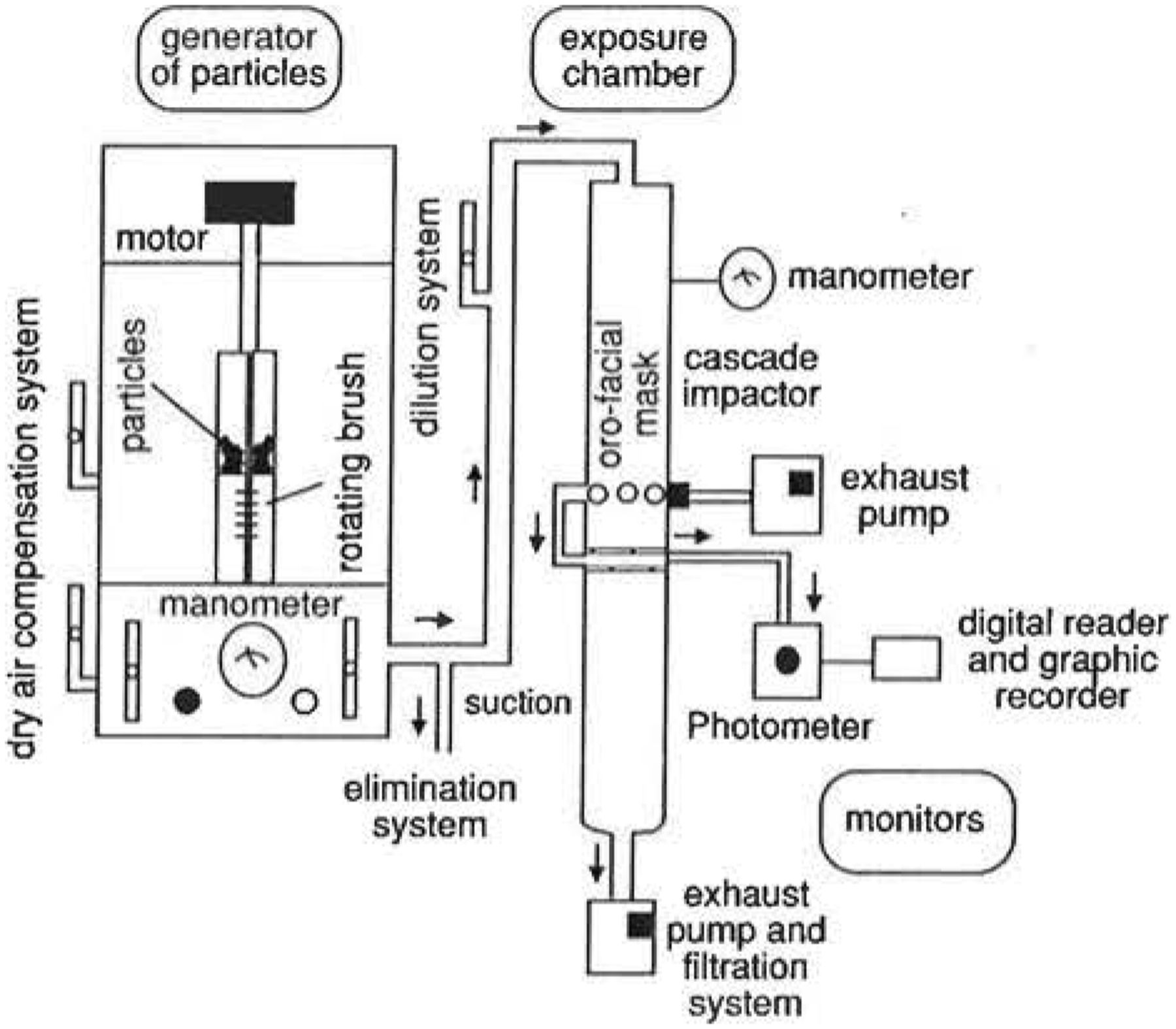
Aerosolization device made in three parts: 1) the particles generator on the left end where the powder is vibrated, taken to the rotating plate by an endless screw and sucked out; 2) the exposure of aerosol delivery chamber, a long plexiglass cylinder with three holes in the centre, one for the facemask, one for the photometer probe and one for the cascade impactor probe; 3) the recording instruments: the photometer and the cascade impactor. Modified from Cloutier and Malo [189], with permission.
Vapours
Challenges with vapours or gases have so far been performed by various investigators in challenge rooms that are similar to each other (fig. 8) [191–193]. The rooms are usually well ventilated with a circuit near the ceiling, so that the air in the antechamber and challenge rooms can be completely changed within a few minutes.

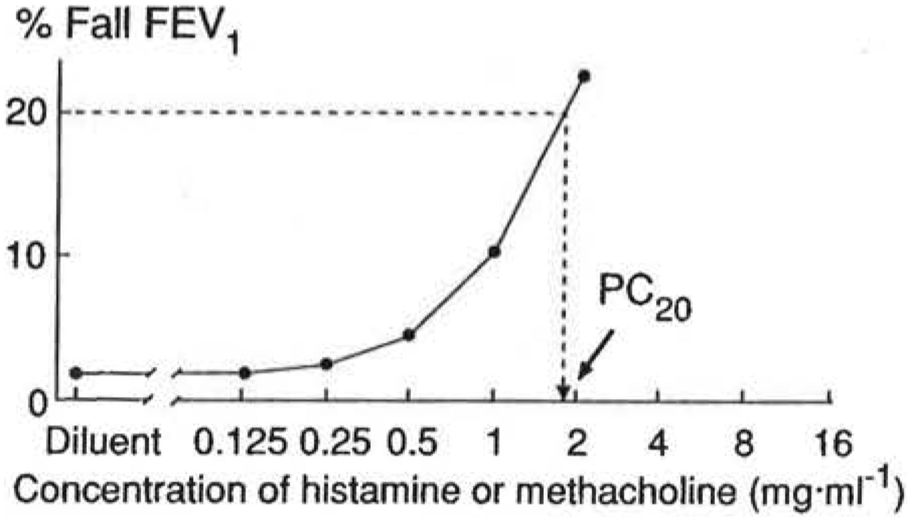
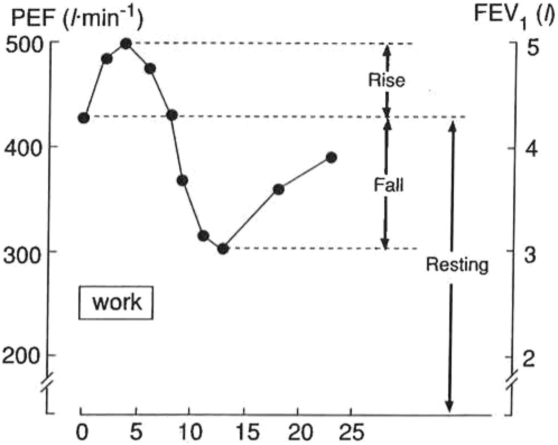
The challenge rooms used at the Hôpital du Sacré-Coeur (Montreal, Canada) for the last few years. There are two antechambers and two challenge rooms. The ventilation system in the ceiling of the rooms is shown by transparency. Two large plexiglass windows allow the technician located in the antechamber at the top of the figure to have a direct access to the ventilation and solenoid valve regulators to control aerosolization within the challenge rooms. For challenges with polyisocyanates, a MDA-7100 monitor is placed in room 1, adjacent to room 2 where the subject sits. The small sampling tube crosses the plexiglass window between rooms 1 and 2.
After the control days required for assessment of reproducibility of lung function measurements, and to exclude airflow obstruction induced by nonspecific mechanisms, subjects are usually exposed to increasing concentrations of the vapour or gas. For polyisocyanates, for example, exposure may be performed in one of the following ways depending on the type of isocyanate:
1 for TDI: 20–100 mL of pure commercial TDI (80% 2.4-TDI and 20% 2.6-TDI) is deposited in a small cup within a bottle, and air is passed through and dispersed in the exposure room;
2 for HDI (hexamethyl diisocyanate), the commercial preparation the subject is exposed to at work (from 20% to 75% of HDI/HDI biuret, depending on the product) is nebulized with the diluent (1:3 concentration) using a plastic or glass nebulizer;
3 for MDI (methylene diphenyl diisocyanate): the commercial preparation containing 40% to 50% of MDI and 50% to 60% of polyethylene polyphenyl isocyanate is heated in a metal cup to approximately 80°C.
The isocyanate concentration may be monitored by automatic monitor (MDA-7050 or MDA-7100). The monitor is usually put in the adjacent challenge room as contamination of the recording paper can occur if the monitor is located in the same room as the source of isocyanate. The tip of the sampling tube of the MDA-7100 monitor crosses the window and is put at a distance equivalent to that which separates the source of polyisocyanates from the subject's mouth. By controlling the ventilation in the room and the output of the nebulizer, the technician can stabilize the concentration of polyisocyanates in the challenge room and keep it below the threshold limit value for short term exposure level (e.g. TLV-STEL which is 20 ppb for isocyanates) [187]. The humidity in the chamber should be kept close to 50% (the reading of polyisocyanates by the MDA-7100 monitor is affected by humidity). A ventilator can homogenize the isocyanate in the air.
A self-contained apparatus including a closed-circuit mixing chamber has recently been developed which allows delivery of controlled concentrations of gaseous isocyanates at the mouth of the subject, without the need of a cumbersome exposure chamber [194]. This alternative approach is promising, and deserves further validation.
3.6.4 Level and duration of exposure
After the control day of assessment of reproducibility of lung function, the subjects are usually exposed to a control chemical (e.g. for TDI challenge it would be the chemical that is usually mixed with polyisocyanates) for a total of 15 min in a challenge room. This allows to verify that the reaction to the «sensitizer» is specific and not due to nonspecific irritation.
The principle is to generate a dose-response curve by increasing the duration and/or the concentration of exposure on different days. The duration of exposure to a very low concentration should be increased progressively for the first day of exposure (e.g. 1, 5, 10, 15 min exposure to 1 ppb TDI). If there is no asthmatic reaction (see below), the duration and/or the concentration of exposure may be increased in different days (e.g. 1, 5, 10, and 15 min to 5, 10, 20 ppb TDI, respectively) during the following 3 days.
In principle, the overall concentration should not exceed the threshold limits value for short-term exposure (TLV-STEL) recommended by the ACGIH [187].
The duration and intensity of exposure to the suspected offending agent should be dictated by clinical history. If there is any indication of a severe immediate reaction, short exposure under controlled conditions should be considered. The level of bronchial obstruction and nonspecific airway responsiveness are also helpful in determining the duration and level of the initial exposure [195].
3.6.5 Expression of the response
FEV1 is more sensitive than the simpler PEF in detecting the response [196]. To be considered positive, investigators generally require a fall of 20% or more in FEV1 at the time of an immediate reaction with progressive recovery in the first hour, or a sustained fall in FEV1 of at least 20% in the case of non-immediate reactions, both in the absence of significant changes on the day of exposure to the control product.
FEV1 is usually assessed for at least 8 h, i.e. every 10 min for the first hour, every 30 min for the second hour and then hourly. Subjects are also asked to continue measuring at least PEF during the evening or during the night in order to investigate whether airway obstruction occurs after the usual period of 8 h monitoring.
An increase in histamine or methacholine airway responsiveness typically occurs after late reactions, but it may also occur after immediate asthmatic reactions [185, 197–201]. Airway responsiveness may also increase without significant changes in FEV1. In these cases a longer exposure to the specific agent can induce bronchoconstrictive reaction [200].
Patterns of reaction
As with bronchial provocation tests with allergens, immediate, late and dual reactions may develop after a specific bronchial provocation test with occupational agents [182, 184].
Atypical reactions, particularly after exposure to polyisocyanates, may also develop [184], mainly of the progressive type (starting within a minute after exposure ended, progressing to a maximum 7 to 8 h later). In addition, there are «square wave» reactions, i.e. no recovery between the immediate and late components of the reaction. Two «progressive recovery» patterns were also seen where subjects had maximum bronchoconstriction immediately after exposure to polyisocyanates [184].
Safety
The same safety procedures that apply to bronchial provocation tests with allergens apply to bronchial provocation tests with occupational sensitizers (§ 2.4.1 and 2.4.2). In particular, it is important to ensure that the asthmatic subject is in a reasonably steady state (FEV1 fluctuations should be less than 10% throughout an observation period of 8 h on the control day), and that medications are kept constant for subjects who require continuous medication (see also § 2.3). It needs to be emphasized that the so-called anti-inflammatory drugs have profound effects on early and late asthmatic responses to sensitizing agents. If PC20 FEV1 to methacholine is <1 mg·mL−1 there is an increased risk of severe reactions to isocyanates [195], which thereby should be considered as a relative contraindication.
False positive and false negative reactions
Even if specific inhalation challenges are the gold standard, false positive or false negative tests can occur.
With immediate reactions, it is sometimes difficult to distinguish between a nonspecific reaction and a reaction similar to one that could occur after exposure to a proper sensitizing agent. Indeed, the pattern of reaction in terms of peak effect and recovery is similar to one that has been described for non-specific reactions such as exercise and hyperventilation [153]. The use of a control «irritant» substance (lactose for powders, the varnish usually mixed with polyisocyanates, etc.) is suggested and using an apparatus that gives information on the concentration and diameter of the particles and polyisocyanates. False positive late reactions can also be observed in subjects in an unstable clinical condition. This is the reason why subjects should always be observed on a control day to make sure that they are in a stable clinical state.
There are several reasons for a false negative test. First of all, subjects can be exposed to the wrong agent [202], or subjects may well have been away from work for too long [203]. If the specific inhalation challenge is negative in the laboratory, provisions should be made to return the subject to his normal workplace while serial monitoring of peak expiratory flow is done. As soon as changes in PEF or bronchial responsiveness is demonstrated, specific inhalation challenges should be repeated in the laboratory with the appropriate agent.
3.7 Experimental challenge tests
Apart from the above standardized tests, a number of relatively new challenging agents have been introduced in research. These are now being used by various laboratories, and are promising approaches for the investigation of specific pathways of pathophysiology of airway narrowing. In view of a potentially wider application of some of these agents in future, the following concise description has been added. It needs to be emphasized that at present none of these challenges have sufficiently been standardized. Therefore, they must be regarded as experimental procedures.
3.7.1 Adenosine 5′-monophosphate
Adenosine 5′-monophosphate (AMP) is considered to be an “indirect” stimulus of airway narrowing, since it exerts its effects through the release of mediators from immunologically activated airway mast cells and probably also through activation of neuronal reflexes [204]. AMP is rapidly metabolised to adenosine, which stimulates several subtypes of purinoceptors. Due to its solubility, AMP enables relatively high doses of adenosine to be given by inhalation in vivo. Asthmatics and atopic non-asthmatic subjects usually respond to AMP with acute airway narrowing, whereas nonatopic normal subjects do not [205, 206]. Repeated challenges with AMP lead to substantial tachyphylaxis and cross-tachyphylaxis with e.g. exercise. Furthermore, complex interaction with hypertonic saline and allergen challenges has been described. AMP is increasingly used in testing the involvement of cellular mechanisms in airway hyperresponsiveness [204].
3.7.2 Leukotrienes
Leukotrienes are pro-inflammatory metabolites of arachidonic acid through the 5-lipoxygenase pathway. Since the cysteinyl leukotrienes (LTC4, LTD4, and LTE4) are considered to be of major importance in bronchial asthma [207], these agents have been given by inhalation in many research studies. They exert their effects through specific receptors, acting on smooth muscle, endothelial cells, nerve fibres, and secretory cells. Therefore, airway narrowing to these substances has a “direct” as well as an “indirect” component. Asthmatic subjects are hyperresponsive to leukotrienes, particularly LTE4 [208], and in some studies leukotrienes have been shown to induce transient airway hyperresponsiveness to other stimuli [209, 210]. A highly efficient challenge methodology has been described [210], which has been shown to produce reproducible results [211]. Leukotriene provocation tests are used in pathophysiological research, particularly in testing the increasingly available leukotriene-antagonists or synthesis inhibitors.
3.7.3 Platelet-activating factor
Platelet-activating factor (PAF) is also a pro-inflammatory mediator of potential importance in asthma [212]. It acts through various subtype PAF-receptors on inflammatory cells, endothelium, and secretory cells. The acute bronchoconstriction after PAF inhalation might be due to the secondary release of leukotrienes [212]. PAF has been shown to induce airway hyperresponsiveness in normal subjects [213], but this could not be confirmed by other investigators [214]. There is rapid tachyphylaxis to PAF inhalation.
3.7.4 Bradykinin
Bradykinin is a potent pro-inflammatory peptide [215] leading to airway narrowing through an “indirect” mechanism. When given by inhalation, it causes acute bronchoconstriction in asthmatics [216], which is most likely due to smooth muscle contraction, vasodilation, oedema, and/or airway secretion secondary to stimulation of C-fibre nerve endings and the release of sensory neuropeptides. Therefore, this challenge is currently used to examine the role of axon reflexes under various experimental conditions, e.g. following allergen challenge in asthma [217].
3.7.5 SO2 and sodium metabisulphite
SO2 is considered to be a stimulus to investigate the role of cholinergic and/or noncholinergic neural pathways in airway narrowing. Instead of administering gaseous SO2 [58, 218] it is much simpler to aerosolize a SO2-generating solution: sodium metabisulphite [219]. The challenge with this preservative causes acute airway narrowing in vivo in a reproducible way [220], and is thereby suitable for research studies. SO2 and metabisulphite challenges may be used to distinguish asthma from COPD [220], but this needs further investigation.
3.7.6 Tachykinins
Tachykinins are sensory neuropeptides localized in airway C-fibre nerve endings, which are believed to play a role in bronchial asthma [222]. These pro-inflammatory peptides are released by local axon-reflexes, causing smooth muscle contraction, vasodilation, microvascular leakage, and hypersecretion, through specific NK-receptors [222]. Therefore, tachykinins can be considered to stimulate both «direct» and «indirect» mechanisms of airway narrowing. Among the tachykinins, neurokinin A and substance P are being used for inhalation challenge testing in research, either by traditional jet nebulizer [223] or by highly efficient nebulization [224]. Relatively high doses are needed to produce bronchoconstriction in normal subjects, because of endogenous neuropeptide-degrading enzyme activity [224]. In male tachykinins have not been shown to induce subsequent airway hyperresponsiveness to other agents.
3.7.7 Propranolol
Another alternative challenging agent is the β-blocker propranolol. When given by inhalation propranolol induces bronchoconstriction in asthmatic patients but not in normal subjects [225, 226]. Many patients with COPD do not respond to propranolol [25, 221]. The mechanism of bronchoconstriction to propranolol is probably related to a blockade of inhibitory β-receptors on cholinergic nerves. This would enhance acetylcholine release, particularly in asthma, because asthmatics may have impaired function of the inhibitory muscarinic autoreceptors [6]. This mechanism explains the effective reversal of propranolol-induced bronchoconstriction by anticholinergic agents [225, 226]. Since propranolol blocks the effect of β-adrenergic bronchodilators, anti-cholinergic drugs are an essential component of the safety precautions during this type of challenge.
4 ANALYSIS AND INTERPRETATION
4.1 Dose-response curves: indices and terminology
The results of bronchoconstrictor challenge tests are preferably analysed from dose-response curves. This allows calculation of the sensitivity to the stimulus (position of the curve) as well as measurement of the change in severity of the response with increasing dose (slope and maximal response) (figs. 1 and 4) [227]. Unfortunately, not every stimulus can easily be given in a dose-response way (e.g. exercise), so that under those conditions the analysis should be limited to the time-response curve following a single dose or multiple doses.
The dose-response curve is currently calculated from log-linear dose-response curves, according to in vitro pharmacology. Concentrations can be used instead of doses. In order to allow comparisons between various agents, it is recommended to use molar units. The response (e.g. in FEV1) can be expressed in percentage fall from baseline value or from predicted value [227]. Since the complete sigmoid dose-response curve can often not be obtained in vivo, the position of the curve can not be expressed as the most commonly used parameter in vitro: the median effective dose (ED50). Instead, the interpolated provocative dose (PD) or concentration (PC) at a given change in lung function is recommended as an index of sensitivity in vivo (fig. 3) [2]. A decreased PD- or PC-value then indicates the presence of hypersensitivity to the stimulus. In the literature, this term is often regarded as synonymous to the term hyperresponsiveness.
The severity of the response is reflected by the shape of the dose-response curve [227]. This can be expressed as the slope of either the linear mid-part of the curve [228] or of a sigmoid curve fit [229]. A steepening of the slope should be referred to as hyperreactivity [62]. This term should, therefore, not be regarded as being synonymous to hyperresponsiveness. If sigmoid dose-response curves can be recorded safely (not exceeding 50% fall in FEV1 in case of normal baseline values), the presence and level of a maximal response plateau provides relevant information on the potential severity of airways obstruction (fig. 1) [3, 42, 227]. The plateau should be calculated by averaging a «stable» response at high doses within a predefined response range [50], or by curve-fitting procedures [50, 229]. Due to irregularly shaped dose-response curves, these criteria for a maximal response may not always be applicable [230]. An absent or elevated maximal response plateau is an indication of excessive airway narrowing [231], which is associated with asthma symptoms [230]. Measurements of excessive airway narrowing are not recommended in patients with lowered levels of baseline FEV1 (§ 2.4.1), except for in specifically designed research protocols under careful guidance of the absolute value of FEV1 during the test [231]. For this reason maximal responses should not be measured for clinical usage, but are important in research studies [3, 227].
Recently, an overall index of position and shape of the dose-response curve in vivo has been introduced for epidemiological purposes by O'Connor et al. [232]. The index is referred to as «dose-response slope» or «two-point slope», and is obtained by calculation of the slope of the straight line between the origin (% decline in FEV1 post-saline) and the response to the final dose (highest dose or dose giving >20% decline in FEV1). The slope is obtained by dividing the difference between post-saline and final FEV1 by the highest dose given, and is expressed in % decline FEV1 per mol methacholine or histamine. This procedure enables one to measure airway responsiveness quantitatively over the full in vivo range, which may be particularly attractive in epidemiological surveys [233–235]. The usefulness of this index requires further investigation (§ 4.3).
4.2 Time-response curves
Even though the early response to allergens or occupational sensitizers can be analysed in a dose-response way as PD- or PC-value (§ 4.1), the most common expression of the results is based on time-response curves, as is the case for exercise tests (fig. 5) [14]. Under those conditions, within- and between-subjects comparison can only be made when exactly the same dose of the stimulus has been given. Usually, time-response curves are analysed by calculating the largest response during a predetermined time-interval. This approach can be applied to both the early and late response. Trapezoidal integration of time-responses curve gives the area under the curve (AUC), which might be a better alternative that needs to be further evaluated (fig. 6) [169].
4.3 Reproducibility
In general, measures of airway responsiveness to histamine and methacholine are reproducible between days when the clinical state of the patient is stable. When repeating challenge tests any potential carry-over effects, such as cumulation, priming, or refractoriness need to be taken into consideration. Reproducibility needs to be calculated according to accepted statistical methods [236–239]. It is strongly dependent on the standardization of the protocol, the experience of the technician and the patient, and on the clinical stability of the patient [46, 97, 99, 240, 241].
When histamine and methacholine inhalation tests, using the tidal breathing and dosimeter methods, are repeated within one week in clinically stable patients, the within-subject standard deviation of log PC20 or log PD20 is between 0.09 and 0.33 [83, 85, 92]. Therefore, in hospital populations under optimal conditions, the 95% confidence interval of PC20 or PD20 based on a single determination is less than one twofold concentration difference. However, these conditions can not always be met, particularly in an epidemiological setting. In those circumstances where the clinical stability of the patients and/or the intertechnician variability can not be controlled, the 95% confidence interval for single measurements of PC20 or PD20 increases towards 2–3 twofold dose difference [46, 97, 99]. This also holds for lack of subject's experience [240], and for non-optimally standardized techniques [90, 241]. When the within-subject variance is related to the between-subject variance in the clinical range (PC20: 0.3–16 mg·mL−1), the intraclass correlation of the PC20 or PD20 has been reported to be around 0.9 in adults [19, 83, 92, 98]. When using the Yan method in children, an intraclass correlation coefficient of 0.78 has been reported [98].
The other variables that can be obtained from the dose-response curve have not been evaluated extensively on their reproducibility. For instance, the repeatability of the «two-point slope» has been reported to be satisfactory [98, 232], although a recent study indicates that it becomes worse in the least responsive subjects in whom the slopes are very shallow [242]. The maximal response plateau has similar reproducibility in clinical [50] and epidemiological studies [230], the 95% confidence interval of single measurements being about 11% in adults as well as in children [243].
Less is also known about the reproducibility of the non-pharmacological challenges. The data on cold/dry air inhalation [128], non-isotonic aerosols [125], and exercise [152, 158, 244] are encouraging, but certainly warrant further studies on the present standardized methodologies. The repeatability of standardized allergen challenges has not formally been investigated. When the level of baseline FEV1, skin test sensitivity and histamine airway responsiveness are not taken into consideration the repeatability of EAR [245, 246] or LAR [247, 248] is disappointing. However, if these pre-test patient characteristics are controlled [167] and the procedure is carefully standardized (§ 3.5), the EAR and LAR seem to be fairly well reproducible (fig. 6) [38, 164, 249]. Obviously, this needs further investigation.
4.4 Baseline lung function
The association between airway responsiveness and baseline airway calibre has long been recognized [250], but the precise mechanisms of this relation have not been determined. Interdependence between these two physiological characteristics might be explained by: (a) geometrical factors determining airway responsiveness, (b) airway responsiveness causing lower levels of baseline lung function, or (c) both variables being commonly dependent on other disease characteristics. These three mechanisms may all be involved, depending on the population of subjects. The association between baseline lung function and airway responsiveness can be observed in cross-sectional analysis in the general population [251]. In clinical populations the association appears to be stronger in COPD than in asthma [25]. Cross-sectional studies in COPD indicate that the level of lung function can account for 24 to 74% of the variance in airway responsiveness [7]. However, cross-sectional and longitudinal measurements in asthma have indicated that baseline lung function is of minor importance in determining airway responsiveness in this group of patients [252, 253], accounting for about 30% of the within-subject variance of PC20 [254]. The reverse effect, namely, airway responsiveness causing an accelerated decline in lung function, does seem to play a role in asthma [32] as well as COPD [255].
These findings hamper the interpretation of airway hyperresponsiveness in patients with airways obstruction. On the other hand, they also show that airway responsiveness and baseline lung function are partially independent variables.
4.5 Clinical relevance of the various challenges
The choice of the bronchoconstrictor depends on pathophysiological, methodological, and clinical aspects (§ 2.5). Regarding the application in clinical practice, first, airway responsiveness measurements can provide useful information in clinical diagnosis (§ 2.1), particularly in patients without otherwise documented variable airways obstruction [16, 17]. Airway hyperresponsiveness is not a diagnosis in itself. It is a composite functional disorder, reflecting the potential of variable airways obstruction. The combination of airway hyperresponsiveness and recent symptoms of wheezing (within the previous year) has been advocated as a «gold standard» for defining asthma in epidemiology [256].
Second, changes in airway responsiveness can be used in monitoring treatment effects [35]. In this way it can be shown that bronchodilators can afford an acute protection against bronchoconstrictor stimuli for their duration of action. In research this is often examined using e.g. histamine or methacholine [231, 257], but in clinical practice it seems to be more relevant to investigate their protection against physical stimuli, such as exercise or cold air [35]. In particular this holds for the acute effects of sodium cromoglycate and nedocromil [35]. Anti-inflammatory drugs like sodium cromoglycate, nedocromil, and particularly inhaled corticosteroids have been shown to reduce airway responsiveness after long-term treatment in asthma [258], but not in patients with COPD in whom it may take substantially longer to reverse airway hyperresponsiveness [259, 260].
When comparing the effects of long-term anti-inflammatory treatment on the various challenges, it appears that the induced changes in the position of the dose-response curve (decrease in sensitivity) differs between pharmacological and physical stimuli [44, 261]. Although changes in the position of curve may not always be the optimal way of expressing protective effects [44], these findings suggest that therapeutical efficacy on airway hyperresponsiveness can not be judged by using one single challenge procedure. Obviously, this needs to be further investigated in combination with other disease characteristics, such as the change in clinical indices and in pathology. Therefore, even though it is clear that longterm therapy can be monitored by airway responsiveness measurements, any superiority of one test over the other is still unclear. Altogether, it can be postulated that airway responsiveness can thus be used as a guide to asthma therapy [26]. However, it still needs to be examined whether adjusting therapy also based on the degree of hyperresponsiveness leads to better prognosis of the disease than therapy according to current international recommendations, based on symptoms and lung function alone [16, 17].
This leads to the following general recommendations on choice between the various challenge tests in clinical practice. Histamine and methacholine challenges are first choice in diagnosis and follow-up of patients with asthma. Even though slight differences have been observed between these two agents (§ 3.1.8), they can be used interchangeably in clinical practice. The main indication of these tests is to exclude asthma when spirometry is normal. The clinical relevance of the physical tests is still limited. They do provide information on «indirect» mechanisms of airway narrowing [43], and the results obtained with these tests might be closer associated with clinical disease indices than those obtained with pharmacological tests [262]. Regarding the differential diagnosis between asthma and COPD, non-isotonic aerosols do not allow a clear distinction between these two entities [25, 221]. However, a positive response to isocapnic hyperventilation [136, 263] or exercise [149] seems to be consistent with the diagnosis of asthma. Further application of physical tests consists of the laboratory investigation of the potential protection afforded by anti-asthma drugs [35].
Challenges with allergens and occupational sensitizers should be considered as research tools. Allergen challenge may only have a role in those few cases were immunotherapy is seriously indicated. Although challenge with occupational sensitizers is considered to be the «gold standard» for the confirmation of occupational asthma [184], these tests are only indicated in case of inconclusive diagnosis based on history, peak expiratory flow and/or histamine responsiveness measurements. These laboratory exposure procedures with sensitizing agents must be restricted to specialized centres.
4.6 Applications in research
The measurement of airway responsiveness has a major impact in pathophysiological, clinical, and epidemiological research in patients with asthma or COPD in vivo [5–7]. Airway hyperresponsiveness may represent a non-invasive indication of various pathological processes in the airway wall [13]. The increase in sensitivity and excessive airway narrowing in response to bronchoconstrictor stimuli are associated with e.g.: epithelial damage, the number and activity of (sub)mucosal inflammatory cells, (non) adrenergical-(non) cholinergical autonomic dysfunction, hyperaemia, plasma exudate, and smooth muscle contractility [3]. The contribution of these mechanisms can be investigated by using various bronchoconstrictor stimuli, either or not combined with specific antagonists or inhibitors of the pathways leading to airway narrowing (see also § 3.7).
One of the best investigational models of airway responsiveness in male in vivo is the induced hyperresponsiveness after exposure to inhaled allergen, occupational sensitizing agents or virus infections [37]. Among these, the allergen-induced hyperresponsiveness following a late asthmatic response has been validated most extensively [264]. This procedure is being widely used in pathophysiological research, and the pharmacological modulation of allergen-induced responses forms a key to the development of anti-asthma drugs. It remains to be established as to whether drug effects on allergen challenges are predictive for their efficacy in clinical asthma.
5 CONCLUSIONS
Even though methodological issues are not a purpose on their own, they are of vital importance in clinical decision making as well as in science. It is obvious that the present document does not solve all the dilemma's in this area. Uncovered and controversial areas still need to be explored. Consequently, improvement and standardization of airway responsiveness measurements should be an ongoing effort. In the following, some of the established issues and a number of remaining questions have been selected.
5.1 Recommendations
5.1.1 General
- measurements of airwayresponsiveness have a good safety record, provided the procedures are carefully standardized;
- the presently standardized laboratory-protocols are the methods of first choice for pharmacological and physical challenges, as well as for challenges with sensitizing agents; they allow comparison of the results with previous data in the literature;
- the term «nonspecific» airway responsiveness is ambiguous, and should be identified with the challenging agent, since the mechanisms of bronchoconstriction varies between the distinct stimuli;
- hyperresponsiveness should be regarded as an abnormality of airway function, which is associated with various features of inflammation;
- newly developed and potentially better methods should be evaluated on their repeatability and their agreement with currently standardized challenges.
5.1.2 Clinical usage
- airway responsiveness measurements with pharmacological agents are particularly useful for the exclusion of asthma in the clinical setting, they are less useful in confirming and distinguishing the diagnoses of asthma and COPD;
- hyperresponsiveness is not synonymous with the diagnosis of asthma, but can provide additional information to clinical symptoms and peak flow measurements on the potential of variable airways obstruction in epidemiological and clinical setting;
- a 10–20% fall in FEV1 in response to isocapnic hyperventilation or exercise is consistent with the diagnosis of asthma; however, a negative response to these stimuli does not exclude asthma;
- the combination of airway hyperresponsiveness and recent wheezing (in the past year) can be regarded as a definition of asthma for epidemiology;
- in clinical practice histamine and methacholine tests are most extensively validated, and thereby the challenges of first choice in adults; in paediatrics exercise tests can be a better alternative;
- physical challenges (exercise, cold air, non-isotonic aerosols) reflect the involvement of a cascade of «indirect» components of airway narrowing, such as cellular and neurogenic mechanisms;
- inhalation tests with common allergens should be regarded as a research tool;
- challenge tests with occupational sensitizers should only be performed in specialized centres;
- the safety requirements for inhalation challenge tests vary between the different types of stimuli, being most stringent for tests with allergens and occupational sensitizers;
5.1.3 Materials and methods
- output should be calibrated for each individual nebulizer; in clinical practice this can be estimated adequately by weighing;
- for research purposes the same nebulizer should be used at exactly the same setting throughout the study;
- there are three standardized methods for pharmacological challenges (tidal breathing, dosimeter, and Yan method); the results have similar reproducibility, and the choice between them depends on the type of study (clinical or epidemiological) and local facilities and experience;
- for the tidal breathing method, 2 min inhalation of doubling doses (nebulizer output 0.13 mL·min−1) is recommended;
- for the dosimeter method, doubling doses should be administered by inhaling 5 puffs of 9 μl per dose;
- FEV1 is the lung function index of first choice during challenge tests in clinical practice.
- hypotonic- and hypertonic aerosol challenge should be performed with ultrasonically nebulized distilled water and 4.5% saline, respectively, with 1.2 mL·min−1 aerosol output and doubling exposure times;
- isocapnic hyperventilation can be performed by doubling steps of minute ventilation with cooled dry air (water content 0 mg·l−1, −15°C) or dry air at room temperature; alternatively dry air at room temperature can be used at 30, 60, and 100% of maximal voluntary ventilation;
- during exercise challenge the subject should attain a level of ventilation between 40%–60% of the subjects predicted maximal voluntary ventilation during the last 4 min of a 6–8 min test;
- in allergen challenge tests the starting allergen dose is calculated from the histamine or methacholine PC20 and the result of allergen prick skin tests («skin test endpoint»);
- until European consensus about the exposure limits during challenge tests with occupational sensitizers is reached, the American limits (TLV-STEL) are recommended.
5.1.4 Analysis and interpretation
- when analyzing dose-response curves obtained by inhalation challenge tests, the position of the curve (PC or PD value) is the index of choice in clinical practice;
- the so-called «two-point slope» enables analysis of dose-response curves in the general population, and is a promising index of airway responsiveness in epidemiological studies; its repeatability needs further studies, particularly in adults;
- there is a complex interaction between baseline lung function and airway responsiveness, that appears to be different between asthma and COPD; therefore, challenge tests are hard to interpret in the presence of airways obstruction.
5.2 Remaining questions
- what is the meaning of airway hyperresponsiveness in clinical diagnoses?
- what is the role of airway responsiveness in the clinical follow-up of patients with asthma?
- can airway responsiveness measurements be a guide to asthma therapy?
- what is the meaning of airway hyperresponsiveness in patients with COPD?
- how should the test results or inhaled doses be size-corrected in children?
- what are the advantages of using physical tests instead of pharmacological tests in clinical practice?
- are monitoring of peak flow and histamine or methacholine challenges true alternatives to challenge with occupational sensitizers in the diagnosis of occupational asthma?
- is an allergen inhalation test an adequate model of clinical asthma in the prediction of treatment effects?
- is maximal bronchoconstriction more relevant than sensitivity as an index of airway responsiveness in clinical practice?
- to what extent do the various tests for hyperresponsiveness relate to the pathology in the airways?
- are physical tests more specific while less sensitive for the diagnosis of asthma?
- ©ERS Journals Ltd
REFERENCES


