
Main menu

  • Home
  • Current issue
  • ERJ Early View
  • Past issues
  • ERS Guidelines
  • Authors/reviewers
    • Instructions for authors
    • Submit a manuscript
    • Open access
    • COVID-19 submission information
    • Peer reviewer login
  • Alerts
  • Subscriptions
  • ERS Publications
    • European Respiratory Journal
    • ERJ Open Research
    • European Respiratory Review
    • Breathe
    • ERS Books
    • ERS publications home

User menu

  • Log in
  • Subscribe
  • Contact Us
  • My Cart
  • Log out

Search

  • Advanced search
  • ERS Publications
    • European Respiratory Journal
    • ERJ Open Research
    • European Respiratory Review
    • Breathe
    • ERS Books
    • ERS publications home

Login

European Respiratory Society

Advanced Search

  • Home
  • Current issue
  • ERJ Early View
  • Past issues
  • ERS Guidelines
  • Authors/reviewers
    • Instructions for authors
    • Submit a manuscript
    • Open access
    • COVID-19 submission information
    • Peer reviewer login
  • Alerts
  • Subscriptions

European Respiratory Society statement on thoracic ultrasound

Christian B. Laursen, Amelia Clive, Rob Hallifax, Pia Iben Pietersen, Rachelle Asciak, Jesper Rømhild Davidsen, Rahul Bhatnagar, Eihab O. Bedawi, Niels Jacobsen, Courtney Coleman, Anthony Edey, Gabriele Via, Giovanni Volpicelli, Gilbert Massard, Francesco Raimondi, Matthew Evison, Lars Konge, Jouke Annema, Najib M. Rahman, Nick Maskell
European Respiratory Journal 2021 57: 2001519; DOI: 10.1183/13993003.01519-2020
Christian B. Laursen
1Dept of Respiratory Medicine, Odense University Hospital, Odense, Denmark
2Dept of Clinical Research, Faculty of Health Sciences, University of Southern Denmark, Odense, Denmark
  • Find this author on Google Scholar
  • Find this author on PubMed
  • Search for this author on this site
  • ORCID record for Christian B. Laursen
  • For correspondence: christian.b.laursen@rsyd.dk
Amelia Clive
3Academic Respiratory Unit, University of Bristol, Bristol, UK
4Southmead Hospital, North Bristol NHS Trust, Bristol, UK
  • Find this author on Google Scholar
  • Find this author on PubMed
  • Search for this author on this site
Rob Hallifax
5Oxford Centre for Respiratory Medicine, Churchill Hospital, Oxford University Hospitals NHS Foundation Trust, Oxford, UK
6Oxford Respiratory Trials Unit, Nuffield Dept of Medicine, University of Oxford, Oxford, UK
  • Find this author on Google Scholar
  • Find this author on PubMed
  • Search for this author on this site
Pia Iben Pietersen
1Dept of Respiratory Medicine, Odense University Hospital, Odense, Denmark
2Dept of Clinical Research, Faculty of Health Sciences, University of Southern Denmark, Odense, Denmark
7Regional Center for Technical Simulation, Odense University Hospital, Odense, Denmark
  • Find this author on Google Scholar
  • Find this author on PubMed
  • Search for this author on this site
Rachelle Asciak
5Oxford Centre for Respiratory Medicine, Churchill Hospital, Oxford University Hospitals NHS Foundation Trust, Oxford, UK
6Oxford Respiratory Trials Unit, Nuffield Dept of Medicine, University of Oxford, Oxford, UK
  • Find this author on Google Scholar
  • Find this author on PubMed
  • Search for this author on this site
Jesper Rømhild Davidsen
1Dept of Respiratory Medicine, Odense University Hospital, Odense, Denmark
2Dept of Clinical Research, Faculty of Health Sciences, University of Southern Denmark, Odense, Denmark
8South Danish Center for Interstitial Lung Diseases (SCILS), Odense University Hospital, Odense, Denmark
  • Find this author on Google Scholar
  • Find this author on PubMed
  • Search for this author on this site
  • ORCID record for Jesper Rømhild Davidsen
Rahul Bhatnagar
3Academic Respiratory Unit, University of Bristol, Bristol, UK
4Southmead Hospital, North Bristol NHS Trust, Bristol, UK
  • Find this author on Google Scholar
  • Find this author on PubMed
  • Search for this author on this site
Eihab O. Bedawi
5Oxford Centre for Respiratory Medicine, Churchill Hospital, Oxford University Hospitals NHS Foundation Trust, Oxford, UK
6Oxford Respiratory Trials Unit, Nuffield Dept of Medicine, University of Oxford, Oxford, UK
  • Find this author on Google Scholar
  • Find this author on PubMed
  • Search for this author on this site
  • ORCID record for Eihab O. Bedawi
Niels Jacobsen
1Dept of Respiratory Medicine, Odense University Hospital, Odense, Denmark
2Dept of Clinical Research, Faculty of Health Sciences, University of Southern Denmark, Odense, Denmark
7Regional Center for Technical Simulation, Odense University Hospital, Odense, Denmark
  • Find this author on Google Scholar
  • Find this author on PubMed
  • Search for this author on this site
Courtney Coleman
9European Lung Foundation, Sheffield, UK
  • Find this author on Google Scholar
  • Find this author on PubMed
  • Search for this author on this site
Anthony Edey
10Dept of Radiology, Southmead Hospital, North Bristol NHS Trust, Bristol, UK
  • Find this author on Google Scholar
  • Find this author on PubMed
  • Search for this author on this site
Gabriele Via
11Cardiac Anesthesia and Intensive Care, Cardiocentro Ticino, Lugano, Switzerland
  • Find this author on Google Scholar
  • Find this author on PubMed
  • Search for this author on this site
Giovanni Volpicelli
12Dept of Emergency Medicine, Ospedale San Luigi Gonzaga, Torino, Italy
  • Find this author on Google Scholar
  • Find this author on PubMed
  • Search for this author on this site
Gilbert Massard
13Faculty of Science, Technology and Medicine, University of Luxembourg, Grand-Duchy of Luxembourg
  • Find this author on Google Scholar
  • Find this author on PubMed
  • Search for this author on this site
Francesco Raimondi
14Division of Neonatology, Section of Pediatrics, Dept of Translational Medical Sciences, Università “Federico II” di Napoli, Naples, Italy
  • Find this author on Google Scholar
  • Find this author on PubMed
  • Search for this author on this site
Matthew Evison
15Wythenshawe Hospital, Manchester University NHS Foundation Trust, Manchester, UK
  • Find this author on Google Scholar
  • Find this author on PubMed
  • Search for this author on this site
Lars Konge
16Copenhagen Academy for Medical Education and Simulation, The Capital Region of Denmark, Centre for HR, University of Copenhagen, Copenhagen, Denmark
  • Find this author on Google Scholar
  • Find this author on PubMed
  • Search for this author on this site
Jouke Annema
17Dept of Respiratory Medicine, Amsterdam UMC, University of Amsterdam, Amsterdam, The Netherlands
  • Find this author on Google Scholar
  • Find this author on PubMed
  • Search for this author on this site
Najib M. Rahman
5Oxford Centre for Respiratory Medicine, Churchill Hospital, Oxford University Hospitals NHS Foundation Trust, Oxford, UK
6Oxford Respiratory Trials Unit, Nuffield Dept of Medicine, University of Oxford, Oxford, UK
18Laboratory of Pleural and Lung Cancer Translational Research, Nuffield Dept of Medicine, University of Oxford, Oxford, UK
19National Institute for Health Research, Oxford Biomedical Research Centre, University of Oxford, Oxford, UK
20Joint last authors
  • Find this author on Google Scholar
  • Find this author on PubMed
  • Search for this author on this site
Nick Maskell
3Academic Respiratory Unit, University of Bristol, Bristol, UK
4Southmead Hospital, North Bristol NHS Trust, Bristol, UK
20Joint last authors
  • Find this author on Google Scholar
  • Find this author on PubMed
  • Search for this author on this site
  • Article
  • Figures & Data
  • Info & Metrics
  • PDF
Loading

Abstract

Thoracic ultrasound is increasingly considered to be an essential tool for the pulmonologist. It is used in diverse clinical scenarios, including as an adjunct to clinical decision making for diagnosis, a real-time guide to procedures and a predictor or measurement of treatment response. The aim of this European Respiratory Society task force was to produce a statement on thoracic ultrasound for pulmonologists using thoracic ultrasound within the field of respiratory medicine. The multidisciplinary panel performed a review of the literature, addressing major areas of thoracic ultrasound practice and application. The selected major areas include equipment and technique, assessment of the chest wall, parietal pleura, pleural effusion, pneumothorax, interstitial syndrome, lung consolidation, diaphragm assessment, intervention guidance, training and the patient perspective. Despite the growing evidence supporting the use of thoracic ultrasound, the published literature still contains a paucity of data in some important fields. Key research questions for each of the major areas were identified, which serve to facilitate future multicentre collaborations and research to further consolidate an evidence-based use of thoracic ultrasound, for the benefit of the many patients being exposed to clinicians using thoracic ultrasound.

Abstract

Continued clinical use and research has established thoracic ultrasound as a key tool and skill for the modern pulmonologist https://bit.ly/3cCL6KS

Introduction

Thoracic ultrasound (TUS) is increasingly considered an essential tool for the pulmonologist [1–3]. Although this technique was for many years considered “of no use” in the lung, many decades of research have demonstrated high clinical utility in a number of areas of pulmonary disease. While the technique of TUS originated with radiologists, it is increasingly being used by pulmonologists “at the bedside”, and in this context is used with several potential aims. These include as an adjunct to clinical decision making for diagnosis, as a real-time guide to procedures and as a predictor or measurement of treatment response [1, 2, 4–6].

This European Respiratory Society (ERS) statement has been written in light of the growing evidence behind the use of TUS across a broad range of respiratory disease areas, as a summary of the evidence to clinicians who wish to understand the current rationale and state of the art of this technique. An evidence-based approach has been used throughout, addressing major areas of TUS practice and application. These include sections on required equipment and technique, assessment of the chest wall, parietal pleura, pleural effusion, pneumothorax, the utility of ultrasound in diffuse lung parenchymal diseases, diaphragm assessment, intervention guidance and suitable training in TUS.

We have highlighted areas of potential future research, as suggested by the current state of the evidence, at the end of each topic, and hope that this will lead to further definitive studies that will further improve our diagnostic and treatment armamentarium and benefit our patients.

Methods

The task force was comprised of clinicians with internationally recognised expertise in TUS. In order to reflect the multidisciplinary use of TUS, the expert group included pulmonologists and relevant experts from other specialties (e.g. radiology, emergency medicine, intensive care, thoracic surgery, paediatrics). This group was supplemented with young ERS members with TUS experience, and representatives of the European Lung Foundation (ELF). An ERS methodologist provided feedback on research strategy, evidence and statement synthesis, and oversight of the task force process.

The task force was initiated in December 2018 and comprised one face-to-face meeting, regular telephone conferences and e-mail correspondence. Initially, the task force established the overall scope and aim of the statement. It was agreed to limit the statement to the general use of TUS in the context of the clinical use by a pulmonologist. The members then agreed on a list of core topics to be addressed in the final statement.

A group of task force members with a designated topic leader was assigned to each topic and was responsible for development of a search strategy, evidence synthesis, and writing of an initial topic section for the statement. MEDLINE and Scopus databases were used for the literature searches, with inclusion of additional studies identified by individual task force members. An ELF representative was assigned to the write a section on the topic “patient perspectives”. The search terms for each topic are provided in the supplementary material. The topic leader identified and used relevant studies and knowledge of current clinical practice to make an initial topic draft, which the topic group then reviewed until a proposed final topic section had been completed. In addition to evidence synthesis, each group was given the task of identifying major gaps in the current evidence and provide key areas for future TUS research. The findings from the literature regarding the patient perspectives topic were shared with patients who had experience of TUS to identify additional perspectives.

Using the proposed topic sections, the task force chairmen composed a first draft of the statement manuscript. Task force members then provided comments and suggestions in the making of the final manuscript based on the draft. The final version of the manuscript was reviewed and approved by all task force members. The statement provides an overview of the evidence and current clinical practice for general TUS performed by pulmonologists, but does not provide recommendations for clinical practice.

According to ERS policies, task force members disclosed potential conflict of interests at the beginning of the task force process and prior to the publication of the statement.

Results

Equipment and technique

Overview of the evidence and current practice

When compared to other forms of clinical ultrasound with an established clinical tradition or more narrow clinical indication, international consensus publications regarding the equipment and technique used for TUS are scarce [1, 3, 7]. The first international consensus paper on point-of-care lung ultrasound provides some essential basic definitions and terminology [1]. However, when compared to recommendations on point-of-care cardiac ultrasound, a general recommendation regarding equipment and technique is not provided [8]. This reflects current clinical practice, in which choice of TUS scanning protocol, equipment and technique varies between specialties and countries. Numerous different protocols, techniques and use of different equipment have been assessed and validated in prospective diagnostic accuracy studies [6, 9–29]. Studies directly assessing or comparing different TUS equipment or techniques are fairly limited. However, these studies have demonstrated that factors such as choice of ultrasound machine (e.g. high-end, hand-held), protocol, transducer and patient positioning have a potential clinical impact [30–39]. Even though studies have addressed important factors, it is not possible to derive a universal and evidence-based TUS approach for any given clinical scenario. Apart from the examination itself, the coronavirus disease 2019 (COVID-19) pandemic has increased awareness of ultrasound operators in ensuring necessary safety precautions, specifically regarding cleansing of equipment and appropriate infection control [40].

Conclusions

Many different approaches and techniques have been described and validated. Comparative studies directly comparing different TUS approaches are limited. Given the many clinical settings and indications, a one-size-fits-all TUS approach is not feasible or meaningful. There remains a need to reach consensus on a general TUS principles and to determine the optimal approach for more specific clinical problems or settings (table 1).

View this table:
  • View inline
  • View popup
TABLE 1

Recommendations for future research: equipment and technique

Chest wall and parietal pleura

Chest wall soft tissues

On TUS, the intercostal muscle and fascia are visualised as echogenic layers under the subcutaneous tissue (figure 1). TUS can be used to identify and characterise superficial chest wall lesions, although generally cross-sectional imaging modalities, such as computed tomography (CT) and magnetic resonance imaging are more accurate [41].

FIGURE 1
  • Download figure
  • Open in new tab
  • Download powerpoint
FIGURE 1

Normal findings and chest wall. a) Normal findings: the muscles, fascia and other soft tissues of the chest wall (CW) are located in the upper part of the image. The surfaces of the ribs (R) can be seen as two horizontal hyperechoic, white, lines with posterior “shadowing” (*). The pleural line (P) is located just below the ribs. The lung tissue is filled with air and can therefore not be seen; therefore, the area (“L”) which can be seen below the pleural line is not the lung tissue, but artefacts. b) V rib fracture: the surface of the rib (R) can be seen as a horizontal, hyperechoic white line. A rib fracture (F) is present. c) Subcutaneous emphysema: air in the chest wall is typically seen as hyperechoic lines or dots with posterior shadowing (*). When the air is placed along a fascia it can be mistaken for the pleural line. However, when compared to (a), the ribs cannot be visualised and the hyperechoic white line caused by the subcutaneous emphysema is much more superficially located (∼1 cm) than the pleural line seen in (a) (∼2.5 cm). d) Benign pleural tumour: a parietal pleural tumour (T) is present. It is seen as a hypoechoic, black, well demarcated structure located above the pleural line (P).

Visceral and parietal pleura

Below the intercostal muscle, the visceral and parietal pleura are visualised as an echogenic “pleural line” visible between and deep to the ribs (figure 1). In healthy individuals, the pleural line slides parallel to the chest wall during respiration generating a “sparkling” appearance (“lung sliding”) and may move in synchrony with cardiac pulsation (“lung pulse”). Lung sliding can be confirmed using M-mode, which gives a characteristic “seashore” sign [1].

Hypoechoic parietal pleural thickening may mimic a pleural effusion on ultrasound and use of colour Doppler may help to differentiate these conditions, with fluid showing disordered colour flow, unlike static, solid pleural thickening. Benign pleural tumours such as fibromas and lipoma are relatively rare, are usually round or ovoid in shape, hypoechoic and homogeneous and do not infiltrate surrounding structures (figure 1) [42]. Asbestos-related pleural plaques have a distinctive TUS appearance and are hypoechoic, elliptical and smoothly limited foci; if calcified, they produce prominent acoustic shadows [42].

Malignant pleural nodularity is a more common finding and can be seen as irregular, well-circumscribed, often heterogeneous lumps arising from the parietal pleura and distorting the normal contour of the visceral pleura. They may be associated with a pleural effusion or chest wall/rib invasion [43].

TUS has been evaluated in the identification of the presence and degree of chest wall invasion of intrathoracic malignancies, and in one study was shown to have a higher sensitivity than CT [44]. The absence of pleural motion next to a peripheral lung lesion may identify parietal pleural invasion, thus refining radiological staging. In one study, the use of qualitative and quantitative colour Doppler sonography was more sensitive and specific than CT for predicting chest wall invasion by lung tumours [45].

Ribs

The ribs are seen as superficial, curvilinear structures, which completely reflect the ultrasound wave, resulting in posterior acoustic shadowing (rib shadows) (figure 1). If an ultrasound probe is placed along the long axis of a rib, the cortex of the bone is visible as a static, bright, echogenic line. When a cortical fracture is present, this line is disrupted by a step or gap and reverberation echoes occur at the point of the fracture (known as the “lighthouse phenomenon”) (figure 1). Fractures may be associated with a visible haematoma, a reactive pleural effusion or subpleural parenchymal changes from lung contusion. A recent systematic review suggests that TUS is more sensitive than chest radiography in diagnosing rib fractures; however, it can be painful and time-consuming, may be technically challenging in obese patients and the first ribs and retroscapular areas are incompletely visible. TUS may have a useful role in assessing focal areas of rib pain [46].

Metastasis of disease to the ribs causes destruction of the bone cortex, resulting in an irregular cortical appearance and loss of the usual rib shadows. In this instance, the infiltrated bone structure may be more visible and appear hypoechoic and heterogenous [47].

Intercostal muscles

The intercostal muscles can be directly visualised between the ribs. Even though the role of TUS assessment of intercostal muscle function is yet to be established, studies have reported several possible clinical implications. In a study by Wallbridge et al. [48] muscle thickness and echogenicity was found to correlate with spirometry assessment of severity in patients with COPD. In addition, intercostal muscle assessment has been reported as a possible tool for assessing respiratory workload in mechanically ventilated patients and for predicting failure of spontaneous breathing trials after supported ventilation [49–51].

Intercostal vessels

Distal to the apex of the rib posteriorly, the intercostal vessels run in the subcostal groove, but their course may be tortuous, particularly in the elderly. Colour flow Doppler using a linear probe allows good visualisation of the vessels and in theory may reduce vascular injury during pleural intervention [52]. However, the technique is operator- and experience-dependent and its reliability and accuracy has been questioned.

Conclusions

TUS is a useful clinical tool for assessing the chest wall and parietal pleura. Further studies are required to ascertain its clinical utility and impact in specific clinical scenarios (table 2).

View this table:
  • View inline
  • View popup
TABLE 2

Recommendations for future research: chest wall and parietal pleura

Pleural effusion

TUS has been used to assess suspected pleural effusion for ≤40 years [53–55]. Recently, a desire for better identification, classification and quantification of pleural fluid, coupled with rapid improvements in technology, has driven the widespread adoption of TUS by many pulmonologists. Indeed, being able to locate fluid with ultrasound to guide intervention is now seen as a core skill for trainees [56].

Benefits of TUS over other modalities

Basic, grey-scale TUS can identify much smaller volumes of fluid in comparison to other modalities, particularly chest radiography [57, 58]. It can do so reliably (meta-analysis data suggests a sensitivity of 93% and specificity of 96%) [59] in real-time at the bedside, with very high spatial resolution. The addition of colour Doppler may enhance assessment and improve differentiation of fluid from pleural thickening [60, 61].

Point-of-care versus diagnostic imaging

TUS is most commonly used as a point-of-care test to guide intervention for pleural effusions, and there is strong evidence to suggest this improves safety and can guide management decisions [62–64]. With sufficient experience, more formal diagnostic imaging is possible. While the ultrasonographic appearance of the fluid in itself cannot be considered diagnostic of the underlying disease, other typical TUS findings may help support a diagnosis (e.g. irregular nodularity on the diaphragm in malignant pleural effusion) (figure 2) [65].

FIGURE 2
  • Download figure
  • Open in new tab
  • Download powerpoint
FIGURE 2

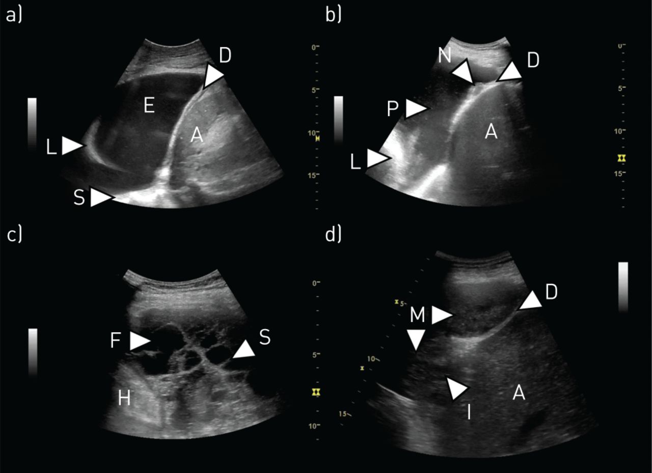
Pleural effusion and pleural malignancy. a) Simple pleural effusion: a simple, anechoic, pleural effusion (E) is present. There are no septations or visible structures floating within the effusion. The effusion can be used as an acoustic window to visualise underlying structures such as the lung (L), diaphragm (D) and thoracic spine (S). Abdominal structures such as the spleen (A) can be visualised below the diaphragm. b) Malignant pleural effusion: image from a patient with a malignant pleural effusion. A large nodule (N) is present on the diaphragm. Hyperechoic “plankton” (P) can be seen floating within the effusion. The compressed lung (L) and the liver (A) can also be seen. c) Complex septated pleural effusion: a complex septated pleural effusion is present, containing areas of anechoic fluid (F) as well as several septa (S). The heart (H) can be seen in the lower left side of the image. d) Malignant pleural mesothelioma: a large malignant pleural mesothelioma (M) is present and seen as a relatively homogeneous structure, resembling that of the liver (A) placed below the diaphragm (D). Invasive growth (I) through the diaphragm and into the liver is present.

Visualising fluid and estimating volume

Pleural effusions are most completely imaged with low-frequency ultrasound transducers, which allow for better understanding of fluid location and depth relative to deeper organs. Such frequencies are usually associated with curvilinear (typically 2–6 MHz) or sector (typically 1–3 MHz) probes [66]. Care must be taken with image processing settings, particularly gain, as these may adversely influence interpretation of fluid characteristics if incorrect [67]. The following four categories, described by Yang et al. [68], are commonly referred to when describing effusion appearance: anechoic, complex nonseptated, complex septated and homogeneous echogenic (figure 2).

Freely mobile fluid will often be most easy to scan with the probe placed on the posterior or lateral chest, with the patient sat upright to allow the effusion to pool inferiorly. In some circumstances, particularly in those who are recumbent on the intensive care unit, finding a suitable window may be more challenging.

Basic estimations of fluid volume may be useful when trying to quantify treatment effects of noninvasive therapy (such as diuretics for cardiac impairment) or when deciding whether to drain an effusion in a ventilated patient. Several simple equations have been devised to try and estimate fluid volume based on ultrasound appearances. Hassan et al. [69] tested the accuracy of five of these in 46 patients, and determined the most accurate to involve the total height of the effusion (H) in cm and the distance from bottom of the lung to apex of the diaphragm (C) in cm:Embedded Image This calculation was found to have an 83% accuracy when predicting fluid volume.

Transudative disease versus exudative disease

Although not specific, presentation with bilateral effusions or associated ascites are strongly associated with transudates [70, 71]. Effusions due to transudative processes tend to have lower concentrations of complex molecules, particularly proteins. On TUS, this often makes the fluid appear anechoic (figure 2). However, this is not specific, with one series finding 14% of transudates to be echogenic [72]. By contrast, effusions which are exudates will almost always demonstrate echogenicity, complexity, or both [72–74]. There is a strong correlation between “swirling” and exudative processes, although again this is not a specific sign (figure 2) [75–77].

Features of malignant disease

There are no effusion features specific to malignant effusions, although many will show exudate characteristics and swirling is also frequently noted [74, 75]. Anechoic appearances have been described in ∼10% of cases [65]. Chronicity or more active malignancy may lead to the formation of fibrous septations and/or loculation [74]. The presence of an effusion in conjunction with pleural or diaphragmatic nodularity is almost always indicative of malignancy, although such signs may be subtle (figure 2) [65].

Features of pleural infection

Septation and/or loculation are suggestive of pleural infection requiring drainage in the appropriate clinical context (figure 2) [63], and may indicate a greater likelihood of failing fibrinolytic therapy [78]. One small series described the “suspended microbubble” sign and found it to be highly sensitive and specific for frank empyema [79]. In tuberculous effusions, complex septated appearances have a positive predictive value of 84% at 12 months for residual pleural thickening [80]. For “simple” parapneumonic effusions, no fluid signs are known to be associated with eventual need for treatment [81], but in a small paediatric series, a greater degree of echogenicity was associated with positive fluid culture, the need for more procedures, and longer duration of treatment [82].

Imaging atelectatic lung within fluid

The presence of even a relatively small effusion may cause compressive atelectasis, and this allows the nonaerated lung to be assessed by ultrasound [83]. Atelectatic segments have been described as resembling a “J” or a “hockey stick” (figure 2), and within them it may be possible to appreciate malignant lesions. In addition, there is evidence to suggest that M-mode measurements taken from the atelectatic lung may be predictive of nonexpandable lung [84].

Conclusions

The immediate and accurate identification of fluid prior to intervention remains the primary purpose of most TUS. Ongoing developments in technology, leading to even greater portability and higher resolution, is likely to improve our ability to identify and characterise effusions, especially at an early stage, as may the use of fluid contrast agents, which remains relatively rare (table 3).

View this table:
  • View inline
  • View popup
TABLE 3

Recommendations for future research: pleural effusion

Pneumothorax

Overview of the evidence and current practice

Pneumothorax has traditionally been identified on erect chest radiography. However, there has been increasing interest in the use of TUS in the identification of pneumothorax, particularly in the context of trauma and critical care [12]. The difficulty with TUS in pneumothorax is due to the high impedance of the tissue–air interface, causing most of the ultrasound waves to be reflected. Therefore, both air in the lung and air in the pleural space create a bright line at the pleural surface. However, there are three specific features of TUS described in pneumothorax: a lack of lung sliding, the absence of “B-lines”, and identification of a “lung point” [85–87].

Lung sliding is a sparkling of the pleural line as the lung moves with respiration [85]. If lung sliding is identified, then pneumothorax can be excluded in that area. Conversely, the absence of lung sliding is not specific for pneumothorax. Lung sliding can also be assessed using M-mode [88]. Lung sliding distal to the pleural line creates a granular pattern distal to the pleural line, referred to as the seashore sign. The absence of lung sliding creates lines known as the “bar-code” or “stratosphere” sign (figure 3). Importantly, loss of lung sliding can be caused by hyperinflation or bullous emphysema in COPD [89] and pleural adhesions.

FIGURE 3
  • Download figure
  • Open in new tab
  • Download powerpoint
FIGURE 3

Pneumothorax and B-lines. a) Normal M-mode findings: at the top of the image, the M-mode line can be seen as being placed vertically through the pleural line (P). In the corresponding M-mode image, the pleural line is seen as a hyperechoic line placed at the same distance from the transducer as can be seen in the two-dimensional (2D) image (∼1.8 cm). The nonmoving structures of the chest wall can in the M-mode image be seen as horizontal lines (Se) above the pleural line. In the presence of lung sliding, the area below the pleural line in the M-mode image will have a grainy appearance. The pattern has been described as resembling a seashore (Sh) and is therefore also known as the “seashore sign”. b) M-mode findings in pneumothorax: if lung sliding and lung pulse is absent, there will be no change in the area below the pleural line in the 2D/B-mode image. When using M-mode this will be seen as horizontal lines (St) being present above and below the pleural line (P). The M-mode pattern has been described as resembling a barcode or a stratosphere, and is therefore also known as “barcode sign” or “stratosphere sign”. The sign can be seen when a pneumothorax is present, but also in other conditions with absence of lung sliding and pulse (e.g. pleural adhesions). c) Multiple B-lines: multiple B-lines (B) can be seen as vertical, hyperechoic lines originating in the pleural line and stretching all the way from the pleural line to the bottom of the 2D/B-mode image. d) Idiopathic pulmonary fibrosis: thoracic ultrasound image of the lower lobe of a patient diagnosed with idiopathic pulmonary fibrosis. Multiple B-lines (B) are present and the pleural line (P) appears severely thickened and fragmented.

B-lines (otherwise known as “comet-tails”) are vertical artefacts projecting distally from the pleural line due to imperfections at the lung surface (figure 3) [86]. The presence of B-lines excludes pneumothorax, but their absence does not confirm it.

The lung point is an ultrasonographic sign which attempts to locate the junction between the pneumothorax and area with no air between the visceral and parietal pleural, i.e. where the visceral and parietal pleural part company [90]. With a stationary probe, the lung point refers to a pattern of repeated transitions between no lung sliding or B-lines (pneumothorax) into a demonstrable area of sliding (lung). It has been suggested that identification of the lung point is 100% specific for pneumothorax [90], and, by marking the lung point at multiple locations on the chest wall, this can be used to determine pneumothorax size [91]. However, a lung point is only seen in partial pneumothoraces and will be dependent on patient position.

TUS for pneumothorax can be challenging in small loculated pneumothoraces, and impossible in the context of significant subcutaneous emphysema where air in the subcutaneous tissue reflects all ultrasound waves (figure 1).

Studies have been published on the utility of TUS in pneumothorax diagnosis for >20 years. The majority of these have been prospective case series comparing imaging modalities in diagnosing pneumothorax in the context of trauma, iatrogenic (post-image guided biopsy) or in critical care, but no randomised controlled trials have assessed clinical effect or outcome. Currently, four meta-analyses have been published pooling data comparing the accuracy of TUS for pneumothorax compared to chest radiography [92–95]. Pooled sensitivity for TUS was 78–90% and pooled specificity was >98%. Chest radiography performed poorly, with a pooled sensitivity of 39–52%, but a similar specificity. However, these results must be taken in context; the vast majority of studies included mainly trauma patients lying in a supine position in the emergency department, which would naturally reduce the sensitivity of the chest radiography comparator.

There was significant heterogeneity among all four meta-analyses, with one meta-analysis suggesting it was due to operator performance [92], but a number of other factors could contribute. Importantly, pneumothoraces in trauma patients missed on supine chest radiography could have been occult. The diagnosis of occult pneumothorax has not been shown to impact clinical outcome and may indeed lead to over-treatment (e.g. pleural drainage may not be required).

The identification of pneumothorax post-lung biopsy was specifically assessed in three studies: Chung et al. [96] performed high-resolution CT (HRCT) scans on 97 patients after fluoroscopic-guided lung biopsy, identifying pneumothorax in 36%. The authors concluded a sensitivity of 80% for TUS, but did not discriminate the size of pneumothorax; thus, these studies are likely to be identifying a number of small, clinically insignificant pneumothoraces on CT (in keeping with the higher than usual pneumothorax rate). Reissig and Kroegel [97] studied 53 patients post-transbronchial biopsy during bronchoscopy, with TUS identifying pneumothorax in all four (7.5%) cases. Sartori et al. [98] also concluded that the sensitivity for TUS was 100% by examining 285 patients post-TUS guided lung biopsy. In this series only eight (2.8%) patients had pneumothorax, all of whom were identified by TUS; although CT was only performed when there was a discrepancy between TUS and chest radiography.

Another application of TUS could be in determining when pneumothoraces have resolved after chest tube drainage. One study suggested that TUS was superior to chest radiography [99], but was limited by being a single-centre study with small patient numbers (n=44).

Conclusions

The utility of TUS in diagnosing pneumothorax has been reported in many prospective case series, mostly in the context of trauma, iatrogenic (post-image guided biopsy) or in critical care. Four meta-analyses suggest that TUS has a better sensitivity for pneumothorax compared to chest radiography, but there are no randomised controlled trials assessing clinical effect or outcome, and the performance may be operator-dependent. The development of well-designed clinical trials will help to guide practice in the future (table 4).

View this table:
  • View inline
  • View popup
TABLE 4

Recommendations for future research: pneumothorax

Interstitial syndrome

Overview of the evidence and current practice

The interstitial syndrome describes a composite TUS finding that represents an increased density of the lung interstitium secondary to a diffuse underlying disease or condition [100, 101]. The space and tissues around the alveolar sacs compose the lung interstitium and includes the alveolar epithelium, pulmonary capillary endothelium, basement membrane, perivascular and perilymphatic tissues. If one or more of these tissues are affected in both lungs, interstitial syndrome may be present.

Presence and quantification of B-lines constitute the cornerstone finding when aiming to identify and confirm interstitial syndrome [102]. B-lines arise due to continuous reflection of the ultrasound beam between increased lung density areas and nonaerated areas near the pleural line [103, 104], and are defined as vertical reverberation artefacts originating from the pleural line in synchrony with lung sliding, extending uninterrupted to the edge of the screen without fading (figure 3) [105]. Several TUS scanning approaches involving a different number of scanning zones to detect interstitial syndrome have been recommended [5, 16, 17, 27, 106–110], but the majority are expansions of Volpicelli et al. [105] defining interstitial syndrome when at least three B-lines in more than two anterior or lateral lung interstitial spaces are present in each hemithorax. In many settings cardiogenic and noncardiogenic pulmonary oedema are the most common causative interstitial syndrome conditions, but other conditions, such as acute respiratory distress syndrome (ARDS) also cause interstitial syndrome [111]. In these conditions, interstitial syndrome arises due to hydrostasis or capillary leak with protein accumulation in the interstitium leading to interstitial and alveolar oedema [112]. A meta-analysis of 1827 patients found TUS to be more sensitive to detect interstitial syndrome in dyspnoeic patients with acute heart failure (AHF) than chest radiography (88% versus 73%), but with comparable specificities (90%) [113]. In a prospective multicentre study including 1005 patients attending the emergency department with acute dyspnoea, Pivetta et al. [114] found that adding TUS to a standard diagnostic regime was superior to detect interstitial syndrome as part of AHF. Two randomised clinical trials support these findings: Laursen et al. [5] compared usual clinical assessment and diagnostics with an approach using point-of-care ultrasound of the lung, heart and deep veins alongside usual clinical assessment and diagnostics. A significantly higher proportion received a correct diagnosis (88.0% versus 63.7%) and treatment (78.0% versus 56.7%) in the ultrasound group compared to the usual clinical assessment and diagnostics group. Pivetta et al. [115] demonstrated that an approach using TUS had a higher diagnostic accuracy than an approach using chest radiography and N-terminal pro-brain natriuretic peptide (area under the curve 0.95 versus 0.87, p<0.01) for the diagnosis of AHF. However, in the context of an intensive care setting, Bataille et al. [116] found that the presence of interstitial syndrome was poor in discriminating between cardiogenic pulmonary oedema and pneumonia, unless supplementary echocardiography was performed. Whether the results of the two trials can be generalised to other settings with more highly selected patients and whether TUS in the case of presence of interstitial syndrome should be routinely combined with echocardiography or focused cardiac ultrasound require further investigation.

In addition to interstitial syndrome, pleural oedema and the development of pleural fibrosis may occur in ARDS, giving rise to pleural irregularity and decreased lung sliding [23, 117]. However, extrapolation of data regarding diagnostic accuracy of interstitial syndrome detected in AHF may not necessarily be applied to interstitial syndrome in ARDS [118]. This also applies to patients undergoing dialysis, although a clear association between interstitial oedema identified by interstitial syndrome and fluid overload has been shown [119, 120]. Hence, besides its relevance as a diagnostic add-on modality in these “wet B-line” conditions, TUS shows operational applicability to monitor interstitial syndrome dynamics and guide treatment [121–124].

In interstitial lung diseases (ILDs), interstitial syndrome arises from ongoing inflammation or formation of fibrosis following collagen accumulation in the interstitium, resulting in distorted lung architecture with compromised alveolar aeration [104]. ILD represents a heterogeneous disease category involving idiopathic and connective tissue disease (CTD)-related subtypes [108, 125]. The applicability of TUS to detect interstitial syndrome based on the number of B-lines has primarily been assessed within CTD-ILDs secondary to scleroderma, rheumatoid arthritis, Sjogren's and antisynthethase syndromes [106, 107, 126, 127]. In several of these studies, an increased number of B-lines correlated with disease severity better than HRCT [108, 126]. Furthermore, the presence of pleural irregularity and increased distance between B-lines were associated with increasing fibrosis and reduced lung physiological parameters such as total lung capacity (TLC) and diffusion capacity of the lung for carbon monoxide (figure 3) [108]. Similar findings are observed in idiopathic ILDs [11]. A recent review proposed that TUS-identified interstitial syndrome can be used to determine the distribution of a usual interstitial pneumonia pattern when comparing to a HRCT [128]. Although some studies have observed high diagnostic accuracies of TUS-related interstitial syndrome in ILDs compared to HRCT [107], it is questionable whether these are representative due to small study cohorts, misclassification of disease and disease behaviour [129, 130].

The use of TUS for assessment of patients with COVID-19 has been reported in some initial studies following the outbreak of the pandemic. The typical pattern being reported as being presence of B-lines, interstitial syndrome and lung consolidation [131–133]. However, the currently published literature is still relatively scarce and further studies are urgently needed [40, 132].

Conclusions

The presence of interstitial syndrome is a dynamic surrogate marker of a disease or condition affecting the lung interstitium. As the genesis of interstitial syndrome does not clearly permit the differentiation between “wet” and “connective” B-lines, the role of TUS is as an integrated clinical add-on modality together with supplemental diagnostic work-up in order to determine an underlying diagnosis, it may also be used to monitor disease behaviour and treatment response. However, available knowledge on the validity of TUS to identify interstitial syndrome in selected disease categories is lacking. This warrants further prospective large-scale studies to determine diagnostic cut-off points for TUS-detected interstitial syndrome before clarifying its clinical use in controlled trials (table 5).

View this table:
  • View inline
  • View popup
TABLE 5

Recommendations for future research: interstitial syndrome

Lung consolidation

Overview of the evidence and current practice

The sonographic pattern of lung consolidation has been defined as a subpleural echo-poor region or one with tissue-like echotexture [1]. Animal and human studies have demonstrated that consolidation as it appears at TUS is the result of an increase in density of lung tissue, eventually resulting in complete de-aeration [24, 134]. This is the sole condition in which the lung can be visualised as a solid organ. Since many conditions (e.g. pneumonia, malignancy, pulmonary embolism, atelectasis, contusion, aspiration) may result in complete de-aeration of the lung tissue, lung consolidation is in itself a nonspecific finding. Lung consolidation should be differentiated from the presence of B-lines in which the density of the lung tissue is increased but the lung parenchyma remains at least partially aerated and therefore does not allow visualisation of the lung parenchyma.

In order to visualise lung consolidation by the use of TUS, the de-aerated lung area needs to be in contact with the chest wall (with or without interposition of fluid) in a “lung zone” which can be assessed transthoracically. Nonetheless, the diagnostic accuracy of TUS for lung consolidation has been shown to be higher than chest radiography when CT is used as the reference standard [135]. However, studies with more robust designs limiting potential biases are still warranted [136].

Studies assessing different aspects of using TUS for diagnosing pneumonia, especially community-acquired pneumonia, have been published since the 1980s, with a steady increase with the more widespread availability of point-of-care ultrasound [26, 137–145]. In a meta-analysis by Orso et al. [146] with a combined sample size of 5108 patients, the pooled diagnostic accuracy of TUS for diagnosing pneumonia in the emergency department was sensitivity 92% (95% CI 87–96%), specificity 94% (95% CI 87–97%) (figure 4). The use of TUS integrated with clinical assessment and other diagnostic modalities including chest radiography seems to increase the overall diagnostic accuracy, but little is known of the clinical impact of TUS for diagnosing pneumonia [5, 147, 148]. Based on a study by Jones et al. [149], it seems that TUS can safely replace chest radiography as the initial imaging modality of pneumonia in children and thus reduce radiation exposure. However, the study did report frequent use of chest radiography in the TUS group. Despite studies favouring the use of TUS over chest radiography for diagnosing pneumonia in adults, the optimal combination of TUS and chest radiography from a diagnostic and safety perspective has not been established. Several confounding factors in the intensive care setting make TUS diagnosis of pneumonia more complex, reducing the diagnostic accuracy; in this setting, conversely, a constellation of TUS additional signs and preliminary microbiological findings yields a high diagnostic accuracy [145].

FIGURE 4
  • Download figure
  • Open in new tab
  • Download powerpoint
FIGURE 4

Consolidation and invasive procedures. a) Pneumonia: a lung consolidation is present (C). Air bronchograms (A) are present and can be seen as hyperechoic dots or lines within the consolidation. The border zone (B) to the air-filled parts of the lung tissue appear serrated and irregular. B-lines can often be seen in the area surrounding the consolidation. The sonomorphology is typical for a pneumonia. b) Lung tumour: a lung tumour (T) is present and can be seen as a rounded, well demarcated structure with a fairly homogenous, tissue-like appearance. Invasive growth (I) into the chest wall is present. c) Ultrasound-guided biopsy of a pleural tumour: a pleural tumour (T) can be seen superficial to the pleural line (P). A needle (N) has been placed in the tumour in order to obtain a biopsy. The needle tip can be seen above the pleural line, minimising the risk of pneumothorax. d) Ultrasound guided biopsy of a lung tumour: a lung tumour (T) is present. A needle (N) has been placed in the tumour in order to obtain a biopsy. The needle tip has been placed in the margin of tumour, but still with some distance to the normal air-filled lung. This has been done in order to avoid obtaining a biopsy from central necrotic areas of the lung and yet still attempting to minimise the risk of pneumothorax.

Despite the fact that using TUS for the diagnosis of pulmonary embolism was described >50 years ago, research in the area has primarily evolved within the past two decades, with several descriptive and diagnostic accuracy studies [15, 29, 150–164]. In a meta-analysis of the diagnostic accuracy of TUS for pulmonary embolism, Squizzato et al. [165] reported a bivariate weighted mean sensitivity and specificity of 87.0% (95% CI 79.5–92.0%) and 81.8% (95% CI 71.0–89.3%), respectively. Based on these findings, TUS seems superior to other forms of mono-organ ultrasound for diagnosing pulmonary embolism [166, 167]. Several studies have advocated a whole-body-ultrasonography approach combining assessment of the lungs, heart and deep veins in patients with suspected pulmonary embolism or respiratory symptoms. This multi-organ approach is superior to what has been described using a mono-organ approach, but randomised trials assessing potential clinical impact and safety aspects are yet to be published [5, 22, 27, 29, 163, 168].

Several studies describing the use of TUS for assessing various other specific causes of lung consolidation (e.g. atelectasis, tumours, contusion) have been published. The findings and utility of TUS for assessing these conditions may be highly clinically relevant (e.g. assessment of invasive growth), but most of the studies are of a descriptive nature or have relatively small sample sizes (figure 4) [19, 44, 169–180].

While most of the previous research has aimed to assess the use of TUS as a diagnostic tool, increasing attention is being directed at its abilities as a monitoring tool [181]. The basic principles on how TUS can be used in real time to monitor a gradual change from normal pattern to interstitial syndrome, lung consolidation and subsequent reversal of these findings have been well described using a whole-lung lavage model [24]. Several TUS monitoring studies have reported promising and clinically relevant results, especially in the intensive care setting of ARDS, ventilator-associated pneumonia and weaning from mechanical ventilation. However, there is still a lack of robust data on TUS-oriented management in improving clinical outcomes [6, 110, 122, 138, 182–188].

Conclusions

Based on currently published studies, TUS has a role as a bedside tool for assessing patients with possible or known lung consolidation, and potentially as a monitoring tool. Future research should focus on the effect of TUS on clinically relevant outcomes and how TUS is ideally used alongside other diagnostic modalities (table 6).

View this table:
  • View inline
  • View popup
TABLE 6

Recommendations for future research: lung consolidation

Diaphragm

Overview of the evidence and current practice

Diaphragm mobility and thickness have been correlated with respiratory muscle strength and lung function in healthy subjects [189, 190]. In 27 patients with hemidiaphragm paralysis, diaphragm mobility during quiet breathing, thickness at functional residual capacity and TLC, and diaphragmatic thickening fraction (TF; diaphragmatic thickness variation during respiration) were decreased on the side of the hemidiaphragm paralysis when compared to the nonparalysed hemidiaphragm [191]. TUS was more sensitive than fluoroscopy to detect hemidiaphragm movement abnormalities, with four out of 30 technical failures for fluoroscopy and no failures for TUS [192]. Even though the diaphragm has traditionally been assessed using conventional B- or M-mode, a few studies have indicated that more advanced techniques (e.g. area method, speckle tracking) might prove more accurate and feasible [193–195].

TUS assessment of the diaphragm in the intensive care unit

Spontaneous breathing trials (SBT) are used to predict weaning outcomes in patients on mechanical ventilation; however, 13–26% of those extubated after successful SBT need rescue noninvasive ventilation or are re-intubated within 48–72 h [196, 197]. Ventilation-induced diaphragm dysfunction is often observed in patients who are difficult to wean off mechanical ventilation, and can be assessed using TUS by measuring diaphragm thickness at end expiration, or more dynamic evaluation of TF or diaphragm excursion (DE) at the zone of apposition of the pleural and peritoneal membranes. Data on TUS parameters and weaning are varied; in a systematic review including 19 studies and 1071 patients on invasive mechanical ventilation for ≤24 h, the area under the operating curve for TF was 0.87, with a pooled diagnostic odds ratio (dOR) 21 (95% CI 11–40), and pooled sensitivity for DE was 75%, dOR 10 (95% CI 4–24) [198]. Another meta-analysis (13 studies and 742 patients) reported pooled sensitivity of 90%, specificity 80% and dOR 32.5 (95% CI 18.6–56.8) for TF, and 80%, 70%, 10.6 (95% CI 4.2–27.1), respectively, for DE [199]. Low TF was a good predictor of weaning outcome with consistency across studies, and higher dOR suggests that TF has better diagnostic accuracy than DE. Both TF and DE are reproducible [196, 198, 200–204]. However, in a large multicentre randomised controlled trial by Vivier et al. [205], diaphragmatic dysfunction identified by TUS was not associated with an increased risk of extubation failure. Hence, further studies are needed to establish the exact role of TUS assessment of the diaphragm in mechanically ventilated patients.

Pleural effusion

The mechanism of breathlessness in pleural effusion is not fully understood. Pleural effusion adversely affects the diaphragm's ability to generate negative pressure, and this is postulated to be a cause of breathlessness. In 14 mechanical ventilation patients on pressure support ventilation with a pleural effusion, respiratory rate decreased, tidal volume increased and diaphragm displacement and thickening increased after pleural fluid aspiration, with correlation between volume of effusion drained and increase in tidal diaphragm thickening [206]. When TUS was performed before and after thoracoscopy (14 out of 19 malignant pleural effusions), larger effusion volumes were associated with impaired diaphragm movement compared to effusions with normal diaphragm movement [207]. After pleural aspiration, patients with paradoxical movement of the hemidiaphragm (n=21) had a small but significant improvement in forced expiratory volume in 1 s, forced vital capacity, arterial oxygen tension, alveolar–arterial oxygen gradient and dyspnoea, whereas those without paradoxical movement of the diaphragm (n=41) did not [208]. In a larger study (n=145) of patients with symptomatic pleural effusions, Muruganandan et al. [209] showed that TUS demonstrating abnormal hemidiaphragm shape and movement prior to thoracentesis were independently associated with relief of breathlessness post-drainage. These results suggest that diaphragm flattening or abnormal movement are strong indications for aspiration to restore normal diaphragm position and shape, and TUS can aid in this assessment.

Other

Diaphragm mobility has been shown to be decreased in COPD patients compared to healthy subjects. DE correlates with lung function [210, 211], and TF in acute exacerbations of COPD complicated by respiratory acidosis correlates with noninvasive ventilation failure, longer stay in the intensive care unit, prolonged mechanical ventilation, need for tracheotomy and mortality [212, 213]. TF predicted nocturnal hypoxaemia in COPD with mild or no daytime hypoxaemia [214].

A review of respiratory muscle imaging modalities in neuromuscular disorders (NMD) identified nine studies (n=292 patients) that used ultrasound [215]. Diaphragm thickness was significantly lower in patients with NMD than in healthy controls [216, 217], and in amyotrophic lateral sclerosis was positively correlated with vital capacity and negatively correlated with arterial carbon dioxide tension, with good interobserver reliability [218].

Conclusions

TUS assessment of the diaphragm has been assessed in various clinical settings. However, studies are mostly small observational studies, the findings of which are not yet well validated, and it is as yet unclear whether TUS findings can be translated to clinically meaningful outcomes. In addition, most studies are either done in healthy volunteers or in small highly selected populations. More data are needed from larger studies (table 7).

View this table:
  • View inline
  • View popup
TABLE 7

Recommendations for future research: diaphragm

Ultrasound-guided procedures

Overview of the evidence and current practice

The increased use of TUS has transformed the scope of procedures the interventional pulmonologist is able to offer. Studies have consistently demonstrated that TUS is safer than clinical examination in direct comparison [219], and reduces risk and cost of iatrogenic complications [220–222]. The current position of most international guidelines is that all pleural procedures (for fluid) should be performed under TUS guidance [3]. Clinical research has highlighted the diagnostic and therapeutic value of pleural interventions in improving key outcomes [223, 224], further challenging physicians to extend their procedural boundaries with the aid of TUS, to meet an increasing patient demand and improve accessibility.

As well as pre-procedure TUS guiding optimal pleural puncture site, real-time ultrasound imaging can facilitate thoracentesis of small effusions, where most experts suggest ≤1 cm depth is required to be safe [42]. In the setting of loculated effusions, the introducer needle of the aspiration catheter can be guided towards the largest collection of fluid, while being used to traverse and break-up septations along its course [225]. Post-procedure ultrasound can rule out pneumothorax with up to 100% negative predictive value [85]. The use of colour Doppler can screen for the intercostal artery at the site of intervention pre-procedure, as well as confirm absence of post-procedure haemorrhage [226, 227].

TUS provides similar procedural benefits in chest tube insertion. TUS is not currently recommended to guide drainage of pneumothorax. Currently used techniques for chest tube insertion are “Seldinger” (or guide-wire), blunt dissection and the trocar method. Most of the data suggests that 12-French (F) drain is an appropriate size for the majority of pleural drainage indications, providing a balance of safety, effectiveness and patient comfort [228, 229]. A retrospective analysis of the largest prospective randomised controlled trial of pleural infection to date (Multicenter Intrapleural Sepsis Trial-1; n=405) [230], showed that there was no significant difference in frequency of death or surgery in patients managed with small-bore (<15 F) chest tubes [229]. In addition, their suitability for intrapleural fibrinolysis makes them an appropriate treatment choice [231]. The optimal size of chest tube for pleurodesis is still an area of controversy, with some studies suggesting that small-bore (<14 F) drains may be less effective [232, 233]. Based on these studies, the pulmonologist will be able to handle the majority of indications for chest tube insertions by the use of TUS-guided insertion of small-bore chest tubes.

Indwelling pleural catheters (IPCs) have had a huge impact on the management of recurrent malignant pleural effusions [234], with ongoing studies looking to delineate their place in benign effusions. While TUS plays an established role in guiding initial insertion, it may potentially guide the selection of patients who may be more suitable for IPCs rather than drainage and pleurodesis. The identification of nonexpandable lung has been traditionally achieved through drainage of pleural fluid followed by a chest radiography demonstrating pneumothorax ex vacuo or using pleural manometry [235]. Data suggest that speckle tracking imaging analysis and M-mode can identify entrapped lung prior to effusion drainage, allowing upfront choice of definitive management option [84].

Local anaesthetic thoracoscopy (LAT) has now become the gold-standard investigation of an undiagnosed unilateral pleural effusion and/or suspected malignancy [236], with an increasing number of centres having routine access [237]. TUS is a vital accessory to LAT, allowing the operator to assess volume of fluid, presence/absence of lung sliding and degree of septation as well as characterising the nature of pleural and diaphragmatic thickening and nodularity. The operator is then able to target entry point for maximal success or conversion to an alternative intervention in the same visit, e.g. if effusion volume is deemed to be inadequate. In this circumstance, TUS can facilitate artificial pneumothorax-induction in suitable patients, using real-time introduction of a Boutin needle or blunt dissection [238, 239]. Another option in this setting, and increasingly conducted by physicians in recent years, is TUS-guided closed pleural biopsy (figure 4). This technique is particularly advantageous in the elderly or frail patient, as a less invasive alternative to LAT. In the hands of an experienced operator, TUS-guided pleural biopsy outcomes are comparable to those conducted by specialised colleagues in radiology [4]. To date, there is no robust evidence to determine whether newer core-cutting needles are superior to traditional reverse-bevel (e.g. Abrams) needles.

TUS-guided lung biopsy conducted by pulmonologists is not only safe and feasible with comparable yields [240, 241], but may also be advantageous over radiologist-led CT-guided biopsy due to shorter procedure times, quicker access and limited risk of complications [242–244] (figure 4). The supplementary use of more advanced ultrasound modalities such as contrast-enhanced ultrasound and elastography could have a role for selecting patients with a high risk of malignancy for subsequent biopsy and to guide the choice of biopsy site, thereby increasing the diagnostic yield of the biopsy procedure [245–249].

Focused ultrasound performed by pulmonologists of structures related to the chest (e.g. neck) and subsequent ultrasound-guided biopsy has been described and potentially provides a rapid, less invasive method for obtaining a diagnosis and staging patients with suspected thoracic malignancy [250–253]. In addition, a potential role has been described in other diseases with extrapulmonary involvement (e.g. sarcoidosis, tuberculosis) [254–256].

Conclusions

TUS is portable, cost-effective and adds diagnostic and therapeutic value in guiding pleural interventions. As an increasing number of practitioners continue to extend the scope and complexity of procedures they undertake, it is important to recognise limitations, both of operator and environment, and remain safe and evidence-based at all times (table 8).

View this table:
  • View inline
  • View popup
TABLE 8

Recommendations for future research: ultrasound-guided procedures

Training

Overview of the evidence and current practice

TUS has no direct complications or risks, but important decisions are made based on TUS and competent operators are essential to achieve a high diagnostic accuracy [97–258]. Structured and evidence-based training and assessment of new operators are necessary to ensure competence [2, 259]. A systematic review in training methods and assessment in TUS was published in 2018 with 16 included articles [259]. Since then, 12 articles have been published and were eligible for inclusion in this statement.

Procedural competence in TUS is often taught at the bedside, during rounds by an experienced colleague, or at courses with a fixed time frame [3, 260–263]. Unfortunately, the clinical setting can be an unsystematic and stressful environment, where learning is dependent on the simultaneous availability of suitable patients and skilled supervisors. Several TUS courses exist, but the fixed time frame makes it impossible to guarantee that all trainees reach competence in scanning a range of different pathologies. Additionally, implementation and integration of the technical procedure is just as important as learning the procedure itself, meaning that feedback and clinical discussions with other ultrasound operators are important.

Simulation or phantom-based training provides a stress-free and standardised learning environment where individual trainees can continue practicing their technical skills and hand–eye coordination until they have acquired necessary competencies [264, 265]. Trainees’ hands-on training time is maximised and the need for expert supervision is reduced, which can make simulation-based training more effective and economically advantageous [266].

Hands-on training, whether on simulators or by scanning simulated patients or healthy volunteers, must be based on solid theoretical knowledge to improve the efficacy of training and must be followed by supervised refinement of skills in the clinic until independent competency is acquired. A fixed time frame or an arbitrary number of performed/supervised procedures do not equal obtained competence; all trainees have different learning paces [267].

The European Federation of Societies for Ultrasound in Medicine and Biology and the Royal College of Radiologists have made recommendations about which topics to include in a theoretical curriculum (table 9) [262, 263]. Several studies assess new operators’ theoretical knowledge by using theoretical tests covering the same topics, or nearly the same topics [260, 268–270]. Many different learning methods have been used, e.g. classroom-based lectures, group sessions, web-based sessions or individual homework with books or papers [270–273]. All studies showed increased knowledge regardless of the learning method used, but only one study presented validity evidence for the theoretical test that was used [274].

View this table:
  • View inline
  • View popup
TABLE 9

Comparing the recommendations on theoretical knowledge needed for completion of level 1 practitioner in thoracic ultrasound by different organisations

Practical hands-on training was included in a majority of studies [260, 261, 269, 271–273, 275–279]. Several hands-on training modalities are represented and probably useful, including animal models, virtual reality simulators, phantoms and humans (healthy volunteers or patients with pulmonary disease/symptoms). However, the study designs and methods were heterogeneous; outcome measures without evidence of validity were used; and results were difficult to compare. Two studies have presented simulator models with validity evidence for practical assessment [280, 281]; several tools for assessment in a clinical setting were identified [282–284]; and a guide for a minimum training standard with both theoretical and practical training by experienced TUS operators is proposed [285].

Gaps in knowledge and/or evidence in training and assessing TUS

Just as physicians are expected to treat and practise according to best medical evidence, educators should use the best available evidence to guide their education in the best possible direction [286]. Geographical, financial and administrative aspects can affect the possibilities to educate on the highest possible level. No studies have assessed the effect of different hands-on training modalities, the educational intervention on an institutional level or used patient outcomes as a primary end-point. These studies are needed to propose recommendations on a European level.

Conclusion

A TUS curriculum should be well planned and evidence-based similar to the requirement for clinical practice. The ERS launched such a training programme in 2020. Heterogeneous caseload, scarcity of expert supervision and different learning paces are major challenges to education in a clinical setting. Suitable, objective assessments with solid evidence of validity are necessary to ensure competence at each step before independent practice moving towards competency-based training and entrusted professional activities (table 10).

View this table:
  • View inline
  • View popup
TABLE 10

Recommendations for future research: training

Patient perspectives

Overview of the evidence

The literature search could not identify any quantitative or qualitative studies specifically addressing this topic. Four articles were included for review addressing patient discomfort and satisfaction with ultrasound in emergency department settings, and included, but were not limited to TUS [287–290]. Key themes identified were high patient satisfaction, low levels of discomfort with bedside ultrasound and patient–provider interactions.

Patient satisfaction

Bedside ultrasound in the emergency department was found to increase patient satisfaction in two studies [287, 288]. Heating the ultrasound gel did not significantly increase patient satisfaction, overall satisfaction with the emergency department visit or patient perceptions of physician professionalism [290]. Patient feedback indicated that bedside scanning is also welcome outside the emergency setting, particularly if the patient is having trouble breathing as it avoids the additional strain of visiting multiple hospital departments. Patients may take comfort from understanding the lower risks of ultrasound when compared to other imaging techniques, such as CT. Exposure to radiation is of concern to patients, particularly if multiple scans are required over time.

Discomfort

The majority of patients do not experience discomfort during point-of-care ultrasound of the heart, lungs and deep veins [289]. An increased level of discomfort was most often due to an underlying condition (e.g. rib fracture) or the result of an intervention (e.g. resuscitation) causing localised pain. Most patients, including those who experience some discomfort, would be willing to accept ultrasound assessment in future. Before performing ultrasound, professionals should inform patients with an underlying condition that they may experience discomfort.

Patient–provider interactions

Bedside ultrasound may improve communication between patients and professionals by offering the chance to explain examination results and provide a clearer understanding of the patient's diagnosis [287, 288]. Professionals’ communication skills play a crucial role in patient experience of imaging. Professionals should be mindful of the language they use and avoid jargon when discussing the procedure and results.

Conclusions

TUS is acceptable to most patients in emergency department settings. Further qualitative studies are needed to fully understand patient experiences and preferences of TUS (table 11).

View this table:
  • View inline
  • View popup
TABLE 11

Recommendations for future research: patient perspectives

Overall conclusions

Continued clinical use and research has established TUS as a key tool and skill for the modern pulmonologist. The increased availability of ultrasound equipment has helped facilitate the implementation and use of TUS across Europe and worldwide. Since, historically, ultrasound examinations have been provided by other specialties, many of the national and international respiratory societies have no tradition or recommendations for the use of TUS. Therefore, in many ways the clinical use of TUS by pulmonologists is far ahead of the guidelines and recommendations. A potential advantage of TUS is the relatively short pathway from research to clinical implementation, with the major disadvantage being a general lack of consensus, and research results being implemented without sufficient scientific evidence to support such implementation. In order to achieve a further “professionalisation” of pulmonologists performing TUS, societal guidelines and recommendations from national and international respiratory societies are called for. The aim of this task force statement was to provide a state-of-the art summary guide for the pulmonologist of the current use of TUS and to identify key future research areas. This first official ERS statement on TUS is an important step to further advance professionalisation of TUS at an international level, which will in turn benefit the many patients being assessed by physicians in this way on a daily basis.

Supplementary material

Supplementary Material

Please note: supplementary material is not edited by the Editorial Office, and is uploaded as it has been supplied by the author.

Appendix: Search terms used for each statement topic ERJ-01519-2020.SUPPLEMENT

Shareable PDF

Supplementary Material

This one-page PDF can be shared freely online.

Shareable PDF ERJ-01519-2020.Shareable

Acknowledgement

We thank David Rigau Comas (Iberoamerican Cochrane Center, Barcelona, Spain) for providing valuable methodological assistance and support during the making of the statement.

Footnotes

  • This article has supplementary material available from erj.ersjournals.com

  • This ERS statement was endorsed by the ERS Executive Committee on 2 September, 2020.

  • Conflict of interest: C.B. Laursen has nothing to disclose.

  • Conflict of interest: A. Clive has nothing to disclose.

  • Conflict of interest: R. Hallifax has nothing to disclose.

  • Conflict of interest: P.I. Pietersen has nothing to disclose.

  • Conflict of interest: R. Asciak has nothing to disclose.

  • Conflict of interest: J.R. Davidsen has nothing to disclose.

  • Conflict of interest: R. Bhatnagar has nothing to disclose.

  • Conflict of interest: E.O. Bedawi has nothing to disclose.

  • Conflict of interest: N. Jacobsen has nothing to disclose.

  • Conflict of interest: C. Coleman is an employee of the European Lung Foundation.

  • Conflict of interest: A. Edey has nothing to disclose.

  • Conflict of interest: G. Via has nothing to disclose.

  • Conflict of interest: G. Volpicelli has nothing to disclose.

  • Conflict of interest: G. Massard has nothing to disclose.

  • Conflict of interest: F. Raimondi has nothing to disclose.

  • Conflict of interest: M. Evison has nothing to disclose.

  • Conflict of interest: L. Konge has nothing to disclose.

  • Conflict of interest: J. Annema has nothing to disclose.

  • Conflict of interest: N.M. Rahman reports equipment provision from Esaote, Italy, outside the submitted work.

  • Conflict of interest: N. Maskell has nothing to disclose.

  • Support statement: This work was supported by the European Respiratory Society (grant: TF-2018-05). Funding information for this article has been deposited with the Crossref Funder Registry.

  • Received May 1, 2020.
  • Accepted August 20, 2020.
  • Copyright ©ERS 2021
https://www.ersjournals.com/user-licence

References

  1. ↵
    1. Volpicelli G,
    2. Elbarbary M,
    3. Blaivas M, et al.
    International evidence-based recommendations for point-of-care lung ultrasound. Intensive Care Med 2012; 38: 577–591.
    OpenUrlCrossRefPubMedWeb of Science
  2. ↵
    1. Laursen CB,
    2. Rahman NM,
    3. Volpicelli G
    , eds. Thoracic Ultrasound (ERS Monograph). Sheffield, European Respiratory Society, 2018.
  3. ↵
    1. Havelock T,
    2. Teoh R,
    3. Laws D, et al.
    Pleural procedures and thoracic ultrasound: British Thoracic Society Pleural Disease Guideline 2010. Thorax 2010; 65: Suppl. 2, ii61–ii76. doi:10.1136/thx.2010.137026
    OpenUrlCrossRefPubMed
  4. ↵
    1. Hallifax RJ,
    2. Corcoran JP,
    3. Ahmed A, et al.
    Physician-based ultrasound-guided biopsy for diagnosing pleural disease. Chest 2014; 146: 1001–1006. doi:10.1378/chest.14-0299
    OpenUrlCrossRefPubMed
  5. ↵
    1. Laursen CB,
    2. Sloth E,
    3. Lassen AT, et al.
    Point-of-care ultrasonography in patients admitted with respiratory symptoms: a single-blind, randomised controlled trial. Lancet Respir Med 2014; 2: 638–646. doi:10.1016/S2213-2600(14)70135-3
    OpenUrl
  6. ↵
    1. Bouhemad B,
    2. Liu ZH,
    3. Arbelot C, et al.
    Ultrasound assessment of antibiotic-induced pulmonary reaeration in ventilator-associated pneumonia. Crit Care Med 2010; 38: 84–92. doi:10.1097/CCM.0b013e3181b08cdb
    OpenUrlCrossRefPubMedWeb of Science
  7. ↵
    1. Mathis G,
    2. Sparchez Z,
    3. Volpicelli G
    . 2010. Chest sonography. In: Dietrich CF, ed. EFSUMB Course Book Available from: www.efsumb.org.
  8. ↵
    1. Via G,
    2. Hussain A,
    3. Wells M, et al.
    International evidence-based recommendations for focused cardiac ultrasound. J Am Soc Echocardiogr 2014; 27: 683.e1–683.e33.
    OpenUrlCrossRefPubMed
  9. ↵
    1. Targhetta R,
    2. Chavagneux R,
    3. Bourgeois JM, et al.
    Ultrasonographic approach to diagnosing hydropneumothorax. Chest 1992; 101: 931–934. doi:10.1378/chest.101.4.931
    OpenUrlCrossRefPubMedWeb of Science
    1. Targhetta R,
    2. Chavagneux R,
    3. Bourgeois JM, et al.
    Sonographic approach to diagnosing pulmonary consolidation. J Ultrasound Med 1992; 11: 667–672. doi:10.7863/jum.1992.11.12.667
    OpenUrlAbstract
  10. ↵
    1. Reissig A,
    2. Kroegel C
    . Transthoracic sonography of diffuse parenchymal lung disease: the role of comet tail artifacts. J Ultrasound Med 2003; 22: 173–180. doi:10.7863/jum.2003.22.2.173
    OpenUrlAbstract/FREE Full Text
  11. ↵
    1. Kirkpatrick AW,
    2. Sirois M,
    3. Laupland KB, et al.
    Hand-held thoracic sonography for detecting post-traumatic pneumothoraces: the extended focused assessment with sonography for trauma (EFAST). J Trauma 2004; 57: 288–295. doi:10.1097/01.TA.0000133565.88871.E4
    OpenUrlCrossRefPubMedWeb of Science
    1. Jambrik Z,
    2. Monti S,
    3. Coppola V, et al.
    Usefulness of ultrasound lung comets as a nonradiologic sign of extravascular lung water. Am J Cardiol 2004; 93: 1265–1270. doi:10.1016/j.amjcard.2004.02.012
    OpenUrlCrossRefPubMedWeb of Science
    1. Blaivas M,
    2. Lyon M,
    3. Duggal S
    . A prospective comparison of supine chest radiography and bedside ultrasound for the diagnosis of traumatic pneumothorax. Acad Emerg Med 2005; 12: 844–849. doi:10.1197/j.aem.2005.05.005
    OpenUrlCrossRefPubMedWeb of Science
  12. ↵
    1. Mathis G,
    2. Blank W,
    3. Reissig A, et al.
    Thoracic ultrasound for diagnosing pulmonary embolism: a prospective multicenter study of 352 patients. Chest 2005; 128: 1531–1538. doi:10.1378/chest.128.3.1531
    OpenUrlCrossRefPubMedWeb of Science
  13. ↵
    1. Agricola E,
    2. Bove T,
    3. Oppizzi M, et al.
    ‘Ultrasound comet-tail images’: a marker of pulmonary edema: a comparative study with wedge pressure and extravascular lung water. Chest 2005; 127: 1690–1695. doi:10.1378/chest.127.5.1690
    OpenUrlCrossRefPubMedWeb of Science
  14. ↵
    1. Volpicelli G,
    2. Mussa A,
    3. Garofalo G, et al.
    Bedside lung ultrasound in the assessment of alveolar-interstitial syndrome. Am J Emerg Med 2006; 24: 689–696. doi:10.1016/j.ajem.2006.02.013
    OpenUrlCrossRefPubMedWeb of Science
    1. Bedetti G,
    2. Gargani L,
    3. Corbisiero A, et al.
    Evaluation of ultrasound lung comets by hand-held echocardiography. Cardiovasc Ultrasound 2006; 4: 34. doi:10.1186/1476-7120-4-34
    OpenUrlCrossRefPubMed
  15. ↵
    1. Soldati G,
    2. Testa A,
    3. Silva FR, et al.
    Chest ultrasonography in lung contusion. Chest 2006; 130: 533–538. doi:10.1378/chest.130.2.533
    OpenUrlCrossRefPubMedWeb of Science
    1. Picano E,
    2. Frassi F,
    3. Agricola E, et al.
    Ultrasound lung comets: a clinically useful sign of extravascular lung water. J Am Soc Echocardiogr 2006; 19: 356–363. doi:10.1016/j.echo.2005.05.019
    OpenUrlCrossRefPubMedWeb of Science
    1. Fagenholz PJ,
    2. Gutman JA,
    3. Murray AF, et al.
    Chest ultrasonography for the diagnosis and monitoring of high-altitude pulmonary edema. Chest 2007; 131: 1013–1018. doi:10.1378/chest.06-1864
    OpenUrlCrossRefPubMedWeb of Science
  16. ↵
    1. Lichtenstein DA,
    2. Mezière GA
    . Relevance of lung ultrasound in the diagnosis of acute respiratory failure: the BLUE protocol. Chest 2008; 134: 117–125. doi:10.1378/chest.07-2800
    OpenUrlCrossRefPubMedWeb of Science
  17. ↵
    1. Copetti R,
    2. Soldati G,
    3. Copetti P
    . Chest sonography: a useful tool to differentiate acute cardiogenic pulmonary edema from acute respiratory distress syndrome. Cardiovasc Ultrasound 2008; 6: 16. doi:10.1186/1476-7120-6-16
    OpenUrlCrossRefPubMed
  18. ↵
    1. Via G,
    2. Lichtenstein D,
    3. Mojoli F, et al.
    Whole lung lavage: a unique model for ultrasound assessment of lung aeration changes. Intensive Care Med 2010; 36: 999–1007. doi:10.1007/s00134-010-1834-4
    OpenUrlCrossRefPubMedWeb of Science
    1. Prosen G,
    2. Klemen P,
    3. Štrnad M, et al.
    Combination of lung ultrasound (a comet-tail sign) and N-terminal pro-brain natriuretic peptide in differentiating acute heart failure from chronic obstructive pulmonary disease and asthma as cause of acute dyspnea in prehospital emergency setting. Crit Care 2011; 15: R114. doi:10.1186/cc10140
    OpenUrlCrossRefPubMed
  19. ↵
    1. Reissig A,
    2. Copetti R,
    3. Mathis G, et al.
    Lung ultrasound in the diagnosis and follow-up of community-acquired pneumonia: a prospective, multicenter, diagnostic accuracy study. Chest 2012; 142: 965–972. doi:10.1378/chest.12-0364
    OpenUrlCrossRefPubMedWeb of Science
  20. ↵
    1. Laursen CB,
    2. Sloth E,
    3. Lambrechtsen J, et al.
    Focused sonography of the heart, lungs, and deep veins identifies missed life-threatening conditions in admitted patients with acute respiratory symptoms. Chest 2013; 144: 1868–1875. doi:10.1378/chest.13-0882
    OpenUrlCrossRefPubMed
    1. Laursen CB,
    2. Hänselmann A,
    3. Posth S, et al.
    Prehospital lung ultrasound for the diagnosis of cardiogenic pulmonary oedema: a pilot study. Scand J Trauma Resusc Emerg Med 2016; 24: 96. doi:10.1186/s13049-016-0288-2
    OpenUrl
  21. ↵
    1. Nazerian P,
    2. Volpicelli G,
    3. Gigli C, et al.
    Diagnostic performance of Wells score combined with point-of-care lung and venous ultrasound in suspected pulmonary embolism. Acad Emerg Med 2017; 24: 270–280. doi:10.1111/acem.13130
    OpenUrlPubMed
  22. ↵
    1. Dalen H,
    2. Gundersen GH,
    3. Skjetne K, et al.
    Feasibility and reliability of pocket-size ultrasound examinations of the pleural cavities and vena cava inferior performed by nurses in an outpatient heart failure clinic. Eur J Cardiovasc Nurs 2015; 14: 286–293. doi:10.1177/1474515114547651
    OpenUrlCrossRefPubMed
    1. Graven T,
    2. Wahba A,
    3. Hammer AM, et al.
    Focused ultrasound of the pleural cavities and the pericardium by nurses after cardiac surgery. Scand Cardiovasc J 2015; 49: 56–63. doi:10.3109/14017431.2015.1009383
    OpenUrl
    1. Volpicelli G,
    2. Cardinale L,
    3. Mussa A, et al.
    Diagnosis of cardiogenic pulmonary edema by sonography limited to the anterior lung. Chest 2009; 135: 883. doi:10.1378/chest.08-1313
    OpenUrlCrossRef
    1. Frasure SE,
    2. Matilsky DK,
    3. Siadecki SD, et al.
    Impact of patient positioning on lung ultrasound findings in acute heart failure. Eur Heart J Acute Cardiovasc Care 2015; 4: 326–332. doi:10.1177/2048872614551505
    OpenUrlCrossRefPubMed
    1. Scali MC,
    2. Zagatina A,
    3. Simova I, et al.
    B-lines with lung ultrasound: the optimal scan technique at rest and during stress. Ultrasound Med Biol 2017; 43: 2558–2566. doi:10.1016/j.ultrasmedbio.2017.07.007
    OpenUrl
    1. Anderson KL,
    2. Fields JM,
    3. Panebianco NL, et al.
    Inter-rater reliability of quantifying pleural B-lines using multiple counting methods. J Ultrasound Med 2013; 32: 115–120. doi:10.7863/jum.2013.32.1.115
    OpenUrlAbstract/FREE Full Text
    1. Ketelaars R,
    2. Gülpinar E,
    3. Roes T, et al.
    Which ultrasound transducer type is best for diagnosing pneumothorax? Crit Ultrasound J 2018; 10: 27. doi:10.1186/s13089-018-0109-0
    OpenUrl
    1. Bobbia X,
    2. Chabannon M,
    3. Chevallier T, et al.
    Assessment of five different probes for lung ultrasound in critically ill patients: a pilot study. Am J Emerg Med 2018; 36: 1265–1269. doi:10.1016/j.ajem.2018.03.077
    OpenUrl
    1. Helland G,
    2. Gaspari R,
    3. Licciardo S, et al.
    Comparison of four views to single-view ultrasound protocols to identify clinically significant pneumothorax. Acad Emerg Med 2016; 23: 1170–1175. doi:10.1111/acem.13054
    OpenUrl
  23. ↵
    1. Tasci O,
    2. Hatipoglu ON,
    3. Cagli B, et al.
    Sonography of the chest using linear-array versus sector transducers: correlation with auscultation, chest radiography, and computed tomography. J Clin Ultrasound 2016; 44: 383–389. doi:10.1002/jcu.22331
    OpenUrl
  24. ↵
    1. World Federation for Ultrasound in Medicine and Biology Safety Committee
    , Abramowicz JS, Basseal JM. World Federation for Ultrasound in Medicine and Biology position statement: how to perform a safe ultrasound examination and clean equipment in the context of COVID-19. Ultrasound Med Biol 2020; 46: 1821–1826. doi:10.1016/j.ultrasmedbio.2020.03.033
    OpenUrl
  25. ↵
    1. Carter BW,
    2. Benveniste MF,
    3. Betancourt SL, et al.
    Imaging evaluation of malignant chest wall neoplasms. Radiographics 2016; 36: 1285–1306. doi:10.1148/rg.2016150208
    OpenUrl
  26. ↵
    1. Marchetti G,
    2. Arondi S,
    3. Baglivo F, et al.
    New insights in the use of pleural ultrasonography for diagnosis and treatment of pleural disease. Clin Respir J 2018; 12: 1993–2005. doi:10.1111/crj.12907
    OpenUrl
  27. ↵
    1. Mullan CP,
    2. Madan R,
    3. Trotman-Dickenson B, et al.
    Radiology of chest wall masses. AJR Am J Roentgenol 2011; 197: W460–W470. doi:10.2214/AJR.10.7259
    OpenUrlCrossRefPubMed
  28. ↵
    1. Bandi V,
    2. Lunn W,
    3. Ernst A, et al.
    Ultrasound vs. CT in detecting chest wall invasion by tumor: a prospective study. Chest 2008; 133: 881–886. doi:10.1378/chest.07-1656
    OpenUrlCrossRefPubMed
  29. ↵
    1. Sripathi S,
    2. Mahajan A
    . Comparative study evaluating the role of color Doppler sonography and computed tomography in predicting chest wall invasion by lung tumors. J Ultrasound Med 2013; 32: 1539–1546. doi:10.7863/ultra.32.9.1539
    OpenUrlAbstract/FREE Full Text
  30. ↵
    1. Battle C,
    2. Hayward S,
    3. Eggert S, et al.
    Comparison of the use of lung ultrasound and chest radiography in the diagnosis of rib fractures: a systematic review. Emerg Med J 2019; 36: 185–190. doi:10.1136/emermed-2017-207416
    OpenUrlAbstract/FREE Full Text
  31. ↵
    1. Tobin C,
    2. Lee YCG,
    3. Gleeson F, et al.
    Pleural Ultrasound for Clinicians: A Text and E-book. Boca Raton, CRC Press, 2014.
  32. ↵
    1. Wallbridge P,
    2. Parry SM,
    3. Das S, et al.
    Parasternal intercostal muscle ultrasound in chronic obstructive pulmonary disease correlates with spirometric severity. Sci Rep 2018; 8: 15274. doi:10.1038/s41598-018-33666-7
    OpenUrlPubMed
  33. ↵
    1. Nakanishi N,
    2. Oto J,
    3. Ueno Y, et al.
    Change in diaphragm and intercostal muscle thickness in mechanically ventilated patients: a prospective observational ultrasonography study. J Intensive Care 2019; 7: 56. doi:10.1186/s40560-019-0410-4
    OpenUrlPubMed
    1. Umbrello M,
    2. Formenti P,
    3. Lusardi AC, et al.
    Oesophageal pressure and respiratory muscle ultrasonographic measurements indicate inspiratory effort during pressure support ventilation. Br J Anaesth 2020; 125: e148–e157. doi:10.1016/j.bja.2020.02.026
    OpenUrl
  34. ↵
    1. Dres M,
    2. Dubé BP,
    3. Goligher E, et al.
    Usefulness of parasternal intercostal muscle ultrasound during weaning from mechanical ventilation. Anesthesiology 2020; 132: 1114–1125. doi:10.1097/ALN.0000000000003191
    OpenUrlPubMed
  35. ↵
    1. Kanai M,
    2. Sekiguchi H
    . Avoiding vessel laceration in thoracentesis: a role of vascular ultrasound with color Doppler. Chest 2015; 147: e5–e7. doi:10.1378/chest.14-0814
    OpenUrl
  36. ↵
    1. Adams FV,
    2. Galati V
    . M-mode ultrasonic localization of pleural effusion. Use in patients with nondiagnostic physical and roentgenographic examinations. JAMA 1978; 239: 1761–1764. doi:10.1001/jama.1978.03280440045014
    OpenUrlCrossRefPubMed
    1. Gryminski J,
    2. Krakówka P,
    3. Lypacewicz G
    . The diagnosis of pleural effusion by ultrasonic and radiologic techniques. Chest 1976; 70: 33–37. doi:10.1378/chest.70.1.33
    OpenUrlCrossRefPubMedWeb of Science
  37. ↵
    1. Doust BD,
    2. Baum JK,
    3. Maklad NF, et al.
    Ultrasonic evaluation of pleural opacities. Radiology 1975; 114: 135–140. doi:10.1148/114.1.135
    OpenUrlCrossRefPubMed
  38. ↵
    1. Joint Royal Colleges of Physicians Training Board
    . Respiratory Medicine. 2019. www.jrcptb.org.uk/specialties/respiratory-medicine Date last accessed: July 2, 2019.
  39. ↵
    1. Kocijancic I
    . Diagnostic imaging of small amounts of pleural fluid: pleural effusion vs. physiologic pleural fluid. Coll Antropol 2007; 31: 1195–1199.
    OpenUrlPubMed
  40. ↵
    1. Kocijancic I,
    2. Vidmar K,
    3. Ivanovi-Herceg Z
    . Chest sonography versus lateral decubitus radiography in the diagnosis of small pleural effusions. J Clin Ultrasound 2003; 31: 69–74. doi:10.1002/jcu.10141
    OpenUrlCrossRefPubMedWeb of Science
  41. ↵
    1. Grimberg A,
    2. Shigueoka DC,
    3. Atallah AN, et al.
    Diagnostic accuracy of sonography for pleural effusion: systematic review. Sao Paulo Med J 2010; 128: 90–95. doi:10.1590/S1516-31802010000200009
    OpenUrlCrossRefPubMed
  42. ↵
    1. Wu RG,
    2. Yuan A,
    3. Liaw YS, et al.
    Image comparison of real-time gray-scale ultrasound and color Doppler ultrasound for use in diagnosis of minimal pleural effusion. Am J Respir Crit Care Med 1994; 150: 510–514. doi:10.1164/ajrccm.150.2.8049838
    OpenUrlCrossRefPubMedWeb of Science
  43. ↵
    1. Kalokairinou-Motogna M,
    2. Maratou K,
    3. Paianid I, et al.
    Application of color Doppler ultrasound in the study of small pleural effusion. Med Ultrason 2010; 12: 12–16.
    OpenUrlPubMed
  44. ↵
    1. Stevic R,
    2. Colic N,
    3. Bascarevic S, et al.
    Sonographic indicators for treatment choice and follow-up in patients with pleural effusion. Can Respir J 2018; 2018: 9761583. doi:10.1155/2018/9761583
    OpenUrl
  45. ↵
    1. Svigals PZ,
    2. Chopra A,
    3. Ravenel JG, et al.
    The accuracy of pleural ultrasonography in diagnosing complicated parapneumonic pleural effusions. Thorax 2017; 72: 94–95. doi:10.1136/thoraxjnl-2016-208904
    OpenUrlAbstract/FREE Full Text
  46. ↵
    1. Diacon AH,
    2. Brutsche MH,
    3. Solèr M
    . Accuracy of pleural puncture sites: a prospective comparison of clinical examination with ultrasound. Chest 2003; 123: 436–441. doi:10.1378/chest.123.2.436
    OpenUrlCrossRefPubMedWeb of Science
  47. ↵
    1. Qureshi NR,
    2. Rahman NM,
    3. Gleeson FV
    . Thoracic ultrasound in the diagnosis of malignant pleural effusion. Thorax 2009; 64: 139–143. doi:10.1136/thx.2008.100545
    OpenUrlAbstract/FREE Full Text
  48. ↵
    1. Qureshi NR
    . Basic physics of diagnostic ultrasound and control (‘knobology‘). In: Tobin CL, Lee YCG, Gleeson FV, et al., eds. Pleural Ultrasound for Clinicians. Boca Raton, CRC Press, 2014; pp. 11–28.
  49. ↵
    1. Chen HJ,
    2. Tu CY,
    3. Ling SJ, et al.
    Sonographic appearances in transudative pleural effusions: not always an anechoic pattern. Ultrasound Med Biol 2008; 34: 362–369. doi:10.1016/j.ultrasmedbio.2007.09.009
    OpenUrlCrossRefPubMedWeb of Science
  50. ↵
    1. Yang PC,
    2. Luh KT,
    3. Chang DB, et al.
    Value of sonography in determining the nature of pleural effusion: analysis of 320 cases. AJR Am J Roentgenol 1992; 159: 29–33. doi:10.2214/ajr.159.1.1609716
    OpenUrlCrossRefPubMedWeb of Science
  51. ↵
    1. Hassan M,
    2. Rizk R,
    3. Essam H, et al.
    Validation of equations for pleural effusion volume estimation by ultrasonography. J Ultrasound 2017; 20: 267–271. doi:10.1007/s40477-017-0266-1
    OpenUrl
  52. ↵
    1. Kataoka H
    . Ultrasound pleural effusion sign as a useful marker for identifying heart failure worsening in established heart failure patients during follow-up. Congest Heart Fail 2012; 18: 272–277. doi:10.1111/j.1751-7133.2012.00285.x
    OpenUrlCrossRefPubMed
  53. ↵
    1. Gurung P,
    2. Goldblatt M,
    3. Huggins JT, et al.
    Pleural fluid analysis and radiographic, sonographic, and echocardiographic characteristics of hepatic hydrothorax. Chest 2011; 140: 448–453. doi:10.1378/chest.10-2134
    OpenUrlCrossRefPubMed
  54. ↵
    1. Sajadieh H,
    2. Afzali F,
    3. Sajadieh V, et al.
    Ultrasound as an alternative to aspiration for determining the nature of pleural effusion, especially in older people. Ann NY Acad Sci 2004; 1019: 585–592. doi:10.1196/annals.1297.110
    OpenUrlCrossRefPubMedWeb of Science
    1. Evans AL,
    2. Gleeson FV
    . Radiology in pleural disease: state of the art. Respirology 2004; 9: 300–312. doi:10.1111/j.1440-1843.2004.00599.x
    OpenUrlCrossRefPubMedWeb of Science
  55. ↵
    1. Görg C,
    2. Restrepo I,
    3. Schwerk WB
    . Sonography of malignant pleural effusion. Eur Radiol 1997; 7: 1195–1198. doi:10.1007/s003300050273
    OpenUrlCrossRefPubMed
  56. ↵
    1. Bugalho A,
    2. Ferreira D,
    3. Dias SS, et al.
    The diagnostic value of transthoracic ultrasonographic features in predicting malignancy in undiagnosed pleural effusions: a prospective observational study. Respiration 2014; 87: 270–278. doi:10.1159/000357266
    OpenUrlCrossRefPubMed
    1. Chian CF,
    2. Su WL,
    3. Soh LH, et al.
    Echogenic swirling pattern as a predictor of malignant pleural effusions in patients with malignancies. Chest 2004; 126: 129–134. doi:10.1378/chest.126.1.129
    OpenUrlCrossRefPubMedWeb of Science
  57. ↵
    1. Lane AB,
    2. Petteys S,
    3. Ginn M, et al.
    Clinical importance of echogenic swirling pleural effusions. J Ultrasound Med 2016; 35: 843–847. doi:10.7863/ultra.15.05009
    OpenUrlAbstract/FREE Full Text
  58. ↵
    1. Park CS,
    2. Chung WM,
    3. Lim MK, et al.
    Transcatheter instillation of urokinase into loculated pleural effusion: analysis of treatment effect. AJR Am J Roentgenol 1996; 167: 649–652. doi:10.2214/ajr.167.3.8751672
    OpenUrlCrossRefPubMedWeb of Science
  59. ↵
    1. Lin FC,
    2. Chou CW,
    3. Chang SC
    . Usefulness of the suspended microbubble sign in differentiating empyemic and nonempyemic hydropneumothorax. J Ultrasound Med 2001; 20: 1341–1345. doi:10.7863/jum.2001.20.12.1341
    OpenUrlAbstract/FREE Full Text
  60. ↵
    1. Lai YF,
    2. Su MC,
    3. Weng HH, et al.
    Sonographic septation: a predictor of sequelae of tuberculous pleurisy after treatment. Thorax 2009; 64: 806–809. doi:10.1136/thx.2008.110197
    OpenUrlAbstract/FREE Full Text
  61. ↵
    1. Kearney SE,
    2. Davies CW,
    3. Davies RJ, et al.
    Computed tomography and ultrasound in parapneumonic effusions and empyema. Clin Radiol 2000; 55: 542–547. doi:10.1053/crad.1999.0480
    OpenUrlCrossRefPubMedWeb of Science
  62. ↵
    1. James CA,
    2. Braswell LE,
    3. Pezeshkmehr AH, et al.
    Stratifying fibrinolytic dosing in pediatric parapneumonic effusion based on ultrasound grade correlation. Pediatr Radiol 2017; 47: 89–95. doi:10.1007/s00247-016-3711-y
    OpenUrl
  63. ↵
    1. Hallifax RJ,
    2. Rahman NM
    . Image interpretation: pleural effusions. In: Tobin CL, Lee YCG, Gleeson FV, et al., eds. Pleural Ultrasound for Clinicians. Boca Raton, CRC Press, 2014; pp. 59–70.
  64. ↵
    1. Salamonsen MR,
    2. Lo AKC,
    3. Ng ACT, et al.
    Novel use of pleural ultrasound can identify malignant entrapped lung prior to effusion drainage. Chest 2014; 146: 1286–1293. doi:10.1378/chest.13-2876
    OpenUrlCrossRefPubMed
  65. ↵
    1. Lichtenstein DA,
    2. Menu Y
    . A bedside ultrasound sign ruling out pneumothorax in the critically ill. Lung sliding. Chest 1995; 108: 1345–1348. doi:10.1378/chest.108.5.1345
    OpenUrlCrossRefPubMedWeb of Science
  66. ↵
    1. Lichtenstein D,
    2. Mezière G,
    3. Biderman P, et al.
    The comet-tail artifact: an ultrasound sign ruling out pneumothorax. Intensive Care Med 1999; 25: 383–388. doi:10.1007/s001340050862
    OpenUrlCrossRefPubMedWeb of Science
  67. ↵
    1. Lichtenstein D,
    2. Mezière G,
    3. Biderman P, et al.
    The ‘lung point': an ultrasound sign specific to pneumothorax. Intensive Care Med 2000; 26: 1434–1440. 10.1007/s001340000627.
    OpenUrlCrossRefPubMedWeb of Science
  68. ↵
    1. Hallifax RJ,
    2. Talwar A,
    3. Wrightson JM, et al.
    State-of-the-art: radiological investigation of pleural disease. Respir Med 2017; 124: 88–99. doi:10.1016/j.rmed.2017.02.013
    OpenUrlPubMed
  69. ↵
    1. Slater A,
    2. Goodwin M,
    3. Anderson KE, et al.
    COPD can mimic the appearance of pneumothorax on thoracic ultrasound. Chest 2006; 129: 545–550. doi:10.1378/chest.129.3.545
    OpenUrlCrossRefPubMedWeb of Science
  70. ↵
    1. Hew M,
    2. Tay TR
    . The efficacy of bedside chest ultrasound: from accuracy to outcomes. Eur Respir Rev 2016; 25: 230–246. doi:10.1183/16000617.0047-2016
    OpenUrlAbstract/FREE Full Text
  71. ↵
    1. Volpicelli G,
    2. Boero E,
    3. Sverzellati N, et al.
    Semi-quantification of pneumothorax volume by lung ultrasound. Intensive Care Med 2014; 40: 1460–1467. doi:10.1007/s00134-014-3402-9
    OpenUrlCrossRefPubMed
  72. ↵
    1. Ding W,
    2. Shen Y,
    3. Yang J, et al.
    Diagnosis of pneumothorax by radiography and ultrasonography: a meta-analysis. Chest 2011; 140: 859–866. doi:10.1378/chest.10-2946
    OpenUrlCrossRefPubMedWeb of Science
    1. Alrajhi K,
    2. Woo MY,
    3. Vaillancourt C
    . Test characteristics of ultrasonography for the detection of pneumothorax: a systematic review and meta-analysis. Chest 2012; 141: 703–708. doi:10.1378/chest.11-0131
    OpenUrlCrossRefPubMedWeb of Science
    1. Alrajab S,
    2. Youssef AM,
    3. Akkus NI, et al.
    Pleural ultrasonography versus chest radiography for the diagnosis of pneumothorax: review of the literature and meta-analysis. Crit Care 2013; 17: R208. doi:10.1186/cc13016
    OpenUrlCrossRefPubMed
  73. ↵
    1. Ebrahimi A,
    2. Yousefifard M,
    3. Mohammad Kazemi H, et al.
    Diagnostic accuracy of chest ultrasonography versus chest radiography for identification of pneumothorax: a systematic review and meta-analysis. Tanaffos 2014; 13: 29–40.
    OpenUrlPubMed
  74. ↵
    1. Chung MJ,
    2. Goo JM,
    3. Im JG, et al.
    Value of high-resolution ultrasound in detecting a pneumothorax. Eur Radiol 2005; 15: 930–935. doi:10.1007/s00330-004-2518-7
    OpenUrlCrossRefPubMedWeb of Science
  75. ↵
    1. Reissig A,
    2. Kroegel C
    . Accuracy of transthoracic sonography in excluding post-interventional pneumothorax and hydropneumothorax. Comparison to chest radiography. Eur J Radiol 2005; 53: 463–470. doi:10.1016/j.ejrad.2004.04.014
    OpenUrlPubMedWeb of Science
  76. ↵
    1. Sartori S,
    2. Tombesi P,
    3. Trevisani L, et al.
    Accuracy of transthoracic sonography in detection of pneumothorax after sonographically guided lung biopsy: prospective comparison with chest radiography. AJR Am J Roentgenol 2007; 188: 37–41. doi:10.2214/AJR.05.1716
    OpenUrlCrossRefPubMedWeb of Science
  77. ↵
    1. Galbois A,
    2. Ait-Oufella H JLB,
    3. Kofman T, et al.
    Pleural ultrasound compared with chest radiographic detection of pneumothorax resolution after drainage. Chest 2010; 138: 648–655. doi:10.1378/chest.09-2224
    OpenUrlCrossRefPubMedWeb of Science
  78. ↵
    1. Lichtenstein D,
    2. Mézière G,
    3. Biderman P, et al.
    The comet-tail artifact. An ultrasound sign of alveolar-interstitial syndrome. Am J Respir Crit Care Med 1997; 156: 1640–1646. doi:10.1164/ajrccm.156.5.96-07096
    OpenUrlCrossRefPubMedWeb of Science
  79. ↵
    1. Lichtenstein DA
    . BLUE-protocol and FALLS-protocol: two applications of lung ultrasound in the critically ill. Chest 2015; 147: 1659–1670. doi:10.1378/chest.14-1313
    OpenUrlCrossRefPubMed
  80. ↵
    1. Soldati G,
    2. Demi M
    . The use of lung ultrasound images for the differential diagnosis of pulmonary and cardiac interstitial pathology. J Ultrasound 2017; 20: 91–96. doi:10.1007/s40477-017-0244-7
    OpenUrl
  81. ↵
    1. Volpicelli G
    . Lung sonography. J Ultrasound Med 2013; 32: 165–171. doi:10.7863/jum.2013.32.1.165
    OpenUrlAbstract/FREE Full Text
  82. ↵
    1. Dietrich CF,
    2. Mathis G,
    3. Blaivas M, et al.
    Lung B-line artefacts and their use. J Thorac Dis 2016; 8: 1356–1365. doi:10.21037/jtd.2016.04.55
    OpenUrlCrossRefPubMed
  83. ↵
    1. Volpicelli G,
    2. Elbarbary M,
    3. Blaivas M, et al.
    International evidence-based recommendations for point-of-care lung ultrasound. Intensive Care Med 2012; 38: 577–591. doi:10.1007/s00134-012-2513-4
    OpenUrlCrossRefPubMedWeb of Science
  84. ↵
    1. Gargani L,
    2. Doveri M,
    3. D'Errico L, et al.
    Ultrasound lung comets in systemic sclerosis: a chest sonography hallmark of pulmonary interstitial fibrosis. Rheumatology 2009; 48: 1382–1387. doi:10.1093/rheumatology/kep263
    OpenUrlCrossRefPubMedWeb of Science
  85. ↵
    1. Barskova T,
    2. Gargani L,
    3. Guiducci S, et al.
    Lung ultrasound for the screening of interstitial lung disease in very early systemic sclerosis. Ann Rheum Dis 2013; 72: 390–395. doi:10.1136/annrheumdis-2011-201072
    OpenUrlAbstract/FREE Full Text
  86. ↵
    1. Hasan AA,
    2. Makhlouf HA
    . B-lines: transthoracic chest ultrasound signs useful in assessment of interstitial lung diseases. Ann Thorac Med 2014; 9: 99–103. doi:10.4103/1817-1737.128856
    OpenUrlCrossRefPubMed
    1. Davidsen JR,
    2. Bendstrup E,
    3. Henriksen DP, et al.
    Lung ultrasound has limited diagnostic value in rare cystic lung diseases: a cross-sectional study. Eur Clin Respir J 2017; 4: 1330111. doi:10.1080/20018525.2017.1330111
    OpenUrl
  87. ↵
    1. Davidsen JR,
    2. Schultz HHL,
    3. Henriksen DP, et al.
    Lung ultrasound in the assessment of pulmonary complications after lung transplantation. Ultraschall Med 2020; 41: 148–156. doi:10.1055/a-0783-2466
    OpenUrlPubMed
  88. ↵
    1. Gargani L
    . Interstitial syndrome. In: Laursen CB, Rahman NM, Volpicelli G, eds. Thoracic Ultrasound (ERS Monograph). Sheffield, European Respiratory Society, 2018; pp. 75–86.
  89. ↵
    1. DeFouw DO,
    2. Berendsen PB
    . Morphological changes in isolated perfused dog lungs after acute hydrostatic edema. Circ Res 1978; 43: 72–82. doi:10.1161/01.RES.43.1.72
    OpenUrlAbstract/FREE Full Text
  90. ↵
    1. Maw AM,
    2. Hassanin A,
    3. Ho PM, et al.
    Diagnostic accuracy of point-of-care lung ultrasonography and chest radiography in adults with symptoms suggestive of acute decompensated heart failure: a systematic review and meta-analysis. JAMA Netw Open 2019; 2: e190703. doi:10.1001/jamanetworkopen.2019.0703
    OpenUrl
  91. ↵
    1. Pivetta E,
    2. Goffi A,
    3. Lupia E, et al.
    Lung ultrasound-implemented diagnosis of acute decompensated heart failure in the ED: a SIMEU multicenter study. Chest 2015; 148: 202–210. doi:10.1378/chest.14-2608
    OpenUrlCrossRefPubMed
  92. ↵
    1. Pivetta E,
    2. Goffi A,
    3. Nazerian P, et al.
    Lung ultrasound integrated with clinical assessment for the diagnosis of acute decompensated heart failure in the emergency department: a randomized controlled trial. Eur J Heart Fail 2019; 21: 754–766. doi:10.1002/ejhf.1379
    OpenUrlPubMed
  93. ↵
    1. Bataille B,
    2. Riu B,
    3. Ferre F, et al.
    Integrated use of bedside lung ultrasound and echocardiography in acute respiratory failure: a prospective observational study in ICU. Chest 2014; 146: 1586–1593. doi:10.1378/chest.14-0681
    OpenUrlCrossRefPubMed
  94. ↵
    1. Pesenti A,
    2. Musch G,
    3. Lichtenstein D, et al.
    Imaging in acute respiratory distress syndrome. Intensive Care Med 2016; 42: 686–698. doi:10.1007/s00134-016-4328-1
    OpenUrlPubMed
  95. ↵
    1. Bello G,
    2. Blanco P
    . Lung ultrasonography for assessing lung aeration in acute respiratory distress syndrome: a narrative review. J Ultrasound Med 2019; 38: 27–37. doi:10.1002/jum.14671
    OpenUrl
  96. ↵
    1. Noble VE,
    2. Murray AF,
    3. Capp R, et al.
    Ultrasound assessment for extravascular lung water in patients undergoing hemodialysis. Time course for resolution. Chest 2009; 135: 1433–1439. doi:10.1378/chest.08-1811
    OpenUrlCrossRefPubMedWeb of Science
  97. ↵
    1. Panuccio V,
    2. Enia G,
    3. Tripepi R, et al.
    Chest ultrasound and hidden lung congestion in peritoneal dialysis patients. Nephrol Dial Transplant 2012; 27: 3601–3605. doi:10.1093/ndt/gfs116
    OpenUrlCrossRefPubMedWeb of Science
  98. ↵
    1. Miglioranza MH,
    2. Picano E,
    3. Badano LP, et al.
    Pulmonary congestion evaluated by lung ultrasound predicts decompensation in heart failure outpatients. Int J Cardiol 2017; 240: 271–278. doi:10.1016/j.ijcard.2017.02.150
    OpenUrl
  99. ↵
    1. Bouhemad B,
    2. Brisson H,
    3. Le-Guen M, et al.
    Bedside ultrasound assessment of positive end-expiratory pressure-induced lung recruitment. Am J Respir Crit Care Med 2011; 183: 341–347. doi:10.1164/rccm.201003-0369OC
    OpenUrlCrossRefPubMedWeb of Science
    1. Cortellaro F,
    2. Ceriani E,
    3. Spinelli M, et al.
    Lung ultrasound for monitoring cardiogenic pulmonary edema. Intern Emerg Med 2017; 12: 1011–1017. doi:10.1007/s11739-016-1510-y
    OpenUrlPubMed
  100. ↵
    1. Trezzi M,
    2. Torzillo D,
    3. Ceriani E, et al.
    Lung ultrasonography for the assessment of rapid extravascular water variation: evidence from hemodialysis patients. Intern Emerg Med 2013; 8: 409–415. doi:10.1007/s11739-011-0625-4
    OpenUrlCrossRefPubMed
  101. ↵
    1. Travis WD,
    2. Costabel U,
    3. Hansell DM, et al.
    An official American Thoracic Society/European Respiratory Society statement: update of the international multidisciplinary classification of the idiopathic interstitial pneumonias. Am J Respir Crit Care Med 2013; 188: 733–748. doi:10.1164/rccm.201308-1483ST
    OpenUrlCrossRefPubMedWeb of Science
  102. ↵
    1. Pinal-Fernandez I,
    2. Pallisa-Nuñez E,
    3. Selva-O'Callaghan A, et al.
    Pleural irregularity, a new ultrasound sign for the study of interstitial lung disease in systemic sclerosis and antisynthetase syndrome. Clin Exp Rheumatol 2015; 33: Suppl. 91, S136–S141.
    OpenUrlPubMed
  103. ↵
    1. Tardella M,
    2. Gutierrez M,
    3. Salaffi F, et al.
    Ultrasound in the assessment of pulmonary fibrosis in connective tissue disorders: correlation with high-resolution computed tomography. J Rheumatol 2012; 39: 1641–1647. doi:10.3899/jrheum.120104
    OpenUrlAbstract/FREE Full Text
  104. ↵
    1. Manolescu D,
    2. Davidescu L,
    3. Traila D, et al.
    The reliability of lung ultrasound in assessment of idiopathic pulmonary fibrosis. Clin Interv Aging 2018; 13: 437–449. doi:10.2147/CIA.S156615
    OpenUrlCrossRef
  105. ↵
    1. Wang Y,
    2. Gargani L,
    3. Barskova T, et al.
    Usefulness of lung ultrasound B-lines in connective tissue disease-associated interstitial lung disease: a literature review. Arthritis Res Ther 2017; 19: 206. doi:10.1186/s13075-017-1409-7
    OpenUrlPubMed
  106. ↵
    1. Soldati G,
    2. Demi M,
    3. Smargiassi A, et al.
    The role of ultrasound lung artifacts in the diagnosis of respiratory diseases. Expert Rev Respir Med 2019; 13: 163–172. doi:10.1080/17476348.2019.1565997
    OpenUrlCrossRef
  107. ↵
    1. Lomoro P,
    2. Verde F,
    3. Zerboni F, et al.
    COVID-19 pneumonia manifestations at the admission on chest ultrasound, radiographs, and CT: single-center study and comprehensive radiologic literature review. Eur J Radiol Open 2020; 7: 100231. doi:10.1016/j.ejro.2020.100231
    OpenUrlPubMed
  108. ↵
    1. Piscaglia F,
    2. Stefanini F,
    3. Cantisani V, et al.
    Benefits, open questions and challenges of the use of ultrasound in the COVID-19 pandemic era. The views of a panel of worldwide international experts. Ultraschall Med 2020; 41: 228–236. doi:10.1055/a-1149-9872
    OpenUrl
  109. ↵
    1. Lu W,
    2. Zhang S,
    3. Chen B, et al.
    A clinical study of noninvasive assessment of lung lesions in patients with coronavirus disease-19 (COVID-19) by bedside ultrasound. Ultraschall Med 2020; 41: 300–307. doi:10.1055/a-1154-8795
    OpenUrlPubMed
  110. ↵
    1. Soldati G,
    2. Inchingolo R,
    3. Smargiassi A, et al.
    Ex vivo lung sonography: morphologic-ultrasound relationship. Ultrasound Med Biol 2012; 38: 1169–1179. doi:10.1016/j.ultrasmedbio.2012.03.001
    OpenUrlCrossRefPubMed
  111. ↵
    1. Nazerian P,
    2. Volpicelli G,
    3. Vanni S, et al.
    Accuracy of lung ultrasound for the diagnosis of consolidations when compared to chest computed tomography. Am J Emerg Med 2015; 33: 620–625. doi:10.1016/j.ajem.2015.01.035
    OpenUrlCrossRefPubMed
  112. ↵
    1. Hew M,
    2. Corcoran JP,
    3. Harriss EK, et al.
    The diagnostic accuracy of chest ultrasound for CT-detected radiographic consolidation in hospitalised adults with acute respiratory failure: a systematic review. BMJ Open 2015; 5: e007838.
    OpenUrlAbstract/FREE Full Text
  113. ↵
    1. Weinberg B,
    2. Diakoumakis EE,
    3. Kass EG, et al.
    The air bronchogram: sonographic demonstration. AJR Am J Roentgenol 1986; 147: 593–595. doi:10.2214/ajr.147.3.593
    OpenUrlCrossRefPubMedWeb of Science
  114. ↵
    1. Gehmacher O,
    2. Mathis G,
    3. Kopf A, et al.
    Ultrasound imaging of pneumonia. Ultrasound Med Biol 1995; 21: 1119–1122. doi:10.1016/0301-5629(95)02003-9
    OpenUrlCrossRefPubMed
    1. Reissig A,
    2. Kroegel C
    . Sonographic diagnosis and follow-up of pneumonia: a prospective study. Respiration 2007; 74: 537–547. doi:10.1159/000100427
    OpenUrlCrossRefPubMedWeb of Science
    1. Copetti R,
    2. Cattarossi L
    . Ultrasound diagnosis of pneumonia in children. Radiol Med 2008; 113: 190–198. doi:10.1007/s11547-008-0247-8
    OpenUrlCrossRefPubMed
    1. Iuri D,
    2. De Candia A,
    3. Bazzocchi M
    . Evaluation of the lung in children with suspected pneumonia: usefulness of ultrasonography. Radiol Med 2009; 114: 321–330. doi:10.1007/s11547-008-0336-8
    OpenUrlCrossRefPubMed
    1. Parlamento S,
    2. Copetti R,
    3. Di Bartolomeo S
    . Evaluation of lung ultrasound for the diagnosis of pneumonia in the ED. Am J Emerg Med 2009; 27: 379–384. doi:10.1016/j.ajem.2008.03.009
    OpenUrlCrossRefPubMed
    1. Sperandeo M,
    2. Carnevale V,
    3. Muscarella S, et al.
    Clinical application of transthoracic ultrasonography in inpatients with pneumonia. Eur J Clin Invest 2011; 41: 1–7. doi:10.1111/j.1365-2362.2010.02367.x
    OpenUrlPubMed
    1. Shah VP,
    2. Tunik MG,
    3. Tsung JW
    . Prospective evaluation of point-of-care ultrasonography for the diagnosis of pneumonia in children and young adults. JAMA Pediatr 2013; 167: 119–125.
    OpenUrl
  115. ↵
    1. Mongodi S,
    2. Via G,
    3. Girard M, et al.
    Lung ultrasound for early diagnosis of ventilator-associated pneumonia. Chest 2016; 149: 969–980. doi:10.1016/j.chest.2015.12.012
    OpenUrl
  116. ↵
    1. Orso D,
    2. Guglielmo N,
    3. Copetti R
    . Lung ultrasound in diagnosing pneumonia in the emergency department: a systematic review and meta-analysis. Eur J Emerg Med 2018; 25: 312–321. doi:10.1097/MEJ.0000000000000517
    OpenUrlPubMed
  117. ↵
    1. Zanobetti M,
    2. Scorpiniti M,
    3. Gigli C, et al.
    Point-of-care ultrasonography for evaluation of acute dyspnea in the ED. Chest 2017; 151: 1295–1301. doi:10.1016/j.chest.2017.02.003
    OpenUrl
  118. ↵
    1. Mantuani D,
    2. Frazee BW,
    3. Fahimi J, et al.
    Point-of-care multi-organ ultrasound improves diagnostic accuracy in adults presenting to the emergency department with acute dyspnea. West J Emerg Med 2016; 17: 46–53. doi:10.5811/westjem.2015.11.28525
    OpenUrlCrossRef
  119. ↵
    1. Jones BP,
    2. Tay ET,
    3. Elikashvili I, et al.
    Feasibility and safety of substituting lung ultrasonography for chest radiography when diagnosing pneumonia in children: a randomized controlled trial. Chest 2016; 150: 131–138. doi:10.1016/j.chest.2016.02.643
    OpenUrlCrossRefPubMed
  120. ↵
    1. Miller LD,
    2. Joyner CR Jr.,
    3. Dudrick SJ, et al.
    Clinical use of ultrasound in the early diagnosis of pulmonary embolism. Ann Surg 1967; 166: 381–393. doi:10.1097/00000658-196709000-00006
    OpenUrlPubMedWeb of Science
    1. Mathis G,
    2. Metzler J,
    3. Feurstein M, et al.
    Lungeninfarkte sind sonographisch zu entdecken. [Lung infarcts detected with ultrasonography]. Ultraschall Med 1990; 11: 281–283. doi:10.1055/s-2007-1011576
    OpenUrlPubMed
    1. Kroschel U,
    2. Seitz K,
    3. Reuss J, et al.
    Sonographische Darstellung von Lungenembolien. Ergebnisse einer prospektiven Studie. [Sonographic imaging of lung emboli. Results of a prospective study]. Ultraschall Med 1991; 12: 263–268. doi:10.1055/s-2007-1005941
    OpenUrlPubMedWeb of Science
    1. Mathis G,
    2. Dirschmid K
    . Pulmonary infarction: sonographic appearance with pathologic correlation. Eur J Radiol 1993; 17: 170–174. doi:10.1016/0720-048X(93)90098-8
    OpenUrlCrossRefPubMedWeb of Science
    1. Mathis G,
    2. Bitschnau R,
    3. Gehmacher O, et al.
    Chest ultrasound in diagnosis of pulmonary embolism in comparison to helical CT. Ultraschall Med 1999; 20: 54–59. doi:10.1055/s-1999-14234
    OpenUrlCrossRefPubMedWeb of Science
    1. Reissig A,
    2. Heyne JP,
    3. Kroegel C
    . Diagnostik der Lungenembolie mittels transthorakaler Sonographie. [Diagnosis of pulmonary embolism by transthoracic sonography. Sono-morphologic characterization of pulmonary lesions and comparison with spiral computed tomography]. Dtsch Med Wochenschr 2000; 125: 1487–1491. doi:10.1055/s-2000-9115
    OpenUrlPubMed
    1. Reissig A,
    2. Heyne JP,
    3. Kroegel C
    . Sonography of lung and pleura in pulmonary embolism: sonomorphologic characterization and comparison with spiral CT scanning. Chest 2001; 120: 1977–1983. doi:10.1378/chest.120.6.1977
    OpenUrlCrossRefPubMedWeb of Science
    1. Lechleitner P,
    2. Riedl B,
    3. Raneburger W, et al.
    Chest sonography in the diagnosis of pulmonary embolism: a comparison with MRI angiography and ventilation perfusion scintigraphy. Ultraschall Med 2002; 23: 373–378. doi:10.1055/s-2002-36173
    OpenUrlCrossRefPubMedWeb of Science
    1. Reissig A,
    2. Kroegel C
    . Transthoracic ultrasound of lung and pleura in the diagnosis of pulmonary embolism: a novel non-invasive bedside approach. Respiration 2003; 70: 441–452. doi:10.1159/000074195
    OpenUrlCrossRefPubMedWeb of Science
    1. Reissig A,
    2. Heyne J-P,
    3. Kroegel C
    . Ancillary lung parenchymal findings at spiral CT scanning in pulmonary embolism. Relationship to chest sonography. Eur J Radiol 2004; 49: 250–257. doi:10.1016/S0720-048X(03)00141-4
    OpenUrlCrossRefPubMedWeb of Science
    1. Volpicelli G,
    2. Caramello V,
    3. Cardinale L, et al.
    Diagnosis of radio-occult pulmonary conditions by real-time chest ultrasonography in patients with pleuritic pain. Ultrasound Med Biol 2008; 34: 1717–1723. doi:10.1016/j.ultrasmedbio.2008.04.006
    OpenUrlCrossRefPubMed
    1. Pfeil A,
    2. Reissig A,
    3. Heyne JP, et al.
    Transthoracic sonography in comparison to multislice computed tomography in detection of peripheral pulmonary embolism. Lung 2010; 188: 43–50. doi:10.1007/s00408-009-9195-x
    OpenUrlCrossRefPubMedWeb of Science
    1. Volpicelli G,
    2. Cardinale L,
    3. Berchialla P, et al.
    A comparison of different diagnostic tests in the bedside evaluation of pleuritic pain in the ED. Am J Emerg Med 2012; 30: 317–324. doi:10.1016/j.ajem.2010.11.035
    OpenUrlCrossRefPubMed
  121. ↵
    1. Nazerian P,
    2. Vanni S,
    3. Volpicelli G, et al.
    Accuracy of point-of-care multiorgan ultrasonography for the diagnosis of pulmonary embolism. Chest 2014; 145: 950–957. doi:10.1378/chest.13-1087
    OpenUrlCrossRefPubMed
  122. ↵
    1. Koenig S,
    2. Chandra S,
    3. Alaverdian A, et al.
    Ultrasound assessment of pulmonary embolism in patients receiving CT pulmonary angiography. Chest 2014; 145: 818–823. doi:10.1378/chest.13-0797
    OpenUrlCrossRefPubMed
  123. ↵
    1. Squizzato A,
    2. Rancan E,
    3. Dentali F, et al.
    Diagnostic accuracy of lung ultrasound for pulmonary embolism: a systematic review and meta-analysis. J Thromb Haemost 2013; 11: 1269–1278. doi:10.1111/jth.12232
    OpenUrlCrossRefPubMed
  124. ↵
    1. Da Costa Rodrigues J,
    2. Alzuphar S,
    3. Combescure C, et al.
    Diagnostic characteristics of lower limb venous compression ultrasonography in suspected pulmonary embolism: a meta-analysis. J Thromb Haemost 2016; 14: 1765–1772. doi:10.1111/jth.13407
    OpenUrl
  125. ↵
    1. Fields JM,
    2. Davis J,
    3. Girson L, et al.
    Transthoracic echocardiography for diagnosing pulmonary embolism: a systematic review and meta-analysis. J Am Soc Echocardiogr 2017; 30: 714–723. doi:10.1016/j.echo.2017.03.004
    OpenUrl
  126. ↵
    1. Volpicelli G,
    2. Lamorte A,
    3. Tullio M, et al.
    Point-of-care multiorgan ultrasonography for the evaluation of undifferentiated hypotension in the emergency department. Intensive Care Med 2013; 39: 1290–1298. doi:10.1007/s00134-013-2919-7
    OpenUrlCrossRefPubMedWeb of Science
  127. ↵
    1. Lin MS,
    2. Hwang JJ,
    3. Chong IW, et al.
    Ultrasonography of chest diseases: analysis of 154 cases. Gaoxiong Yi Xue Ke Xue Za Zhi 1992; 8: 525–534.
    OpenUrlPubMed
    1. Yang PC,
    2. Luh KT,
    3. Chang DB, et al.
    Ultrasonographic evaluation of pulmonary consolidation. Am Rev Respir Dis 1992; 146: 757–762. doi:10.1164/ajrccm/146.3.757
    OpenUrlCrossRefPubMedWeb of Science
    1. Lichtenstein DA,
    2. Lascols N,
    3. Prin S, et al.
    The ‘lung pulse': an early ultrasound sign of complete atelectasis. Intensive Care Med 2003; 29: 2187–2192. doi:10.1007/s00134-003-1930-9.
    OpenUrlCrossRefPubMedWeb of Science
    1. Karabinis A,
    2. Saranteas T,
    3. Karakitsos D, et al.
    The ‘cardiac-lung mass’ artifact: an echocardiographic sign of lung atelectasis and/or pleural effusion. Crit Care 2008; 12: R122. doi:10.1186/cc7021.
    OpenUrlPubMed
    1. Cavaliere F,
    2. Biasucci D,
    3. Costa R, et al.
    Chest ultrasounds to guide manual reexpansion of a postoperative pulmonary atelectasis: a case report. Minerva Anestesiol 2011; 77: 750–753.
    OpenUrlPubMed
    1. Chen HJ,
    2. Yu YH,
    3. Tu CY, et al.
    Ultrasound in peripheral pulmonary air-fluid lesions. Color Doppler imaging as an aid in differentiating empyema and abscess. Chest 2009; 135: 1426–1432. doi:10.1378/chest.08-2188
    OpenUrlCrossRefPubMed
    1. Suzuki N,
    2. Saitoh T,
    3. Kitamura S
    . Tumor invasion of the chest wall in lung cancer: diagnosis with US. Radiology 1993; 187: 39–42. doi:10.1148/radiology.187.1.8451433
    OpenUrlCrossRefPubMedWeb of Science
    1. Yuan A,
    2. Chang DB,
    3. Yu CJ, et al.
    Color Doppler sonography of benign and malignant pulmonary masses. AJR Am J Roentgenol 1994; 163: 545–549. doi:10.2214/ajr.163.3.8079841
    OpenUrlCrossRefPubMedWeb of Science
    1. Chira R,
    2. Chira A,
    3. Mircea PA
    . Intrathoracic tumours in contact with the chest wall – ultrasonographic and computed tomography comparative evaluation. Med Ultrason 2012; 14: 115–119.
    OpenUrlPubMed
    1. Hyacinthe AC,
    2. Broux C,
    3. Francony G, et al.
    Diagnostic accuracy of ultrasonography in the acute assessment of common thoracic lesions after trauma. Chest 2012; 141: 1177–1183. doi:10.1378/chest.11-0208
    OpenUrlCrossRefPubMedWeb of Science
    1. Wüstner A,
    2. Gehmacher O,
    3. Hämmerle S, et al.
    Ultraschalldiagnostik beim stumpfem Thoraxtrauma. [Ultrasound diagnosis in blunt thoracic trauma]. Ultraschall Med 2005; 26: 285–290. doi:10.1055/s-2005-858524
    OpenUrlCrossRefPubMedWeb of Science
  128. ↵
    1. Lin FC,
    2. Chou CW,
    3. Chang SC
    . Differentiating pyopneumothorax and peripheral lung abscess: chest ultrasonography. Am J Med Sci 2004; 327: 330–335. doi:10.1097/00000441-200406000-00006
    OpenUrlCrossRefPubMedWeb of Science
  129. ↵
    1. Via G,
    2. Storti E,
    3. Gulati G, et al.
    Lung ultrasound in the ICU: from diagnostic instrument to respiratory monitoring tool. Minerva Anestesiol 2012; 78: 1282–1296.
    OpenUrlPubMed
  130. ↵
    1. Arbelot C,
    2. Ferrari F,
    3. Bouhemad B, et al.
    Lung ultrasound in acute respiratory distress syndrome and acute lung injury. Curr Opin Crit Care 2008; 14: 70–74. doi:10.1097/MCC.0b013e3282f43d05
    OpenUrlCrossRefPubMedWeb of Science
    1. Soummer A,
    2. Perbet S,
    3. Brisson H, et al.
    Ultrasound assessment of lung aeration loss during a successful weaning trial predicts postextubation distress. Crit Care Med 2012; 40: 2064–2072. doi:10.1097/CCM.0b013e31824e68ae
    OpenUrlCrossRefPubMedWeb of Science
    1. Haddam M,
    2. Zieleskiewicz L,
    3. Perbet S, et al.
    Lung ultrasonography for assessment of oxygenation response to prone position ventilation in ARDS. Intensive Care Med 2016; 42: 1546–1556. doi:10.1007/s00134-016-4411-7
    OpenUrlCrossRefPubMed
    1. Prat G,
    2. Guinard S,
    3. Bizien N, et al.
    Can lung ultrasonography predict prone positioning response in acute respiratory distress syndrome patients? J Crit Care 2016; 32: 36–41. doi:10.1016/j.jcrc.2015.12.015
    OpenUrlCrossRef
    1. Mongodi S,
    2. Bouhemad B,
    3. Orlando A, et al.
    Modified lung ultrasound score for assessing and monitoring pulmonary aeration. Ultraschall Med 2017; 38: 530–537. doi:10.1055/s-0042-120260
    OpenUrl
    1. Chiumello D,
    2. Mongodi S,
    3. Algieri I, et al.
    Assessment of lung aeration and recruitment by CT scan and ultrasound in acute respiratory distress syndrome patients. Crit Care Med 2018; 46: 1761–1768. doi:10.1097/CCM.0000000000003340
    OpenUrlCrossRefPubMed
  131. ↵
    1. Haji K,
    2. Haji D,
    3. Canty DJ, et al.
    The impact of heart, lung and diaphragmatic ultrasound on prediction of failed extubation from mechanical ventilation in critically ill patients: a prospective observational pilot study. Crit Ultrasound J 2018; 10: 13. doi:10.1186/s13089-018-0096-1
    OpenUrl
  132. ↵
    1. Cardenas LZ,
    2. Santana PV,
    3. Caruso P, et al.
    Diaphragmatic ultrasound correlates with inspiratory muscle strength and pulmonary function in healthy subjects. Ultrasound Med Biol 2018; 44: 786–793. doi:10.1016/j.ultrasmedbio.2017.11.020
    OpenUrlCrossRefPubMed
  133. ↵
    1. Holtzhausen S,
    2. Unger M,
    3. Lupton-Smith A, et al.
    An investigation into the use of ultrasound as a surrogate measure of diaphragm function. Heart Lung 2018; 47: 418–424. doi:10.1016/j.hrtlng.2018.04.010
    OpenUrl
  134. ↵
    1. Caleffi-Pereira M,
    2. Pletsch-Assunção R,
    3. Cardenas LZ, et al.
    Unilateral diaphragm paralysis: a dysfunction restricted not just to one hemidiaphragm. BMC Pulm Med 2018; 18: 126. doi:10.1186/s12890-018-0698-1
    OpenUrl
  135. ↵
    1. Houston JG,
    2. Fleet M,
    3. Cowan MD, et al.
    Comparison of ultrasound with fluoroscopy in the assessment of suspected hemidiaphragmatic movement abnormality. Clin Radiol 1995; 50: 95–98. doi:10.1016/S0009-9260(05)82987-3
    OpenUrlCrossRefPubMedWeb of Science
  136. ↵
    1. Skaarup SH,
    2. Løkke A,
    3. Laursen CB
    . The Area method: a new method for ultrasound assessment of diaphragmatic movement. Crit Ultrasound J 2018; 10: 15. doi:10.1186/s13089-018-0092-5
    OpenUrl
    1. Ye X,
    2. Xiao H,
    3. Bai W, et al.
    Two-dimensional strain ultrasound speckle tracking as a novel approach for the evaluation of right hemidiaphragmatic longitudinal deformation. Exp Ther Med 2013; 6: 368–372. doi:10.3892/etm.2013.1133
    OpenUrl
  137. ↵
    1. Orde SR,
    2. Boon AJ,
    3. Firth DG, et al.
    Diaphragm assessment by two dimensional speckle tracking imaging in normal subjects. BMC Anesthesiol 2016; 16: 43. doi:10.1186/s12871-016-0201-6
    OpenUrlCrossRefPubMed
  138. ↵
    1. Ouanes-Besbes L,
    2. Dachraoui F,
    3. Ouanes I, et al.
    NT-proBNP levels at spontaneous breathing trial help in the prediction of post-extubation respiratory distress. Intensive Care Med 2012; 38: 788–795. doi:10.1007/s00134-012-2524-1
    OpenUrlPubMed
  139. ↵
    1. Frutos-Vivar F,
    2. Ferguson ND,
    3. Esteban A, et al.
    Risk factors for extubation failure in patients following a successful spontaneous breathing trial. Chest 2006; 130: 1664–1671. doi:10.1378/chest.130.6.1664
    OpenUrlCrossRefPubMedWeb of Science
  140. ↵
    1. Llamas-Álvarez AM,
    2. Tenza-Lozano EM,
    3. Latour-Pérez J
    . Diaphragm and lung ultrasound to predict weaning outcome: systematic review and meta-analysis. Chest 2017; 152: 1140–1150. doi:10.1016/j.chest.2017.08.028
    OpenUrlCrossRefPubMed
  141. ↵
    1. Li C,
    2. Li X,
    3. Han H, et al.
    Diaphragmatic ultrasonography for predicting ventilator weaning: a meta-analysis. Medicine 2018; 97: e10968. doi:10.1097/MD.0000000000010968
    OpenUrlCrossRefPubMed
  142. ↵
    1. Scarlata S,
    2. Mancini D,
    3. Laudisio A, et al.
    Reproducibility and clinical correlates of supine diaphragmatic motion measured by M-mode ultrasonography in healthy volunteers. Respiration 2018; 96: 259–266. doi:10.1159/000489229
    OpenUrl
    1. Brown C,
    2. Tseng SC,
    3. Mitchell K, et al.
    Body position affects ultrasonographic measurement of diaphragm contractility. Cardiopulm Phys Ther J 2018; 29: 166–172.
    OpenUrl
    1. Scarlata S,
    2. Mancini D,
    3. Laudisio A, et al.
    Reproducibility of diaphragmatic thickness measured by M-mode ultrasonography in healthy volunteers. Respir Physiol Neurobiol 2019; 260: 58–62. doi:10.1016/j.resp.2018.12.004
    OpenUrl
    1. Pirompanich P,
    2. Romsaiyut S
    . Use of diaphragm thickening fraction combined with rapid shallow breathing index for predicting success of weaning from mechanical ventilator in medical patients. J Intensive Care 2018; 6: 6. doi:10.1186/s40560-018-0277-9
    OpenUrl
  143. ↵
    1. Dhungana A,
    2. Khilnani G,
    3. Hadda V, et al.
    Reproducibility of diaphragm thickness measurements by ultrasonography in patients on mechanical ventilation. World J Crit Care Med 2017; 6: 185–189. doi:10.5492/wjccm.v6.i4.185
    OpenUrl
  144. ↵
    1. Vivier E,
    2. Muller M,
    3. Putegnat JB, et al.
    Inability of diaphragm ultrasound to predict extubation failure: a multicenter study. Chest 2019; 155: 1131–1139. doi:10.1016/j.chest.2019.03.004
    OpenUrl
  145. ↵
    1. Umbrello M,
    2. Mistraletti G,
    3. Galimberti A, et al.
    Drainage of pleural effusion improves diaphragmatic function in mechanically ventilated patients. Crit Care Resusc 2017; 19: 64–70.
    OpenUrl
  146. ↵
    1. Garske LA,
    2. Kunarajah K,
    3. Zimmerman PV, et al.
    In patients with unilateral pleural effusion, restricted lung inflation is the principal predictor of increased dyspnoea. PLoS One 2018; 13: e0202621. doi:10.1371/journal.pone.0202621
    OpenUrl
  147. ↵
    1. Wang LM,
    2. Cherng JM,
    3. Wang JS
    . Improved lung function after thoracocentesis in patients with paradoxical movement of a hemidiaphragm secondary to a large pleural effusion. Respirology 2007; 12: 719–723. doi:10.1111/j.1440-1843.2007.01124.x
    OpenUrlCrossRefPubMedWeb of Science
  148. ↵
    1. Muruganandan S,
    2. Azzopardi M,
    3. Thomas R, et al.
    The Pleural Effusion And Symptom Evaluation (PLEASE) study of breathlessness in patients with a symptomatic pleural effusion. Eur Respir J 2020; 55: 1900980. doi:10.1183/13993003.00980-2019
    OpenUrlAbstract/FREE Full Text
  149. ↵
    1. Hida T,
    2. Yamada Y,
    3. Ueyama M, et al.
    Decreased and slower diaphragmatic motion during forced breathing in severe COPD patients: time-resolved quantitative analysis using dynamic chest radiography with a flat panel detector system. Eur J Radiol 2019; 112: 28–36. doi:10.1016/j.ejrad.2018.12.023
    OpenUrl
  150. ↵
    1. Souza RMP,
    2. Cardim AB,
    3. Maia TO, et al.
    Inspiratory muscle strength, diaphragmatic mobility, and body composition in chronic obstructive pulmonary disease. Physiother Res Int 2019; 24: e1766. doi:10.1002/pri.1766
    OpenUrlPubMed
  151. ↵
    1. Antenora F,
    2. Fantini R,
    3. Iattoni A, et al.
    Prevalence and outcomes of diaphragmatic dysfunction assessed by ultrasound technology during acute exacerbation of COPD: a pilot study. Respirology 2017; 22: 338–344. doi:10.1111/resp.12916
    OpenUrl
  152. ↵
    1. Marchioni A,
    2. Castaniere I,
    3. Tonelli R, et al.
    Ultrasound-assessed diaphragmatic impairment is a predictor of outcomes in patients with acute exacerbation of chronic obstructive pulmonary disease undergoing noninvasive ventilation. Crit Care 2018; 22: 109. doi:10.1186/s13054-018-2033-x
    OpenUrlPubMed
  153. ↵
    1. Okura K,
    2. Kawagoshi A,
    3. Iwakura M, et al.
    Contractile capability of the diaphragm assessed by ultrasonography predicts nocturnal oxygen saturation in COPD. Respirology 2017; 22: 301–306. doi:10.1111/resp.12897
    OpenUrlPubMed
  154. ↵
    1. Harlaar L,
    2. Ciet P,
    3. van der Ploeg AT, et al.
    Imaging of respiratory muscles in neuromuscular disease: a review. Neuromuscul Disord 2018; 28: 246–256. doi:10.1016/j.nmd.2017.11.010
    OpenUrl
  155. ↵
    1. Laviola M,
    2. Priori R,
    3. D'Angelo MG, et al.
    Assessment of diaphragmatic thickness by ultrasonography in Duchenne muscular dystrophy (DMD) patients. PLoS One 2018; 13: e0200582. doi:10.1371/journal.pone.0200582
    OpenUrl
  156. ↵
    1. Hiwatani Y,
    2. Sakata M,
    3. Miwa H
    . Ultrasonography of the diaphragm in amyotrophic lateral sclerosis: clinical significance in assessment of respiratory functions. Amyotroph Lateral Scler Frontotemporal Degener 2013; 14: 127–131. doi:10.3109/17482968.2012.729595
    OpenUrl
  157. ↵
    1. Amundsen BH,
    2. Helle-Valle T,
    3. Edvardsen T, et al.
    Noninvasive myocardial strain measurement by speckle tracking echocardiography: validation against sonomicrometry and tagged magnetic resonance imaging. J Am Coll Cardiol 2006; 47: 789–793. doi:10.1016/j.jacc.2005.10.040
    OpenUrlFREE Full Text
  158. ↵
    1. Diacon AH,
    2. Brutsche MH,
    3. Solèr M
    . Accuracy of pleural puncture sites: a prospective comparison of clinical examination with ultrasound. Chest 2003; 123: 436–441. doi:10.1378/chest.123.2.436
    OpenUrlCrossRefPubMedWeb of Science
  159. ↵
    1. Duncan DR,
    2. Morgenthaler TI,
    3. Ryu JH, et al.
    Reducing iatrogenic risk in thoracentesis: establishing best practice via experiential training in a zero-risk environment. Chest 2009; 135: 1315–1320. doi:10.1378/chest.08-1227
    OpenUrlCrossRefPubMedWeb of Science
    1. Mercaldi CJ,
    2. Lanes SF
    . Ultrasound guidance decreases complications and improves the cost of care among patients undergoing thoracentesis and paracentesis. Chest 2013; 143: 532–538. doi:10.1378/chest.12-0447
    OpenUrlCrossRefPubMedWeb of Science
  160. ↵
    1. Millington SJ,
    2. Koenig S
    . Better with ultrasound: pleural procedures in critically ill patients. Chest 2018; 153: 224–232. doi:10.1016/j.chest.2017.06.043
    OpenUrl
  161. ↵
    1. Bhatnagar R,
    2. Corcoran JP,
    3. Maldonado F, et al.
    Advanced medical interventions in pleural disease. Eur Respir Rev 2016; 25: 199–213. doi:10.1183/16000617.0020-2016
    OpenUrlAbstract/FREE Full Text
  162. ↵
    1. Psallidas I,
    2. Yousuf A,
    3. Talwar A, et al.
    Assessment of patient-reported outcome measures in pleural interventions. BMJ Open Respir Res 2017; 4: e000171. doi:10.1136/bmjresp-2016-000171
    OpenUrlAbstract/FREE Full Text
  163. ↵
    1. Shojaee S,
    2. Argento AC
    . Ultrasound-guided pleural access. Semin Respir Crit Care Med 2014; 35: 693–705. doi:10.1055/s-0034-1395794
    OpenUrlCrossRefPubMed
  164. ↵
    1. Salamonsen M,
    2. Dobeli K,
    3. McGrath D, et al.
    Physician-performed ultrasound can accurately screen for a vulnerable intercostal artery prior to chest drainage procedures. Respirology 2013; 18: 942–947. doi:10.1111/resp.12088
    OpenUrlCrossRefPubMed
  165. ↵
    1. Corcoran JP,
    2. Psallidas I,
    3. Wrightson JM, et al.
    Pleural procedural complications: prevention and management. J Thorac Dis 2015; 7: 1058–1067.
    OpenUrl
  166. ↵
    1. Rahman NM,
    2. Pepperell J,
    3. Rehal S, et al.
    Effect of opioids vs NSAIDs and larger vs smaller chest tube size on pain control and pleurodesis efficacy among patients with malignant pleural effusion: the TIME1 randomized clinical trial. JAMA 2015; 314: 2641–2653. doi:10.1001/jama.2015.16840
    OpenUrlCrossRefPubMed
  167. ↵
    1. Rahman NM,
    2. Maskell NA,
    3. Davies CW, et al.
    The relationship between chest tube size and clinical outcome in pleural infection. Chest 2010; 137: 536–543. doi:10.1378/chest.09-1044
    OpenUrlCrossRefPubMed
  168. ↵
    1. Maskell NA,
    2. Davies CW,
    3. Nunn AJ, et al.
    U.K. controlled trial of intrapleural streptokinase for pleural infection. N Engl J Med 2005; 352: 865–874. doi:10.1056/NEJMoa042473
    OpenUrlCrossRefPubMedWeb of Science
  169. ↵
    1. Rahman NM,
    2. Maskell NA,
    3. West A, et al.
    Intrapleural use of tissue plasminogen activator and DNase in pleural infection. N Engl J Med 2011; 365: 518–526. doi:10.1056/NEJMoa1012740
    OpenUrlCrossRefPubMedWeb of Science
  170. ↵
    1. Hallifax RJ,
    2. Psallidas I,
    3. Rahman NM
    . Chest drain size: the debate continues. Curr Pulmonol Rep 2017; 6: 26–29. doi:10.1007/s13665-017-0162-3
    OpenUrlCrossRef
  171. ↵
    1. Thethi I,
    2. Ramirez S,
    3. Shen W, et al.
    Effect of chest tube size on pleurodesis efficacy in malignant pleural effusion: a meta-analysis of randomized controlled trials. J Thorac Dis 2018; 10: 355–362. doi:10.21037/jtd.2017.11.134
    OpenUrl
  172. ↵
    1. Bhatnagar R,
    2. Keenan EK,
    3. Morley AJ, et al.
    Outpatient talc administration by indwelling pleural catheter for malignant effusion. N Engl J Med 2018; 378: 1313–1322. doi:10.1056/NEJMoa1716883
    OpenUrlPubMed
  173. ↵
    1. Feller-Kopman D,
    2. Parker MJ,
    3. Schwartzstein RM
    . Assessment of pleural pressure in the evaluation of pleural effusions. Chest 2009; 135: 201–209. doi:10.1378/chest.08-1788
    OpenUrlCrossRefPubMedWeb of Science
  174. ↵
    1. Rahman NM,
    2. Ali NJ,
    3. Brown G, et al.
    Local anaesthetic thoracoscopy: British Thoracic Society Pleural Disease Guideline 2010. Thorax 2010; 65: Suppl. 2, ii54–ii60.
    OpenUrlFREE Full Text
  175. ↵
    1. de Fonseka D,
    2. Bhatnagar R,
    3. Maskell NA
    . Local anaesthetic (medical) thoracoscopy services in the UK. Respiration 2018; 96: 560–563. doi:10.1159/000491674
    OpenUrl
  176. ↵
    1. Corcoran JP,
    2. Psallidas I,
    3. Hallifax RJ, et al.
    Ultrasound-guided pneumothorax induction prior to local anaesthetic thoracoscopy. Thorax 2015; 70: 906–908. doi:10.1136/thoraxjnl-2014-206676
    OpenUrlAbstract/FREE Full Text
  177. ↵
    1. Marchetti G,
    2. Valsecchi A,
    3. Indellicati D, et al.
    Ultrasound-guided medical thoracoscopy in the absence of pleural effusion. Chest 2015; 147: 1008–1012. doi:10.1378/chest.14-0637
    OpenUrlCrossRefPubMed
  178. ↵
    1. Meena N,
    2. Bartter T
    . Ultrasound-guided percutaneous needle aspiration by pulmonologists: a study of factors with impact on procedural yield and complications. J Bronchology Interv Pulmonol 2015; 22: 204–208.
    OpenUrl
  179. ↵
    1. Laursen CB,
    2. Naur TM,
    3. Bodtger U, et al.
    Ultrasound-guided lung biopsy in the hands of respiratory physicians: diagnostic yield and complications in 215 consecutive patients in three centers. J Bronchology Interv Pulmonol 2016; 23: 220–228. doi:10.1097/LBR.0000000000000297
    OpenUrl
  180. ↵
    1. Lee MH,
    2. Lubner MG,
    3. Hinshaw JL, et al.
    Ultrasound guidance versus CT guidance for peripheral lung biopsy: performance according to lesion size and pleural contact. AJR Am J Roentgenol 2018; 210: W110–W117. doi:10.2214/AJR.17.18014
    OpenUrl
    1. Sconfienza LM,
    2. Mauri G,
    3. Grossi F, et al.
    Pleural and peripheral lung lesions: comparison of US- and CT-guided biopsy. Radiology 2013; 266: 930–935. doi:10.1148/radiol.12112077
    OpenUrlCrossRefPubMed
  181. ↵
    1. Yamamoto N,
    2. Watanabe T,
    3. Yamada K, et al.
    Efficacy and safety of ultrasound (US) guided percutaneous needle biopsy for peripheral lung or pleural lesion: comparison with computed tomography (CT) guided needle biopsy. J Thorac Dis 2019; 11: 936–943. doi:10.21037/jtd.2019.01.88
    OpenUrl
  182. ↵
    1. Görg C,
    2. Kring R,
    3. Bert T
    . Transcutaneous contrast-enhanced sonography of peripheral lung lesions. AJR Am J Roentgenol 2006; 187: W420–W429. doi:10.2214/AJR.05.0890
    OpenUrlCrossRefPubMed
    1. Cao BS,
    2. Wu JH,
    3. Li XL, et al.
    Sonographically guided transthoracic biopsy of peripheral lung and mediastinal lesions: role of contrast-enhanced sonography. J Ultrasound Med 2011; 30: 1479–1490. doi:10.7863/jum.2011.30.11.1479
    OpenUrlAbstract/FREE Full Text
    1. Laursen CB,
    2. Graumann O,
    3. Møller TV, et al.
    Contrast-enhanced ultrasound-guided transthoracic lung biopsy. Am J Respir Crit Care Med 2016; 194: e5–e6. doi:10.1164/rccm.201603-0500IM
    OpenUrl
    1. Faruk T,
    2. Islam MK,
    3. Arefin S, et al.
    The journey of elastography: background, current status, and future possibilities in breast cancer diagnosis. Clin Breast Cancer 2015; 15: 313–324. doi:10.1016/j.clbc.2015.01.002
    OpenUrl
  183. ↵
    1. Sperandeo M,
    2. Trovato FM,
    3. Dimitri L, et al.
    Lung transthoracic ultrasound elastography imaging and guided biopsies of subpleural cancer: a preliminary report. Acta Radiol 2015; 56: 798–805. doi:10.1177/0284185114538424
    OpenUrlCrossRefPubMed
  184. ↵
    1. Fultz PJ,
    2. Feins RH,
    3. Strang JG, et al.
    Detection and diagnosis of nonpalpable supraclavicular lymph nodes in lung cancer at CT and US. Radiology 2002; 222: 245–251. doi:10.1148/radiol.2221010431
    OpenUrlPubMedWeb of Science
    1. van Overhagen H,
    2. Brakel K,
    3. Heijenbrok MW, et al.
    Metastases in supraclavicular lymph nodes in lung cancer: assessment with palpation, US, and CT. Radiology 2004; 232: 75–80. doi:10.1148/radiol.2321030663
    OpenUrlPubMed
    1. Prosch H,
    2. Strasser G,
    3. Sonka C, et al.
    Cervical ultrasound (US) and US-guided lymph node biopsy as a routine procedure for staging of lung cancer. Ultraschall Med 2007; 28: 598–603. doi:10.1055/s-2007-963215
    OpenUrlPubMed
  185. ↵
    1. Ahmed M,
    2. Daneshvar C,
    3. Breen D
    . Ultrasound-guided cervical lymph node sampling performed by respiratory physicians. Biomed Hub 2019; 4: 1–6. doi:10.1159/000501119
    OpenUrlCrossRefPubMed
  186. ↵
    1. Ahmed M,
    2. Daneshvar C,
    3. Breen D
    . Neck ultrasound for the detection of cervical lymphadenopathy in sarcoidosis: an alternative to endobronchial ultrasound. J Bronchology Interv Pulmonol 2019; 26: 225–227. doi:10.1097/LBR.0000000000000588
    OpenUrl
    1. Fahim A,
    2. Qasim MM,
    3. Rosewarne D
    . Neck as mediastinal extension: diagnosis of sarcoidosis by core biopsy of cervical lymph nodes. Clin Respir J 2020; 14: 16–20. doi:10.1111/crj.13094
    OpenUrl
  187. ↵
    1. Kim DW,
    2. Jung SJ,
    3. Ha TK, et al.
    Individual and combined diagnostic accuracy of ultrasound diagnosis, ultrasound-guided fine-needle aspiration and polymerase chain reaction in identifying tuberculous lymph nodes in the neck. Ultrasound Med Biol 2013; 39: 2308–2314. doi:10.1016/j.ultrasmedbio.2013.07.004
    OpenUrlPubMed
    1. Chavez MA,
    2. Shams N,
    3. Ellington LE, et al.
    Lung ultrasound for the diagnosis of pneumonia in adults: a systematic review and meta-analysis. Respir Res 2014; 15: 50. doi:10.1186/1465-9921-15-50
    OpenUrlCrossRefPubMed
  188. ↵
    1. Staub LJ,
    2. Mazzali Biscaro RR,
    3. Kaszubowski E, et al.
    Lung ultrasound for the emergency diagnosis of pneumonia, acute heart failure, and exacerbations of chronic obstructive pulmonary disease/asthma in adults: a systematic review and meta-analysis. J Emerg Med 2019; 56: 53–69. doi:10.1016/j.jemermed.2018.09.009
    OpenUrlCrossRefPubMed
  189. ↵
    1. Pietersen PI,
    2. Madsen KR,
    3. Graumann O, et al.
    Lung ultrasound training: a systematic review of published literature in clinical lung ultrasound training. Crit Ultrasound J 2018; 10: 23. doi:10.1186/s13089-018-0103-6
    OpenUrlCrossRefPubMed
  190. ↵
    1. Breitkreutz R,
    2. Dutiné M,
    3. Scheiermann P, et al.
    Thorax, trachea, and lung ultrasonography in emergency and critical care medicine: assessment of an objective structured training concept. Emerg Med Int 2013; 2013: 312758.
    OpenUrlPubMed
  191. ↵
    1. Hulett CS,
    2. Pathak V,
    3. Katz JN, et al.
    Development and preliminary assessment of a critical care ultrasound course in an adult pulmonary and critical care fellowship program. Ann Am Thorac Soc 2014; 11: 784–788. doi:10.1513/AnnalsATS.201312-459BC
    OpenUrl
  192. ↵
    1. The Royal College of Radiologists
    . Appendix 6: thoracic ultrasound. In: Ultrasound Training Recommendations for Medical and Surgical Specialties, 3rd edn. London, The Royal College of Radiologists, 2017; pp. 58–62.
  193. ↵
    1. European Federation of Societies for Ultrasound in Medicine and Biology
    . Minimum Tranining Requirements for the Practice of Medical Ultrasound in Europe. Appendix 11: Thoracic Ultrasound. 2009. http://efsumb.org/guidelines/2009-04-14apx11.pdf
  194. ↵
    1. Konge L
    . Lonn L. Simulation-based training of surgical skills. Perspect Med Educ 2016; 5: 3–4. doi:10.1007/s40037-016-0251-y
    OpenUrl
  195. ↵
    1. McGaghie WC,
    2. Issenberg SB,
    3. Petrusa ER, et al.
    A critical review of simulation-based medical education research: 2003–2009. Med Educ 2010; 44: 50–63. doi:10.1111/j.1365-2923.2009.03547.x
    OpenUrlCrossRefPubMedWeb of Science
  196. ↵
    1. Konge L,
    2. Clementsen PF,
    3. Ringsted C, et al.
    Simulator training for endobronchial ultrasound: a randomised controlled trial. Eur Respir J 2015; 46: 1140–1149. doi:10.1183/13993003.02352-2015
    OpenUrlAbstract/FREE Full Text
  197. ↵
    1. Kemp SV,
    2. El Batrawy SH,
    3. Harrison RN, et al.
    Learning curves for endobronchial ultrasound using cusum analysis. Thorax 2010; 65: 534–538. doi:10.1136/thx.2009.127274
    OpenUrlAbstract/FREE Full Text
  198. ↵
    1. Noble VE,
    2. Lamhaut L,
    3. Capp R, et al.
    Evaluation of a thoracic ultrasound training module for the detection of pneumothorax and pulmonary edema by prehospital physician care providers. BMC Med Educ 2009; 9: 3. doi:10.1186/1472-6920-9-3
    OpenUrlCrossRefPubMed
  199. ↵
    1. Oveland NP,
    2. Sloth E,
    3. Andersen G, et al.
    A porcine pneumothorax model for teaching ultrasound diagnostics. Acad Emerg Med 2012; 19: 586–592. doi:10.1111/j.1553-2712.2012.01349.x
    OpenUrlPubMed
  200. ↵
    1. Cuca C,
    2. Scheiermann P,
    3. Hempel D, et al.
    Assessment of a new e-learning system on thorax, trachea, and lung ultrasound. Emerg Med Int 2013; 2013: 145361. doi:10.1155/2013/145361
    OpenUrlCrossRefPubMed
  201. ↵
    1. See KC,
    2. Ong V,
    3. Wong SH, et al.
    Lung ultrasound training: curriculum implementation and learning trajectory among respiratory therapists. Intensive Care Med 2016; 42: 63–71. doi:10.1007/s00134-015-4102-9
    OpenUrlPubMed
    1. Sanchez-de-Toledo J,
    2. Renter-Valdovinos L,
    3. Esteves M, et al.
    Teaching chest ultrasound in an experimental porcine model. Pediatr Emerg Care 2016; 32: 768–772. doi:10.1097/PEC.0000000000000432
    OpenUrl
  202. ↵
    1. Edrich T,
    2. Stopfkuchen-Evans M,
    3. Scheiermann P, et al.
    A comparison of web-based with traditional classroom-based training of lung ultrasound for the exclusion of pneumothorax. Anesth Analg 2016; 123: 123–128. doi:10.1213/ANE.0000000000001383
    OpenUrlPubMed
  203. ↵
    1. Pietersen PI,
    2. Konge L,
    3. Madsen KR, et al.
    Development of and gathering validity evidence for a theoretical test in thoracic ultrasound. Respiration 2019; 98: 221–229. doi:10.1159/000500146
    OpenUrl
  204. ↵
    1. Connolly K,
    2. Beier L,
    3. Langdorf MI, et al.
    Ultrafest: a novel approach to ultrasound in medical education leads to improvement in written and clinical examinations. West J Emerg Med 2015; 16: 143–148. doi:10.5811/westjem.2014.11.23746
    OpenUrlCrossRefPubMed
    1. Dinh VA,
    2. Giri PC,
    3. Rathinavel I, et al.
    Impact of a 2-day critical care ultrasound course during fellowship training: a pilot study. Crit Care Res Pract 2015; 2015: 675041.
    OpenUrl
    1. Heiberg J,
    2. Hansen LS,
    3. Wemmelund K, et al.
    Point-of-care clinical ultrasound for medical students. Ultrasound Int Open 2015; 1: E58–E66. doi:10.1055/s-0035-1565173
    OpenUrl
    1. Greenstein YY,
    2. Littauer R,
    3. Narasimhan M, et al.
    Effectiveness of a critical care ultrasonography course. Chest 2017; 151: 34–40. doi:10.1016/j.chest.2016.08.1465
    OpenUrlCrossRef
  205. ↵
    1. Abbasi S,
    2. Farsi D,
    3. Hafezimoghadam P, et al.
    Accuracy of emergency physician-performed ultrasound in detecting traumatic pneumothorax after a 2-h training course. Eur J Emerg Med 2013; 20: 173–177. doi:10.1097/MEJ.0b013e328356f754
    OpenUrlCrossRefPubMed
  206. ↵
    1. Salamonsen M,
    2. McGrath D,
    3. Steiler G, et al.
    A new instrument to assess physician skill at thoracic ultrasound, including pleural effusion markup. Chest 2013; 144: 930–934. doi:10.1378/chest.12-2728
    OpenUrlCrossRefPubMed
  207. ↵
    1. Pietersen PI,
    2. Konge L,
    3. Graumann O, et al.
    Developing and gathering validity evidence for a simulation-based test of competencies in lung ultrasound. Respiration 2019; 97: 329–336.
    OpenUrl
  208. ↵
    1. Millington SJ,
    2. Arntfield RT,
    3. Guo RJ, et al.
    The Assessment of Competency in Thoracic Sonography (ACTS) scale: validation of a tool for point-of-care ultrasound. Crit Ultrasound J 2017; 9: 25. doi:10.1186/s13089-017-0081-0
    OpenUrl
    1. Skaarup SH,
    2. Laursen CB,
    3. Bjerrum AS, et al.
    Objective and structured assessment of lung ultrasound competence. A multispecialty Delphi consensus and construct validity study. Ann Am Thorac Soc 2017; 14: 555–560. doi:10.1513/AnnalsATS.201611-894OC
    OpenUrl
  209. ↵
    1. Williamson JP,
    2. Twaddell SH,
    3. Lee YC, et al.
    Thoracic ultrasound recognition of competence: a position paper of the Thoracic Society of Australia and New Zealand. Respirology 2017; 22: 405–408. doi:10.1111/resp.12977
    OpenUrl
  210. ↵
    1. Evison M,
    2. Blyth KG,
    3. Bhatnagar R, et al.
    Providing safe and effective pleural medicine services in the UK: an aspirational statement from UK pleural physicians. BMJ Open Respir Res 2018; 5: e000307. doi:10.1136/bmjresp-2018-000307
    OpenUrlAbstract/FREE Full Text
  211. ↵
    1. Downing SM,
    2. Yudkowsky R
    . Assessment in Health Professions Education. New York, Routledge, 2009.
  212. ↵
    1. Howard ZD,
    2. Noble VE,
    3. Marill KA, et al.
    Bedside ultrasound maximizes patient satisfaction. J Emerg Med 2014; 46: 46–53. doi:10.1016/j.jemermed.2013.05.044
    OpenUrlCrossRefPubMed
  213. ↵
    1. Lindelius A,
    2. Törngren S,
    3. Nilsson L, et al.
    Randomized clinical trial of bedside ultrasound among patients with abdominal pain in the emergency department: impact on patient satisfaction and health care consumption. Scand J Trauma Resusc Emerg Med 2009; 17: 60. doi:10.1186/1757-7241-17-60
    OpenUrlPubMed
  214. ↵
    1. Laursen CB,
    2. Sloth E,
    3. Lassen AT, et al.
    Does point-of-care ultrasonography cause discomfort in patients admitted with respiratory symptoms? Scand J Trauma Resusc Emerg Med 2015; 23: 46. doi:10.1186/s13049-015-0127-x
    OpenUrl
  215. ↵
    1. Krainin BM,
    2. Thaut LC,
    3. April MD, et al.
    Heated ultrasound gel and patient satisfaction with bedside ultrasound studies: the HUGS trial. West J Emerg Med 2017; 18: 1061–1067. doi:10.5811/westjem.2017.8.35606
    OpenUrl
PreviousNext
Back to top
View this article with LENS
Vol 57 Issue 3 Table of Contents
European Respiratory Journal: 57 (3)
  • Table of Contents
  • Index by author
Email

Thank you for your interest in spreading the word on European Respiratory Society .

NOTE: We only request your email address so that the person you are recommending the page to knows that you wanted them to see it, and that it is not junk mail. We do not capture any email address.

Enter multiple addresses on separate lines or separate them with commas.
European Respiratory Society statement on thoracic ultrasound
(Your Name) has sent you a message from European Respiratory Society
(Your Name) thought you would like to see the European Respiratory Society web site.
CAPTCHA
This question is for testing whether or not you are a human visitor and to prevent automated spam submissions.
Print
Citation Tools
European Respiratory Society statement on thoracic ultrasound
Christian B. Laursen, Amelia Clive, Rob Hallifax, Pia Iben Pietersen, Rachelle Asciak, Jesper Rømhild Davidsen, Rahul Bhatnagar, Eihab O. Bedawi, Niels Jacobsen, Courtney Coleman, Anthony Edey, Gabriele Via, Giovanni Volpicelli, Gilbert Massard, Francesco Raimondi, Matthew Evison, Lars Konge, Jouke Annema, Najib M. Rahman, Nick Maskell
European Respiratory Journal Mar 2021, 57 (3) 2001519; DOI: 10.1183/13993003.01519-2020

Citation Manager Formats

  • BibTeX
  • Bookends
  • EasyBib
  • EndNote (tagged)
  • EndNote 8 (xml)
  • Medlars
  • Mendeley
  • Papers
  • RefWorks Tagged
  • Ref Manager
  • RIS
  • Zotero

Share
European Respiratory Society statement on thoracic ultrasound
Christian B. Laursen, Amelia Clive, Rob Hallifax, Pia Iben Pietersen, Rachelle Asciak, Jesper Rømhild Davidsen, Rahul Bhatnagar, Eihab O. Bedawi, Niels Jacobsen, Courtney Coleman, Anthony Edey, Gabriele Via, Giovanni Volpicelli, Gilbert Massard, Francesco Raimondi, Matthew Evison, Lars Konge, Jouke Annema, Najib M. Rahman, Nick Maskell
European Respiratory Journal Mar 2021, 57 (3) 2001519; DOI: 10.1183/13993003.01519-2020
Reddit logo Technorati logo Twitter logo Connotea logo Facebook logo Mendeley logo
Full Text (PDF)

Jump To

  • Article
    • Abstract
    • Abstract
    • Introduction
    • Methods
    • Results
    • Overall conclusions
    • Supplementary material
    • Shareable PDF
    • Acknowledgement
    • Footnotes
    • References
  • Figures & Data
  • Info & Metrics
  • PDF
  • Tweet Widget
  • Facebook Like
  • Google Plus One

More in this TOC Section

ERS Official Documents

  • ERS statement on familial pulmonary fibrosis
  • ERS guideline on quality in lung cancer care
  • ERS/ESTS statement on the management of pleural infection in adults
Show more ERS Official Documents

ERS Statement

  • ERS statement on familial pulmonary fibrosis
  • ERS statement defining exacerbations in bronchiectasis for clinical trials
  • ERS statement on long COVID follow-up
Show more ERS Statement

Related Articles

Navigate

  • Home
  • Current issue
  • Archive

About the ERJ

  • Journal information
  • Editorial board
  • Press
  • Permissions and reprints
  • Advertising

The European Respiratory Society

  • Society home
  • myERS
  • Privacy policy
  • Accessibility

ERS publications

  • European Respiratory Journal
  • ERJ Open Research
  • European Respiratory Review
  • Breathe
  • ERS books online
  • ERS Bookshop

Help

  • Feedback

For authors

  • Instructions for authors
  • Publication ethics and malpractice
  • Submit a manuscript

For readers

  • Alerts
  • Subjects
  • Podcasts
  • RSS

Subscriptions

  • Accessing the ERS publications

Contact us

European Respiratory Society
442 Glossop Road
Sheffield S10 2PX
United Kingdom
Tel: +44 114 2672860
Email: journals@ersnet.org

ISSN

Print ISSN:  0903-1936
Online ISSN: 1399-3003

Copyright © 2023 by the European Respiratory Society