


Abstract
While both chronic congestive heart failure (CHF) and chronic obstructive pulmonary disease (COPD) impose a substantial disease burden and share aetiological and epidemiological associations, they have largely been studied separately. The aim of our study was to assess the prevalence and the prognostic implications of the coexistence of left ventricular dysfunction in COPD patients and airway obstruction in CHF patients.
We used a prospective cohort study including stable ≥60-yr-old patients with echocardiographically confirmed CHF (n=201) and stable ≥60-yr-old patients with clinically and spirometry-confirmed COPD (n=218). All CHF patients underwent routine spirometry, and all COPD patients underwent routine echocardiographic assessment and B-type natriuretic peptide (BNP) measurement. Patients were followed for 2 yrs.
The prevalence of airway obstruction among CHF patients was 37.3% and the prevalence of ventricular dysfunction among COPD patients was 17%. The presence of ventricular dysfunction in patients with COPD tended to increase the risk of mortality during follow-up (hazard ratio 2.34, 95% CI 0.99–5.54; p=0.053). The presence of airway obstruction in patients with CHF did not influence survival.
CHF and COPD frequently coexist, and ventricular dysfunction worsens survival in patients with COPD. Considering the high prevalence and the prognostic implications of ventricular dysfunction, routine assessment with either BNP or echocardiogram should be considered in COPD patients.
- Chronic obstructive pulmonary disease management
- chronic obstructive pulmonary disease pharmacology
- epidemiology
- heart failure
Chronic congestive heart failure (CHF) and chronic obstructive pulmonary disease (COPD) contribute enormously to the global burden of disease [1]. Despite the fact that both diseases represent a major challenge for healthcare providers and share some common aetiological and epidemiological factors, there is a lack of relevant studies addressing the often ignored combination of CHF and COPD and still fewer addressing the simple clinical questions of interest to physicians [2–11]. Unfortunately, most studies of the coexistence of COPD and CHF have established diagnosis criteria in a retrospective way and, in most cases, the diagnoses were not based on spirometry and echocardiography [2, 3, 6, 7, 9, 10].
The present study is the first and main report of the REPENSAR (Registro de Enfermedad Pulmonar obstructiva crónica E insuficiencia cardíaca en contextos Asistenciales Reales; in Spanish, “rethink”) registry, a prospective evaluation of stable patients with echocardiographic confirmation of clinically diagnosed heart failure according to the European Society of Cardiology (ESC) criteria [12], and stable patients with clinical and spirometrically confirmed COPD diagnosis according to the Global Initiative for Chronic Obstructive Lung Disease (GOLD) criteria [13]. In the present study, patients with all GOLD stages were prospectively included. The main objectives of the study were to assess: 1) the prevalence of COPD among patients with CHF; 2) the prevalence of ventricular dysfunction assessed by echocardiography in patients with COPD; 3) the degree of awareness of the other condition (CHF or COPD) among their treating physicians; 4) the prognostic influence of airway obstruction among patients with CHF; and 5) the prognostic influence of ventricular dysfunction among patients with COPD.
METHODS
The REPENSAR study was a prospective registry study conducted at public and private hospitals (Hospital Alemán, Hospital Argerich, CEMIC, Hospital María Ferrer, Complejo Médico Policial Churruca-Visca and Centro Gallego de Buenos Aires) in Buenos Aires, Argentina. The study protocol was approved by the institutional review board committees of each participating institution, and all patients provided signed informed consent before recruitment.
Patients
At the time of being included in the study, all patients were under care in outpatient clinics specialising in cardiology or respiratory medicine. All patients were recruited consecutively and met the following criteria.
COPD
Patients were ≥60 yrs of age and had a diagnosis of COPD by both clinical and spirometric GOLD criteria [13]. Predicted normal values for spirometry were those according to the criteria of the American Thoracic Society (ATS)/European Respiratory Society (ERS) Task Force on Standardisation of Lung Function Testing [14].
CHF
Patients were ≥60 yrs and met both clinical and echocardiographic criteria [12] for CHF according to ESC criteria. For the clinical criteria, patients must have had an established clinical history of CHF and should have been clinically stable at the time of enrolment. For the echocardiographic data, patients must have had an impaired ejection fraction (EF) ≤40% measured with echocardiography.
Measurement of baseline and follow-up variables
Baseline assessments included a detailed clinical examination, clinical history, 12-lead electrocardiogram, and measurements of haemoglobin, haematocrit and creatinine. A 2-M and Doppler echocardiogram (for CHF patients) and spirometry (for COPD patients) were also part of the baseline assessments. All patients were asked about the presence of relevant clinical antecedents and smoking status.
Follow-up variables (performed after the declaration of awareness of the other condition by treating physicians) included a complete 2-M and Doppler echocardiogram and blood test for the assessment of N-terminal pro-B-type natriuretic peptide (NT-proBNP) in patients enrolled with COPD, and spirometry in patients recruited with CHF.
Echocardiograms
All echocardiograms were performed by two diagnostic cardiac sonographers, who used the same echocardiographic instrument (HP-2500; Hewlett-Packard, Palo Alto, CA, USA) according to a standardised protocol [15]. Echocardiograms were interpreted by two echocardiologists who were blinded to clinical data.
We used validated criteria for diagnosis and assessment of left ventricular dysfunction in patients enrolled with COPD [16–18]. Briefly, in each participant, measurement of EF was performed by M-mode echocardiography using the modified Quinones formula, by the quantitative two-dimensional (2-D) biplane Simpson method, and by the semiquantitative 2-D visual estimate method. Each participant underwent pulsed-wave Doppler examination of mitral inflow before and during Valsalva manoeuvre, and of pulmonary venous inflow and Doppler tissue imaging of the mitral annulus. Diastolic function was categorised according to the progression of diastolic dysfunction: normal; mild, defined as impaired relaxation without evidence of increased filling pressures; moderate, defined as impaired relaxation associated with moderate elevation of filling pressures or pseudonormal filling; and severe, defined as advanced reduction in compliance or reversible or fixed restrictive filling as previously described and validated. Participants were required to have two Doppler criteria consistent with moderate or severe diastolic dysfunction to be so classified [16, 18].
The aforementioned GOLD criteria [13] and spirometric standards were used for the diagnosis of COPD in patients enrolled with CHF.
Spirometry
The spirometric test was peformed according to ATS/ERS recommendations before and 15 min after the administration of 400 μg of inhaled albuterol. A mass flow sensor or a Fleisch pneumotachograph were used for measuring flows, while volume was obtained by integration of flow signal. Forced vital capacity (FVC), forced expiratory volume in 1 s (FEV1) and other spirometric measurements were obtained according to ATS/ERS standardisation. Patients were instructed about the washout time from bronchodilators and to restrain from smoking at least 24 h before the procedure.
The spirometric criterion for COPD is a post-bronchodilator (400 μg of inhaled albuterol) FEV1/FVC ratio <70%.
NT-proBNP
Blood samples for measurement of concentrations of NT-proBNP were collected on admission in tubes containing EDTA. Plasma NT-proBNP concentrations were determined by electrochemiluminescence immunoassay (Elecsys 2010; Roche Diagnostics, Indianopolis, IN, USA). The inter- and intra-assay coefficients of variation were both <3.1%. The sensitivity of the assay was 0.6 pmol·L−1.
Assessment of physician's awareness for the diagnosis of the other condition
Before proceeding with outcome measurements, all attending physicians were asked to complete a self-administered questionnaire to characterise the likelihood that his/her patient would have the other condition (i.e. physicians who cared for patients with COPD indicated whether they thought each patient had CHF, and physicians who cared for patients with CHF indicated whether they thought each patient had COPD). There were five possible answers: 1) I am sure the patient has the other condition; 2) I am sure the patient does not have the other condition; and 3) I am not sure but I consider this probability low, 4) moderate or 5) high. In all cases, the questionnaire was completed before the results of the outcomes tests.
Follow-up
Hospitalisations and vital status were obtained directly by attending physicians. A specific case report form was designed and collected for all patients. When the attending physician was not able to contact a patient (mostly because the patient failed to attend a specific visit), the coordinator centre proceeded to contact either the patient or his/her family.
Statistical methods
A simulation of binomial distribution based on the Armitage model with a hypothetical prevalence ranging from 0.05 to 0.3 allowed us to recruit a total number of 400 patients to accurately determine real prevalence.
Characteristics of all patients were reported as percentages and mean±sd, and were compared with Pearson’s Chi-squared and Mann–Whitney U-test for categorical and continuous variables, respectively.
The overall prevalence of ventricular dysfunction was reported in all participants enrolled with COPD. Similarly, we reported the overall prevalence of airway obstruction among patients with CHF.
Associations between the presence of ventricular dysfunction and airway obstruction with clinical, biochemical and neurohormonal variables were investigated using the Chi-squared test for univariate associations and logistical regression, controlling for potential confounding variables.
Survival status was estimated using the Kaplan–Meier method. The association with ventricular dysfunction in patients with COPD and airway obstruction in patients with CHF was assessed using the log rank test. Additionally, we performed a Cox proportional hazards regression model to adjust the association of ventricular dysfunction and airway obstruction with all-cause mortality and survival free of hospitalisations. All multivariate analyses were adjusted for the following covariates: age, sex, body mass index, heart rate, diabetes, hypertension, systolic and diastolic arterial pressure, presence of coronary disease, previous stroke, peripheral vascular disease, renal failure, hypercholesterolaemia, atrial fibrillation and malignancy.
All events were assessed personally or by telephone by the attending physicians. In the few cases in which this was not possible, the coordinating centre proceeded to contact the patient or his/her family to make the follow-up. All the hospitalisations were validated with the corresponding clinical records. All deaths were validated with the corresponding death certificates.
All the analyses were performed using SPSS 10.0 (SPSS Inc., Chicago, IL, USA) and SAS Statistical Package Release 9.1 (SAS Institute, Cary, NC, USA).
RESULTS
Baseline characteristics
A total of 419 patients were included in the study. Of these, 218 (52%) were included for COPD and 201 (48%) for CHF. Table 1 shows the characteristics of patients enrolled with CHF, COPD and all patients. The mean age of the population was 72.4±12.1 yrs; patients with CHF were older than those with COPD (75.3 versus 69.8 yrs, p<0.0001). Around 70% of patients of each group were males. Both groups of patients had a heavy smoking history that was remarkably high among those with COPD (nearly 60 pack-yrs). Cardiovascular risk factors were concentrated and significantly more prevalent in patients with CHF; however, hypertension, diabetes, hypercholesterolaemia, documented previous coronary disease, stroke and peripheral vascular disease were also common in patients with COPD.
Among patients with CHF, 78% received β-blockers and 83% received either an angiotensin-converting enzyme inhibitor or an antagonist receptor blocker. As would be expected, significantly fewer patients with COPD received cardiovascular treatments than those with CHF.
Disease severity at baseline is also shown in table 1. Among patients with COPD, the mean±sd post-bronchodilator FEV1 was 1.25±0.49 L, which represents 39±16% of the predicted value. The post-bronchodilator FEV1/FVC was 51±13%.
Patients with COPD were classified at baseline as having GOLD I in 55 (25.2%) patients, GOLD II in 112 (51.4%) patients, GOLD III in 35 (16%) patients, and GOLD IV in 16 (7.3%) patients (data not shown).
Awareness of treating physicians
Figures 1 and 2 show the degree of awareness of physicians treating CHF patients (cardiologists) and those treating COPD patients (pulmonologists) that their patients would have the other condition.

Declared probability by pneumologists and its relationship with the actual finding of chronic heart failure (CHF).

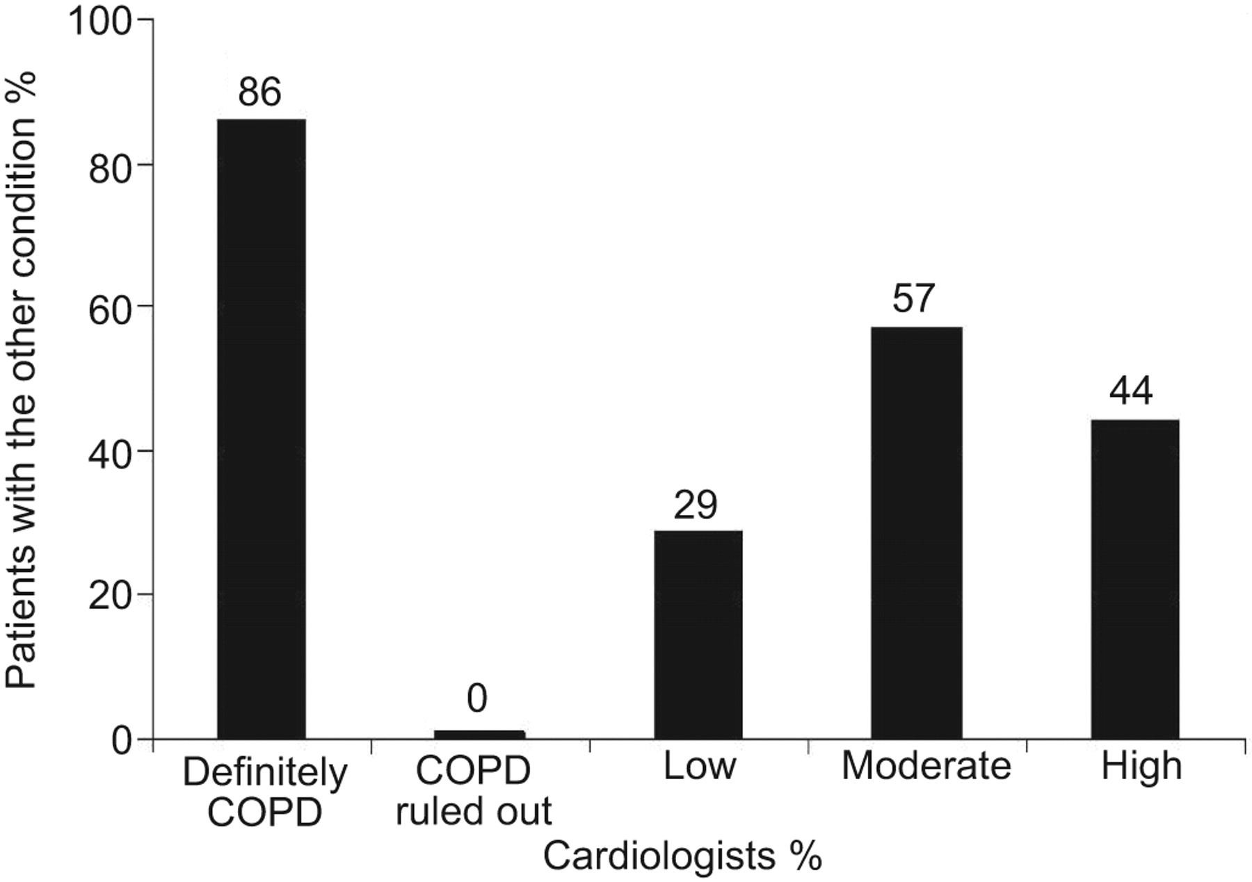
Declared probability of cardiologists and its relationship with the actual finding of chronic obstructive pulmonary disease (COPD).
Awareness of pulmonologists (fig. 1)
Before conducting the protocol echocardiogram: 5% of pulmonologists declared that they were “sure” (based on the results of a previous echocardiogram) that his/her patient would have left ventricular dysfunction; 7.4% declared that they were sure that his/her patient would not have ventricular dysfunction; 63.9% declared that they were not sure but considered the possibility low; 14.8% declared that they were not sure but thought that the possibility was moderate; and 9.7% declared that they were not sure but thought that the possibility was high.
Awareness of cardiologists (fig. 2)
Before conducting the protocol spirometry: 3.5% of cardiologists declared that they were “sure“ (based on the results of a previous spirometry) that his/her patient would have COPD; 3% declared that they were sure that his/her patient would not have COPD; 64.7% declared that they were not sure but considered the possibility low; 20.9% declared that they were not sure but thought that the possibility was moderate; and 8% declared that they were not sure but thought that the possibility was high.
Spirometry results in patients with CHF
Table 2 shows the baseline spirometry results of the patients with CHF. 75 (37.3%) patients with CHF had airway obstruction. Of these, 62 (28.4%) qualified as moderate-to-severe obstruction.
The presence of airway obstruction had no statistical association with demographic characteristics, cardiovascular risk factors, previous coronary disease or stroke, signs or symptoms, aetiology of heart failure, echocardiographic values, or laboratory values. The only variable that was associated with the presence of airway obstruction was the number of pack-yrs (28.8 yrs in patients without airway obstruction versus 38.9 in those with obstruction; p<0.0001).
Relationship between awareness and presence of airway obstruction
The presence of airway obstruction was associated with the a priori declared probability of the attending cardiologists. Spirometric documentation of airway obstruction was observed in 86% of cases in which cardiologists declared they were “sure” and in 0% of those declaring they were sure that their patient would not have airway obstruction. When cardiologists declared that they were not sure, the presence of airway obstruction was 29, 57 and 44% when the cardiologists judged the possibility as low, moderate, or high, respectively (fig. 2).
Survival of patients with CHF associated with the presence of airway obstruction
Median follow-up time was 575 days. There was a total of 61 deaths during that time. 37 of these were in patients included because of heart failure and 24 were in those included because of COPD. Only three patients were completely lost during follow-up. Of the remaining 416 patients, 35 (8.4%) had a follow-up period of ≤30 days.
Figure 3 shows the survival curve of CHF patients with and without airway obstruction. There were no significant differences in survival between groups. The adjusted risk of death in patients with CHF and concomitant airway obstruction was 0.77 (95% CI 0.37–1.58), with a p-value of 0.474. Although patients with airway obstruction had a higher incidence of hospitalisation during follow-up, these differences did not reach statistical significance (adjusted hazard ratio (HR) 1.16 (95% CI 0.71–1.89); p=0.546).

Kaplan–Meier estimates of survival of patients with chronic heart failure with airway obstruction (–––––) and without airway obstruction (-------). Hazard ratio 0.77 (95% CI 0.37–1.58); p=0.474.
Using a more specific threshold for clinically important COPD, such as post-bronchodilatatory FEV1/FVC below the least mean square (adjusted HR for mortality 1.16 (95% CI 0.20–2.13); p=0.483; adjusted HR for hospitalisations 1.37 (95% CI 0.75–2.53)) and FEV1 <60% (adjusted HR for mortality 1.11 (95% CI 0.49–2.55); p=0.795; adjusted HR for hospitalisations 1.40 (95% CI 0.91–2.16)) resulted in a better prediction of the impact of COPD.
Echocardiographic results in patients with COPD
The procedure was considered technically acceptable in all COPD patients.
There were no major discrepancies among echocardiogram operators in the calculation of the EF value and there were no discrepancies at all in the determination of the proportion of patients who were classified as having left ventricular dysfunction (EF ≤40%).
37 (17%) out of 218 patients with COPD had left ventricular dysfunction. Of these, 30 had systolic dysfunction (EF ≤40%) and seven patients were classified as also having severe diastolic dysfunction.
The presence of ventricular dysfunction in patients with COPD was not statistically associated with demographics, cardiovascular risk factors (hypertension, diabetes, high cholesterol), laboratory values or GOLD classification. 26 out of 37 patients with COPD and left ventricular dysfunction were classified GOLD III (n=16) or IV (n=10). Only the presence of previously known coronary disease was significantly associated with the presence of ventricular dysfunction. 15 (40.5%) out of 37 patients with ventricular dysfunction had previous coronary disease, while 20 (12.2%) out of 164 without ventricular dysfunction had previous ischaemic disease (p<0.0001).
Median values of NT-proBNP were 160 pg·mL−1 in patients with COPD. Values of NT-proBNP were strongly associated with the presence of ventricular dysfunction. Median values of NT-proBNP in patients without ventricular dysfunction (103 (95% CI 49.17–273.15) pg·mL−1) were significantly lower than those detected in patients with ventricular dysfunction (677 (95% CI 384.1–1682.25) pg·mL−1; p<0.0001).
In a multivariable regression, both previous coronary disease (HR 3.14 (95% CI 1.14–8.41)) and NT-proBNP >160 pg·mL−1 (HR 10.79 (95% CI 2.96–39.34)) were the two variables statistically associated with the presence of ventricular dysfunction.
Relationship between awareness and presence of left ventricular dysfunction
The presence of ventricular dysfunction was associated with the a priori declared probability of the attending pneumologists. Echocardiographic documentation of ventricular dysfunction was observed in 75% of cases in which pneumologists declared they were “sure” and in 7% of those declaring they were sure that their patient would not have ventricular dysfunction. Conversely, when pneumologists declared that they were not sure, the presence of ventricular dysfunction was 5, 24 and 45% when the pneumologists judged the possibility as low, moderate or high, respectively (fig. 1).
Survival of patients with COPD associated with the presence of left ventricular dysfunction
The presence of ventricular dysfunction increased the probability of dying during follow-up (HR 2.34 (95% CI 0.99–5.54); p=0.053) (fig. 4). There were no differences in the rate of rehospitalisations between those patients with and without ventricular dysfunction (HR 1.69 (95% CI 0.85–3.37); p=0.136).

Kaplan–Meier estimates of survival of patients with chronic obstructive pulmonary disease with left ventricular dysfunction (–––––) and without left ventricular dysfunction (-------). Hazard ratio 2.34 (95% CI 0.99–5.54); p=0.053.
DISCUSSION
Several clinical studies and literature reviews [2–11] emphasise the importance of and need for studies of the prevalence of coexistent CHF and COPD, as well as the prognostic value of each condition in the presence of the other. Although several papers have been published on this topic, to our knowledge none has systematically explored the presence of airway obstruction with spirometry in stable patients with CHF, or ventricular function with echocardiography and neurohormone levels in stable patients with COPD, and followed the patients for a significant period of time.
Awareness
The first finding of the registry is the documentation that treating cardiovascular and pulmonary subspecialists, at least in a tertiary care setting in Argentina, apparently did not recognise the comorbidity of CHF and COPD as a problem. The degree of awareness (i.e. the certainty of a positive or negative diagnosis before the study) was very low among both groups of subspecialists. Only 6.5% of cardiologists and 12% of pulmonologists had certified or ruled out the other disease before the REPENSAR study. This is remarkable, particularly in the case of the pulmonologists. Patients with COPD enrolled in the study were elderly, with a heavy smoking history, a high prevalence of other cardiovascular risk factors, and dyspnoea as the dominant symptom. Among a population with these characteristics, the presence of ventricular dysfunction is high and certainly a clear indicator of poor prognosis [19–21]. Considering that an early diagnosis of ventricular dysfunction allows treatment and risk reduction for avoidable mortality [22], the rate of awareness was low.
Conversely, although the awareness of cardiologists was also low, it should be considered that the effectiveness of routine assessment of airway obstruction as part of CHF treatment has not been adequately demonstrated [23].
Prevalences
The prevalence of ventricular dysfunction in patients with COPD was higher than expected based on estimates in the general population adjusted for age and sex [19–22, 24]. Most patients had severe systolic dysfunction, and a small proportion also had severe diastolic dysfunction. Other published papers reported a prevalence of ventricular dysfunction in patients with COPD ranging from 9 to 52% [2]. However, most of these studies used a nonappropriate definition of both COPD and CHF, as they were not based on GOLD criteria and reproducible echocardiographic parameters.
The presence of airway obstruction in patients with CHF was high. Interestingly, most of these obstructions were classified as moderate to severe. This is important when considering that there is a danger of overdiagnosing COPD in older adults because GOLD criteria do not adequately adjust for age [25]. However, this appears not to be an issue in our study, as most patients with airway obstruction had moderate-to-severe disease. Although previous studies reported the prevalence of COPD in patients with CHF ranging from 19 to 48% [2], these reports were also biased by inappropriate definitions of COPD and the selected patient populations [7–9].
Prognostic value
The presence of ventricular dysfunction in patients with COPD-impaired survival during follow-up. The adjusted probability of death in patients with COPD and ventricular dysfunction was more than two-fold the risk of patients without ventricular dysfunction. Interestingly, this increased mortality risk was seen without a clear increase in the rate of rehospitalisations for any reason. These data suggest there may be an excess of risk outside the hospital that could be attributed to a risk of sudden death. Some reports emphasised an increased risk of death in patients with COPD after initiation of pharmacological treatment of COPD, including short-acting β2-agonists [26] and ipratropium bromide [27]. In this sense, uncovering hidden ventricular dysfunction in patients with COPD would be of particular benefit, as routine use of β2-agonists and/or ipratropium bromide could be deleterious and possibly contraindicated if ventricular dysfunction is present [28, 29]. It is thus necessary to generate prospective evaluations of efficacy and safety of these treatments in this specific subset of patients.
Our data support the necessity for systematic evaluation for the presence of ventricular dysfunction in patients with severe or very severe COPD. A recent population-based study also highlighted the correlation between the extent of emphysema and impaired left ventricular filling, reduced stroke volume and lower cardiac output [30]. Additionally, a recent update [11] of an European survey [4, 5] found that a new diagnosis of heart failure was associated with an increased mortality similar to those reported in our survey.
In contrast, the presence of airway obstruction in patients with CHF did not confer a statistical excess risk of death or hospitalisations during the 2-yr follow-up period. Although the reasons for this lack of association remain unknown, at least two factors could explain this finding. First, the population enrolled for CHF had a high-risk profile at baseline, making it improbable that any added condition would significantly change the prognosis. Secondly, it is possible that the study lacked statistical power to detect differences in this regard. In particular, 2 yrs of follow-up could not be sufficient enough to detect any significant change in prognosis.
Role of NT-proBNP in patients with COPD
The assessment of NT-proBNP was useful for the detection of ventricular dysfunction in patients with COPD. A cut-off point of 160 pg·mL−1 increased >10-fold the probability of finding ventricular dysfunction with echocardiography. This finding suggests that if a Doppler echocardiogram is unavailable, the routine assessment of NT-proBNP could be considered a useful tool with which to select patients for referral for echocardiology.
Implications
The registry emphasises the necessity to intensify research on the safety and efficacy of pharmacological agents among real-world populations. The efficacy and the safety of COPD treatments have been tested largely in patients with no or minimal comorbidity. The results of our registry emphasise the importance to rethink (“repensar”) these fundamental clinical questions that should and could be answered in a normal and pragmatic clinical context.
Additionally, these data suggest that, among elderly patients with COPD, an early and systematic assessment of cardiac function should be strongly considered. Prospective interventions should be carried out to answer this important research question. Conversely, routine assessment of pulmonary function does not seem to be necessary in all patients with CHF.
Study limitations
The people recruited in this survey were seen by specialists of university and teaching hospitals and may not represent the general population demographics.
Although we were able to classify the presence or absence of gross ventricular dysfunction (left ventricular ejection fraction ≤40% or severe diastolic dysfunction) in all patients, this by no means implied that the technical characteristics of the procedure were optimal in all subjects, as patients with COPD usually provide technical challenges to accurately measure ventricular function.
Additionally, not all echocardiographic procedures could be carried out in every patient. Although the written protocol suggested the utilisation of the described procedures in all patients, the operators discretionally used the different methods accordingly to the technical feasibility. We did not record the proportion of patients who underwent each procedure.
Acknowledgments
We thank R. Del Olmo, G. Menga (both from Hospital María Ferrer, Buenos Aires, Argentina), G. Ciambrone, S. Donato, N. Rizzo, I. Nogués, F. Novo and C. Higa (Division of Cardiology, Hospital Alemán, Buenos Aires) for their help in recruiting patients for this study. We are also grateful to A. Rincheski and N. Domínguez (both Hospital María Ferrer, Buenos Aires) for performing the respiratory tests. We thank A. Moralez Lezica, F. Deketele, C. Rivas (all from the Division of Cardiology, Hospital Alemán, Buenos Aires) and M. Munin (Echocardiogram Section, Dept of Internal Medicine, CEMIC, Buenos Aires) and for their comments on the manuscript and echocardiogram interpretation.
Footnotes
Earn CME accreditation by answering questions about this article. You will find these at the back of the printed copy of this issue or online at www.erj.ersjournals.com/misc/cmeinfo.xhtml
Statement of Interest
None declared.
- Received March 11, 2011.
- Accepted May 16, 2011.
- ©ERS 2012
REFERENCES


