


Abstract
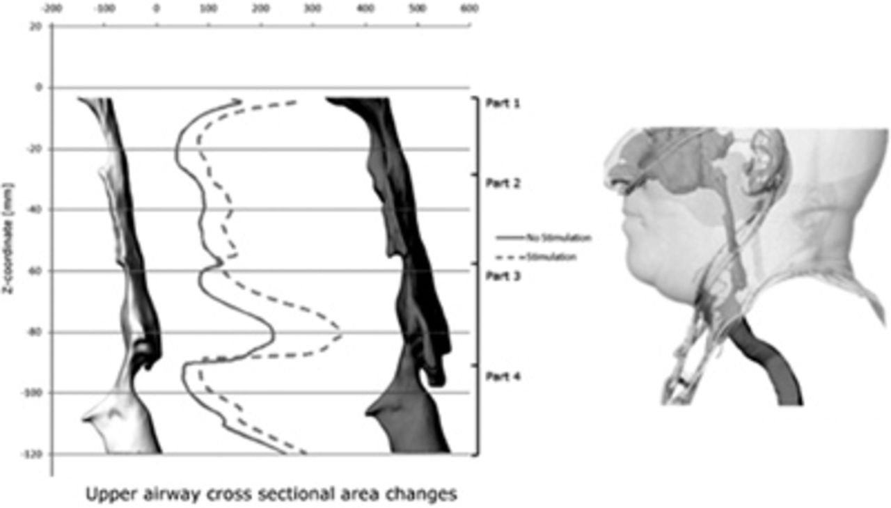
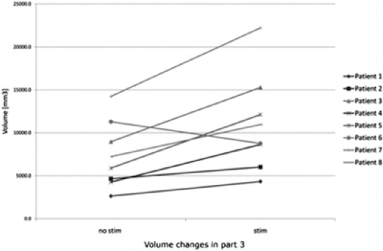
This study assessed the in-vivo effect of stimulation of the hypoglossal nerve on the upper airway morphology using imaging techniques in combination with computational fluid dynamics (CFD) tools. In 8 patients the upper airway was imaged using CT before and during stimulation of the hypoglossal nerve. Changes in airway volume and resistance were determined using segmentation and CFD. A statistically significant (p = 0.035) enlargement of the volume for the upper airway at the level of the tongue-base was observed. A significant correlation (R2= 0.89, p=0.002) was found between the change in hydraulic radius and the volume changes at the level of the palate. The distance from the tongue base to the mandibula measured just above the epiglottis correlated significantly with the change in upper airway resistance (R2=0.91, p=0.014).
It could be concluded that stimulating the hypoglossal nerve changes the upper airway morphology. A relatively complex motion of the tongue is observed with an enlargement of the airway lumen predominantly near the tongue base. Depending on the volume of the oral cavity, the enlargement is homogeneous or a decrease in cross sectional area occurs at the palatal level. The distance between the tongue base and the mandibula appears to be a good surrogate for changes in upper airway resistance.
- © 2011 ERS


