


Figures
- Figure 1–
Early tracheal mucus obstruction in β-epithelial Na+ channel (ENaC)-transgenic (TG) mice. Multiplanar reconstructions (MPRs) in a) coronal and b) sagittal planes of the neck of a 5-day-old wild-type (WT) mouse showing the larynx with its lateral recessus (1), head (2) and spine (3). The MPRs of a representative βENaC-TG mouse show obstruction of the sublaryngeal trachea (arrow) in the d) coronal and e) sagittal views. The mouse died spontaneously on day 6. Notably, this is an example of how three-dimensional reconstruction can help to discriminate between the artificial blurring of the larynx in the WT mouse by partial volume effect and obstruction in the βENaC-TG mouse. Histology (Alcian blue periodic acid–Schiff staining) showing mucus obstruction of the trachea in f) a representative βENaC-TG mouse killed at the age of 3 days but not in c) a WT mouse. Scale bars = 1 mm. g) Longitudinal monitoring of the frequency of narrowing or complete obstruction of the trachea in βENaC-TG mice by volumetric computed tomography. Note that tracheal obstruction was not observed in WT mice. h) Tracheal obstruction in deceased and long-term surviving βENaC-TG mice. Data are shown as percentage of mice with a lesion present at each time-point. n=12–20 mice per group. *: p<0.05 versus age-matched WT mice (Fisher’s exact test); #: p<0.05 versus age-matched βENaC-TG survivors (Fisher’s exact test).
- Figure 2–
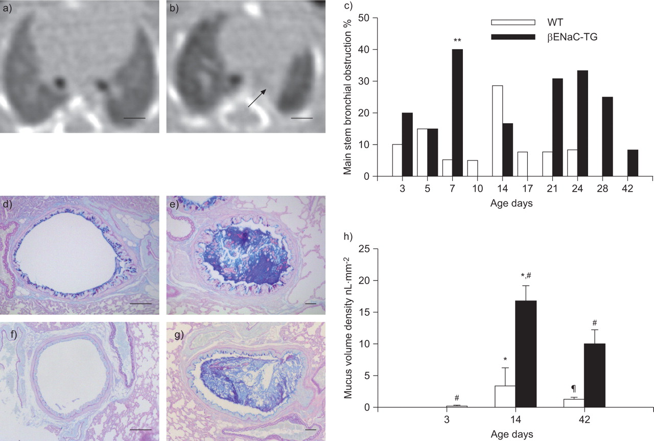
Mucus obstruction in main stem bronchi from wild-type (WT) and β-epithelial Na+ channel (ENaC)-transgenic (TG) mice. a) Volumetric computed tomography (VCT) slice of a WT mouse on day 7. b) Obstruction of the left main stem bronchus (arrow) in a βENaC-TG mouse on day 7. Images are aligned at the level before the right main stem bronchus enters the lung. Scale bars = 1 mm. c) Summary of longitudinal assessment of mucus obstruction in main stem bronchi in WT and βENaC-TG mice by VCT. Data are presented as the percentage of mice with lesion present. n=11–20 mice per group. **: p<0.01 versus age-matched WT (Fisher’s exact test). Representative airway histology (Alcian blue periodic acid–Schiff staining) from d, f) WT and e, g) βENaC-TG mice killed at the age of d, e) 2 and f, g) 6 weeks. Lungs were sectioned at the level of the proximal main axial airway near the hilus, showing transient goblet cell metaplasia and intraluminal mucus in d) 2-week-old but not f) 6-week-old WT mice, and persistent mucus obstruction in e, g) βENaC-TG mice. Scale bars = 100 μm. h) Mucus content in the proximal airways. Data are presented as median±sem. n=5–9 mice per group. *: p<0.05 versus mice of same genotype on day 3 (Kruskal–Wallis ANOVA with post hoc Dunn’s method); #: p<0.017 versus age-matched WT (Bonferroni-corrected Mann–Whitney U-test); ¶: p<0.05 versus mice of same genotype day 14 (Kruskal–Wallis ANOVA with post hoc Dunn’s method).
- Figure 3–
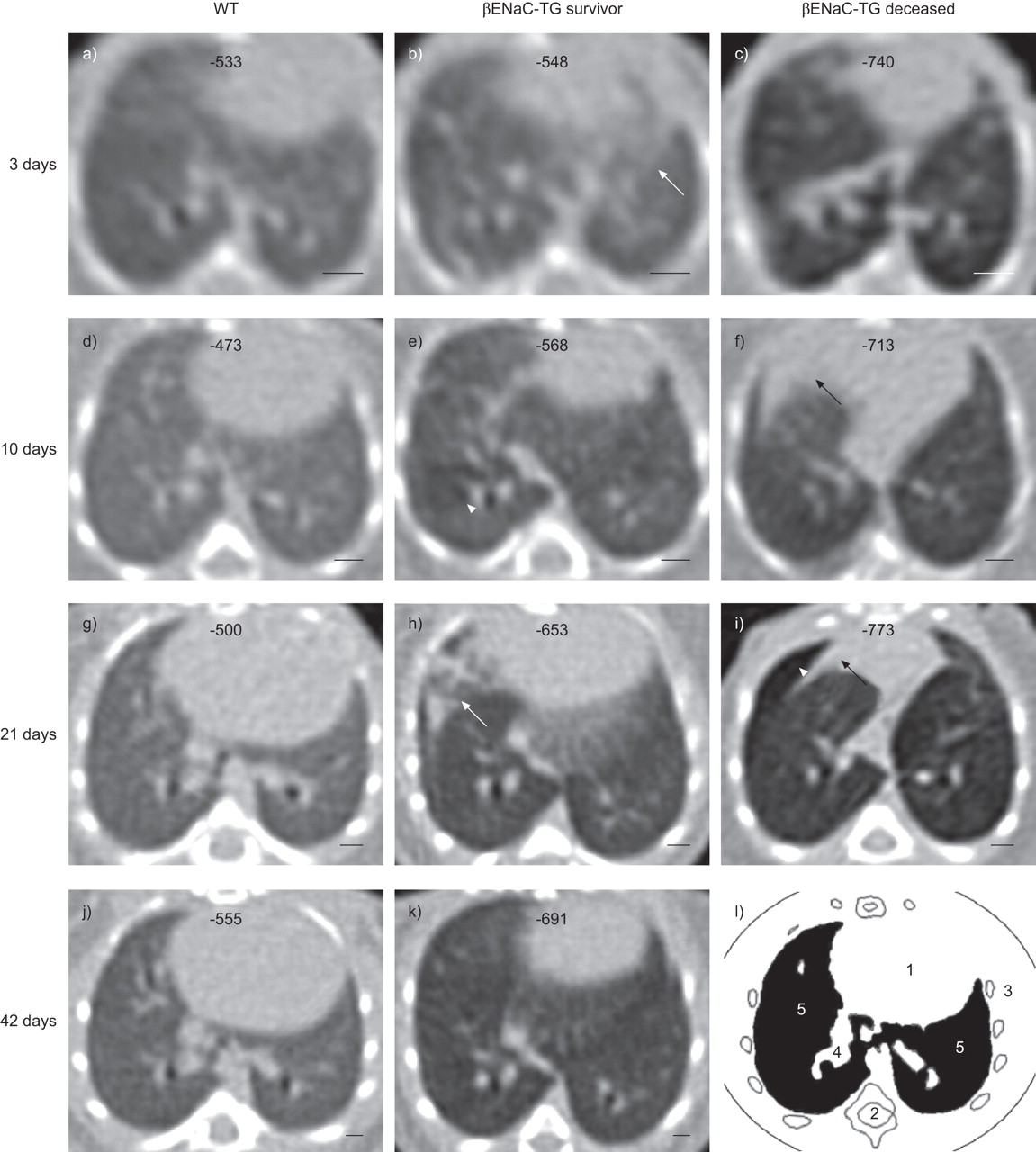
Representative longitudinal volumetric computed tomography (VCT) images of the chest of wild-type (WT) and β-epithelial Na+ channel (ENaC)-transgenic (TG) mice. VCT images of the lung were taken sequentially starting at the age of 3 days in a, d, g, j) a WT mouse, b, e, h, k) a surviving βENaC-TG littermate and c, f, i) a βENaC-TG mouse that died spontaneously on day 21. Note the development of an overall inhomogeneous lung parenchyma in βENaC-TG mice compared with the homogeneous texture in the WT mouse, and a difference in emphysema severity in the two βENaC-TG mice. These images also give examples of other characteristic pathologies, such as diffuse infiltrates (white arrows), atelectasis (black arrows) and air trapping (white arrowheads). Corresponding mean lung densities (in Hounsfield units) for each mouse are given at the top of each image. Images were aligned at the level of the branching of the right inferior lobe bronchus. Scale bars = 1 mm. l) Landmark structures of the mouse chest: heart (1), spine (2), chest wall with costae (3), bronchovascular branches (4) and lung parenchyma (5).
- Figure 4–
Occurrence of diffuse pulmonary infiltrates and atelectasis from neonatal to adult ages in β-epithelial Na+ channel (ENaC)-transgenic (TG) mice and wild-type (WT) littermates. a) Volumetric computed tomography slice at the level of the branching of the inferior lobe bronchus in a 7-day-old WT mouse. b) 3-day-old βENaC-TG mouse with diffuse infiltrates, predominantly of the right thorax (white arrow). c) Morphologically changed diffuse infiltrates (white arrow) and atelectasis of the intermediate lobe (black arrow) in the same βENaC-TG mouse as in (b) but observed on day 7. d) Note the opacity of the left lung including the bronchus (black arrow) and emphysema of the right lung in a long-term surviving βENaC-TG mouse observed on day 21. Images were selected to represent relevant pathologies. Scale bars = 1 mm. Frequency of e) diffuse infiltrates and f) atelectasis in βENaC-TG compared with WT mice. Data are given as the percentage of mice with lesion present. n=11–20 mice per group. *: p<0.05 versus age-matched WT (Fisher’s exact test); ***: p<0.001 versus age-matched WT (Fisher’s exact test).
- Figure 5–
Summary of longitudinal assessment for a) air trapping and b) diffuse parenchymal inhomogeneity in β-epithelial Na+ channel (ENaC)-transgenic (TG) and wild-type (WT) mice. a) Percentage of βENaC-TG mice showing air trapping. Note that air trapping was not observed in WT mice. b) Inhomogeneous lung parenchyma was found in most βENaC-TG mice, but was rarely detected in WT littermates. Data are presented as the percentage of mice with lesion present. n=11–21 mice per group. *: p<0.05 versus age-matched WT (Fisher’s exact test); #: p<0.005 versus age-matched WT (Bonferroni-corrected Fisher’s exact test).
- Figure 6–
Development of emphysema in β-epithelial Na+ channel (ENaC)-transgenic (TG) mice. a) Summary of longitudinal measurements of the density of lung parenchyma, in Hounsfield units (HU), in neonatal to adult wild-type (WT) and βENaC-TG mice. —: median; box: interquartile range; whiskers: 10th and 90th percentiles. n=11–20 mice per group. #: p<0.005 versus age-matched WT (Bonferroni-corrected Mann–Whitney-U test); ¶: p<0.05 versus WT on day 42 (Kruskal–Wallis ANOVA with post hoc Dunn’s test); +: p<0.05 versus βENaC-TG on day 42 (Kruskal–Wallis ANOVA with post hoc Dunn’s test); *: p<0.05 versus WT on day 3 (Kruskal–Wallis ANOVA with post hoc Dunn’s test). b, c) Representative lung histology (haematoxylin and eosin staining) from 2-week-old mice showing enlargement of alveoli in c) βENaC-TG compared with b) WT mice. Scale bars = 100 μm. d) Mean linear intercepts were determined in WT and βENaC-TG mice at the ages of 3 days, 2 weeks and 6 weeks. Data are presented as mean±sem. n=5–8 mice per group. **: p<0.01 versus βENaC-TG on day 3 (one-way ANOVA with post hoc Bonferroni-corrected unpaired t-test); ***: p<0.001 versus WT on day 3 (one-way ANOVA with post hoc Bonferroni-corrected unpaired t-test); §: p<0.017 versus age-matched WT (Bonferroni-corrected unpaired t-test). e) Total lung capacity (TLC) and f) pressure–volume curves were determined in 6-week-old βENaC-TG mice and WT littermates. Data are presented as mean±sem. n=18–20 per group. ƒ: p<0.05 versus WT (unpaired t-test); ##: p<0.01 versus corresponding WT (Bonferroni-corrected unpaired t-test).
Development of emphysema in β-epithelial Na+ channel (ENaC)-transgenic (TG) mice. a) Summary of longitudinal measurements of the density of lung parenchyma, in Hounsfield units (HU), in neonatal to adult wild-type (WT) and βENaC-TG mice. —: median; box: interquartile range; whiskers: 10th and 90th percentiles. n=11–20 mice per group. #: p<0.005 versus age-matched WT (Bonferroni-corrected Mann–Whitney-U test); ¶: p<0.05 versus WT on day 42 (Kruskal–Wallis ANOVA with post hoc Dunn’s test); +: p<0.05 versus βENaC-TG on day 42 (Kruskal–Wallis ANOVA with post hoc Dunn’s test); *: p<0.05 versus WT on day 3 (Kruskal–Wallis ANOVA with post hoc Dunn’s test). b, c) Representative lung histology (haematoxylin and eosin staining) from 2-week-old mice showing enlargement of alveoli in c) βENaC-TG compared with b) WT mice. Scale bars = 100 μm. d) Mean linear intercepts were determined in WT and βENaC-TG mice at the ages of 3 days, 2 weeks and 6 weeks. Data are presented as mean±sem. n=5–8 mice per group. **: p<0.01 versus βENaC-TG on day 3 (one-way ANOVA with post hoc Bonferroni-corrected unpaired t-test); ***: p<0.001 versus WT on day 3 (one-way ANOVA with post hoc Bonferroni-corrected unpaired t-test); §: p<0.017 versus age-matched WT (Bonferroni-corrected unpaired t-test). e) Total lung capacity (TLC) and f) pressure–volume curves were determined in 6-week-old βENaC-TG mice and WT littermates. Data are presented as mean±sem. n=18–20 per group. ƒ: p<0.05 versus WT (unpaired t-test); ##: p<0.01 versus corresponding WT (Bonferroni-corrected unpaired t-test).


Additional Files
Supplementary material
Files in this Data Supplement:
- Supplementary figure 2 - Mucus obstruction in main stem bronchi from wild-type (WT) mice. Volumetric computed tomography slice of a WT mouse on day 14, which depicts subtotal narrowing of the left main stem bronchus (black arrow). The image is aligned at the level before the right main stem bronchus enters the lung. Scale bar=1 mm.


