


Figures
- Fig. 1—
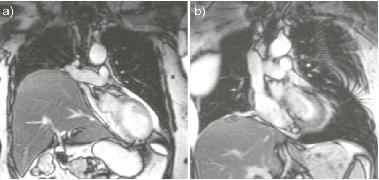
Coronal magnetic resonance image of patient C, a) before and b) after plication of the right hemidiaphragm. After plication, the pressure on right atrium and right ventricle is released, and thereby a functional repair of the patent foramen ovale and right-to-left shunt is accomplished. Settings on the Siemens 1.5T MRI system (Siemens Medical Solutions, Erlangen, Germany) were: electrocardiogram-triggered single-shot Steady State Free Precession imaging, trigger delay 440 ms, acquisition window 418 ms, slice thickness 5.5 mm.
- Fig. 2—
Coronal magnetic resonance image of patient C, a) before and b) after plication of the right hemidiaphragm. After plication, the pressure on right atrium and right ventricle is released, and thereby a functional repair of the patent foramen ovale and right-to-left shunt is accomplished. Settings on the Siemens 1.5T MRI system (Siemens Medical Solutions, Erlangen, Germany) were: electrocardiogram-triggered single-shot Steady State Free Precession imaging, trigger delay 440 ms, acquisition window 418 ms, slice thickness 5.5 mm.


