


Abstract
Repetitive inspiratory effort against an obstructed airway and intermittent hypoxia may be deleterious to the inspiratory muscles in patients with obstructive sleep apnoea (OSA).
We investigated muscular dysfunction by comparing the strength, endurance and fatigability of inspiratory muscles and knee extensors in patients with newly diagnosed severe OSA compared with matched controls. The measurements included strength and endurance tests of both muscles, and a fatigue trial with simultaneous surface electromyography of the diaphragm and the vastus lateralis during voluntary contractions and in response to magnetic stimulation. To our knowledge, this is the first investigation to assess peripheral muscle performance in severe OSA patients versus controls.
Patients in the OSA group exhibited significantly lower strength and endurance in both muscles than the control group. The fatigue index decreased significantly exclusively in the inspiratory muscles of OSA patients. Magnetic stimulation-evoked compound muscle action potential latencies increased and the amplitudes decreased significantly in the diaphragm, but not in the vastus lateralis after a fatigue test in the OSA group.
In conclusion, a significantly lower functional performance was shown for both inspiratory muscles and knee extensors in the OSA group. However, higher fatigability was only seen in the inspiratory muscles of patients with severe OSA.
- Inspiratory
- knee extensors
- magnetic stimulation
- nerve conduction delay
- obstructive sleep apnoea
- surface electromyography
Repetitive obstruction of the upper airway and apnoea/hypopnoea during sleep is a feature of obstructive sleep apnoea (OSA) syndrome 1. An obstructed airway and the subsequent asphyxia may lead to increased inspiratory efforts and, hence, the chronic overload of inspiratory muscles 2. The chronic overuse of the diaphragm was reported to put OSA subjects at risk of inspiratory muscles fatigue 3. However, whether diaphragm fatigue actually occurs in OSA patients is still controversial 4–6. Griggs et al. 4 have shown that pleural pressure relaxation rates during voluntary sniff maneuvers were prolonged in the morning compared with the preceding night prior to sleep in OSA patients. However, Montserrant et al. 5 reported lack of evidence for diaphragmatic fatigue during the night in these patients. They explained that the fatiguing levels of inspiratory effort generated throughout the night did not continue for sufficiently long periods to develop impaired contractility.
The unique repetitive deoxygenation–reoxygenation pattern in OSA patients may induce free radical production and oxidative stress, which causes muscle damage 7. OSA has been considered a systemic oxidative disorder 8. Therefore, it is logical to hypothesise that systemic skeletal muscle dysfunction may develop in patients with OSA, in addition to disorders of the upper airway muscles. However, studies addressing this issue were conducted in animal models and have shown conflicting results 9, 10.
The purpose of this study was to investigate the effect of OSA on muscular strength, endurance, and the fatigability of inspiratory muscles and peripheral knee extensors in patients with severe OSA compared with those in the control group. Comparisons made between these two types of muscle were aimed to determine whether the effects of OSA were generalised or specific to the muscles subjected to increased use.
METHODS
15 males aged 40–65 yrs with newly diagnosed severe OSA who had an apnoea/hypopnoea index (AHI) of ≥30 events·h−1 and an Epworth sleepiness scale (ESS) score of ≥10 were recruited at the Sleep Research Centre (National Taiwan University Hospital, Taipei, Taiwan). The control group consisted of 15 age-, height- and weight-matched subjects (AHI <5 events·h−1) not diagnosed with OSA from the Sleep Research Centre. Subjects were excluded if they presented with any active medical diseases, nervous system diseases, major pulmonary dysfunction, morbid obesity, diabetes under the control of oral hypoglycemic agents, alcoholism (≥50 g per day) or recent infection. All the participants at the time of the study were not receiving pharmacological or mechanical treatment. The study was approved by the Institutional Ethics Committee (National Taiwan University Hospital, Taipei, Taiwan), and all subjects were informed of the procedures in detail and gave their written informed consent prior to enrolment. This study is a registered clinical trial at ClinicalTrials.gov (NCT00813852) .
Diagnosis of OSA
The definitive diagnosis of OSA depended on an in-laboratory overnight polysomnography recording system (Embla, Medcare, Iceland) demonstrating an elevated AHI, which was calculated as the total episodes of hypopnoeas and apnoeas per hour of sleep according to the criteria of the American Academy of Sleep Medicine 1.
The level of daytime sleepiness was assessed using the ESS score in the morning after nocturnal polysomnography. Normal values ranged from 2 to 10, with scores ≥10 indicating daytime sleepiness 11.
Anthropometrics, pulmonary function tests and physical activity evaluation
Body weight and height, and circumferences of the neck, waist, and hip were measured. Body mass index (BMI) and waist-to-hip ratio were also calculated. Pulmonary function tests were performed by using a computerised spirometer (Chestgraph HI-701; Chest MI Inc., Tokyo, Japan) with the subjects in a sitting position, according to the standardised procedure 12. The forced expiratory volume in 1 s (FEV1), forced vital capacity (FVC) and FEV1/FVC ratio were recorded.
Physical activity was evaluated with a 7-day recall questionnaire, which was designed to determine calories expended on all activities during the previous 7-day period 13. The total energy expenditure was the sum of energy consumed by all activities.
Functional performance and fatigue index of inspiratory muscles
The maximal inspiratory pressure (PI,max) indicative of muscle strength was measured from residual volume with an aneroid pressure gauge (Model 4103; Boehringer Ingelheim, Norristown, PA, USA) using standard procedures 12.
Maximal voluntary ventilation (MVV) manoeuvres were used as the index of inspiratory muscle endurance and for the fatigue-induced protocol according to the diagnostic recommendations by the Chestgraph HI-701 spirometer 12. Ventilation time was calculated by the computer automatically. A previous study by our group reported that diaphragmatic fatigue could be induced by the two-set MVV manoeuvres with a 5-min rest in between 14. The fatigue index was defined as the post-MVV manoeuvres PI,max divided by the pre-MVV manoeuvres PI,max, which represents how much the muscle tension declined upon repetitive contraction. For routine muscle fatigue measurements, the decrease in force generated from a maximal volitional contraction is often used as a fatigue index.
Functional performance and fatigue index of knee extensors
The Cybex 6000 (Lumex Inc., Ronkonkoma, NY, USA) was used to measure the strength and endurance of the right knee extensors. Each subject was instructed to perform five maximal isometric knee extension contractions at a 60° knee flexion angle. Each contraction lasted for 5 s, with a resting period of at least 15 s between trials 15. The means of five maximal isometric contraction peak torques (Newton-metre; N-m) were calculated for data analysis.
The endurance test consisted of 30 cycles of alternative knee extension/flexion isokinetic contractions at the speed of 180°·s−1. Before each test, subjects performed three submaximal trials to become familiarised with the test. The muscular endurance was evaluated by the total work (N-m) generated during 30 cycles of contractions as the total area under the torque curve for knee extension movement, which would be automatically provided by the Cybex system 15.
After 30 cycles of isokinetic contractions, each participant performed three maximal isometric knee extension contractions at 60° knee flexion angle again. The fatigue index was defined as the mean of the peak torques of isometric contractions at post-endurance test divided by the pre-endurance value.
Magnetic stimulation
Cervical magnetic stimulation (CMS) was performed by a Magstim 220 stimulator (Magstim, Whitland, Wales, UK) equipped with a circular doughnut-shaped 90-mm coil producing a maximum output of 2.5 Tesla (Magstim) using standard techniques as described previously 16. Stimulations were delivered with the maximal output of the stimulator. Subjects were asked to keep their neck slightly bent forward for stimulating the phrenic nerve 16, while lying on the Cybex table with the coil placed below the inguinal ligament and 2 cm laterally from the femoral artery for stimulating the femoral nerve 17.
Surface electromyopgraphy recordings for the diaphragm and vastus lateralis
The surface electromyopgraphy (EMG) signals were simultaneously and continuously acquired by means of bipolar electrodes from the right diaphragm and the right vastus lateralis (VL). For the diaphragm, the electrodes were placed in the seventh or eighth intercostal space on the right side of the body at the midclavicular line 18, 19. The electrodes were also placed on the skin overlying the lower third of the VL muscle (10–12 cm above the knee joint) and the electrodes were aligned longitudinally to the direction of the muscle fibres. The distance between the two electrodes was kept below 2 cm and the reference electrodes were placed on the sternum and patella. The skin surface was well prepared and cleaned with alcohol to obtain a low inter-electrode resistance prior to positioning the electrodes.
Experimental protocol
All the subjects performed five maximal voluntary contractions (MVCs), followed by five magnetic stimulations. Subsequently, inspiratory muscle fatigue was induced by two MVV manoeuvres with a 5-min rest between each trial, while knee extensor fatigue was induced by 30 repetitions of isokinetic contractions. Immediately after the fatigue protocols, another five magnetic stimulations were performed as described previously and were followed by five MVCs (∼1 min after the fatigue test). The testing protocol is illustrated in figure 1⇓.

Schematic diagram demonstrating the protocol for the fatigue test. CMAP: compound muscle action potential.
EMG data analysis
All EMG signals were amplified (EMG100A; Biopac Systems Co., Goleta, CA, USA), band-pass filtered between 10 Hz to 1,000 Hz, and digitised at a sampling rate of 2 kHz. Subsequently, the final signal was acquired and analysed off-line later using commercially available software (AcqKnowledge 3.9.1 software for Windows; Microsoft Office). The parameters of the surface EMG (sEMG) signals during MVCs included root mean square (RMS) values of the amplitude and median power frequency (MPF). The means of the first three voluntary sEMG signals (pre- and post-MVV PI,max, pre- and post-test maximal isometric knee contractions) (fig. 2⇓), and the first three and the last three breaths during MVV manoeuvres and during 30-cycle isokinetic endurance test were calculated. RMS was used to quantify the diaphragm activation, i.e. combined processes of diaphragm motor unit recruitment and motor firing rate 20. The influence of the electrocardiogram on sEMG signals was minimised by recording from the right side of the body. To further minimise any signal contamination by electrocardiogram on the diaphragmatic sEMG signals during PI,max effort, RMS was measured from the segments between QRS complexes.

Examples of a, b) raw electromyopgraphy recordings and c, d) root mean square during maximal voluntary contractions in one patient with severe obstructive sleep apnoea. Diaphragm a) before and b) after fatigue test. Vastus lateralis c) before and d) after fatigue test.
The conduction time of the phrenic nerve and femoral nerve were measured as the time elapsed between the stimuli delivered by CMS and the onset of the compound muscle action potential (CMAP), which was specifically marked by the first deviation of the signal from baseline (CMAP latencies) (fig. 3⇓). CMAP latencies and amplitudes provided thereafter corresponded to the average of five stimulations. The voluntary EMG and the diaphragm CMAPs are myoelectric signals that represent different meanings: the voluntary signal represents the summated electrical activity generated by asynchronously firing motor units, whereas the diaphragm CMAPs represents the summated synchronised electrical activity generated by all motor units after a supramaximal stimulus of the phrenic nerve 21.

Example of electromyopgraphy recordings of a) the right diaphragm and b) vastus lateralis for motor responses (compound muscle action potential) with magnetic stimulation in a patient with severe obstructive sleep apnoea.
Statistical analysis
Statistical analyses were performed using SPSS version 11.5 for Windows (SPSS Inc., Chicago, IL, USA). Data were expressed as mean±sd. Comparisons of basic characteristics between the two groups were made by independent unpaired t-tests. The MVC force and sEMG parameters were analysed using a repeated measured ANOVA to assess the difference between the baseline and the post-fatigue test in each group. An α-value was set at 0.05.
RESULTS
Baseline characteristics of the study group
There was no significant difference in the basic demographic characteristics, current smokers, and energy expenditure of daily activities between the two groups except that the OSA group had significantly higher values of AHI and ESS (table 1⇓). All the participants had normal FVC, FEV1 and FEV1/FVC ratio. However, the patients with severe OSA had significantly lower values of both FVC and FEV1 than the controls.
Baseline characteristics of the subjects
Functional performance and sEMG parameters at baseline
Patients with severe OSA had significantly lower baseline strength (PI,max and peak torque values for the knee extensors) and endurance than the controls at baseline (p<0.05; table 2⇓). Diaphragmatic sEMG revealed significantly lower MPF in patients with severe OSA than those in the control group (p<0.05). sEMG recordings for VL revealed significantly lower RMS in patients with severe OSA than those in the control group (p<0.05).The evoked sEMG for the diaphragm response to CMS showed a significantly longer CMAP latency (7.28±0.66 ms) and lower CMAP amplitude (1.4±0.5 mV) in patients with severe OSA than those in the control group (p<0.05). It also showed a significantly longer CMAP latency (5.41±0.74 ms) of evoked sEMG for the VL response to magnetic stimulation in patients with severe OSA than those in the control group (p<0.05).
Strength, endurance, and surface electromyographic parameters of inspiratory muscles and knee extensors before and after the fatigue protocol
Functional performance and sEMG parameters after the fatigue test
A similar trend was found for the strength decline and voluntary sEMG parameters of inspiratory muscles and knee extensors in the two groups (table 2⇑). As the muscles of OSA patients were weaker than those of the controls, the level of fatiguing tasks might be relatively higher for the OSA group. The fatigue index derived during maximal voluntary efforts was significantly different between the two groups in the inspiratory muscles (0.86 versus 0.92; p<0.05), but not in the knee extensors (0.94 versus 0.95; p>0.05).
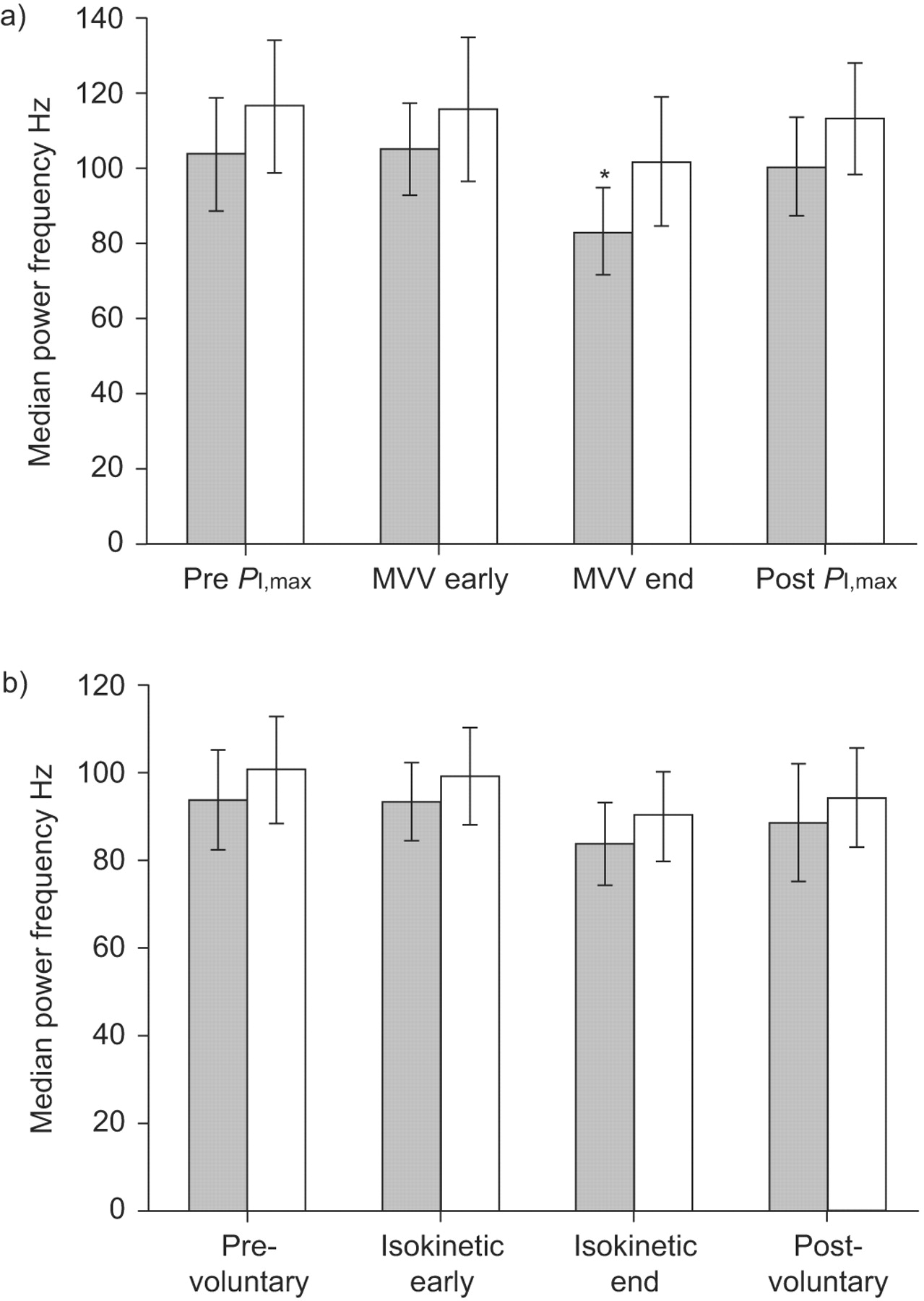
The diaphragmatic voluntary sEMG MPF declined significantly from baseline to the end of the second MVV manoeuvre and returned slightly at post-MVV manoeuvres PI,max (p<0.05; fig. 4⇓). A similar trend was shown in VL; however, no statistical significance was detected.
Group mean values of median power frequency of the a) right diaphragm and b) vastus lateralis for voluntary contractions before, during and after fatigue test by maximal voluntary ventilation (MVV) manoeuvres for inspiratory muscles and isokinetic test for knee extensors. PI,max: maximal inspiratory pressure. ▓: obstructive sleep apnoea; □: control. *: p<0.05, significant group effect by using repeated measures ANOVA.
The diaphragmatic CMAP latency and amplitude showed significant changes after the MVV manoeuvres in the severe OSA group (latency: 7.28±0.66 versus 7.48±0.65 ms; amplitude: 1.4±0.5 versus 1.2±0.5 mV; p<0.05). However, there were no significant changes in CMAP latency and amplitude of the VL after the endurance test.
DISCUSSION
This study demonstrated that severe OSA patients have significantly lower functional performance in strength and endurance, and a prolonged CMAP latency in response to magnetic stimulation of both the inspiratory muscles and knee extensors, but only the inspiratory muscles showed significantly increased fatigability in performance and sEMG assessments. This is the first investigation that we know of in assessing peripheral muscle performance in severe OSA patients versus controls.
Methodological considerations
Diaphragm muscle activity can be assessed either as a pressure or an EMG response which was recorded with surface, intramuscular needle or oesophageal electrodes. However, transdiaphragmatic pressure method and oesophageal EMG have the disadvantage of their invasiveness and discomfort. Use of intramuscular needle electrodes avoids cross-talk but contains disadvantages of invasiveness and sampling bias. sEMG recordings provide a popular, routine tool to investigate chest wall muscle function but can be confounded by noise and cross-talk 22. This is a potential limitation of sEMG. Thus, electrodes were placed in the way that minimises cross-talk, as suggested by previous researches 18, 19. Subjects were well supported during testing to minimise the activities of the adjacent trunk muscles.
It was well established that force production by the diaphragm could be significantly reduced by fatigue induced by periods of high-intensity voluntary isocapnic ventilation 23. During a 2-min MVV manoeuvre there was a progressive reduction in ventilation and transdiaphragmatic pressure generation associated with the development of fatigue of the diaphragm 23. Isocapnic maximal sustained ventilation for 8 min has also been suggested as fatiguing procedures 12. Our preliminary study has also demonstrated that two-set 15-s MVV manoeuvres could induce diaphragmatic fatigue in young healthy participants 14.
Our study employed MVV manoeuvres and 30 cycles of isokinetic knee contractions for determining the endurance of inspiratory muscles and knee extensors, respectively, which, strictly speaking, were not standardised endurance tests by the definition. However, they are widely utilised for clinically evaluating functional muscle endurance 12, 15.
Functional performance of inspiratory muscles and knee extensors
In this study, inspiratory muscles (diaphragm) and knee extensors (VL) were selected to explore the effect of the functional changes observed in OSA patients. The chronic overloading of inspiratory muscles against an obstructed upper airway could lead to structural and metabolic adaptations. Peripheral muscle was chosen as a control because it was considered not to be overloaded during sleep. Several studies have compared the inspiratory muscles and peripheral muscles in patients with OSA.
Mezzanotte et al. 24 reported a significantly lower PI,max in severe OSA subjects, but Shepherd et al. 25 reported that the PI,max was not different between the subjects whose AHI was <20 events·h−1 and >20 events·h−1. In this study, we found significantly lowered values of PI,max in patients with severe OSA compared to those without OSA. The weakened diaphragm would reduce the collapsing force during apnoeas thereby offsetting the large negative pressure generated by the diaphragm. However, the cause–effect relationship could not be determined in the present study.
With regard to the performance of the lower extremity muscles among OSA patients, little investigation has been performed in humans. Sauleda et al. 26 obtained a needle biopsy of quadriceps femoris in severe OSA patients and found structural and bioenergetic changes in the skeletal muscles. Nevertheless, some studies have shown conflicting results regarding limb muscle performance in OSA animals 9, 10. Our study is the first investigation that we know of in showing lower strength and endurance of knee extensors in the OSA group, but not fatigability. Future studies are needed to confirm this observation.
It is noteworthy that factors such as nutrition, obesity state and physical activity have profound effects on muscle performance 27. In our study, the groups were matched for the anthropometric parameters; therefore, the difference in muscular strength could not be attributed to them. However, the relatively lower physical activity levels might be related to the lower baseline muscular strength of both examined muscles in the severe OSA group. In addition, the possibility that lower values of PI,max and peak torques represent difference in effort and cooperation between two groups could not be excluded, even though we gave maximal encouragement to each participant during all the tests.
Fatigability of the inspiratory muscles
Previous studies have shown conflicting results regarding whether inspiratory muscles fatigue during OSA 4–6. One possible reason is the fatigue index may not distinguish between different types of fatigue, i.e. central fatigue and peripheral neuromuscular fatigue. In contrast to the conventional fatigue test induced by MVCs, externally applied electrical or magnetic stimulations for peripheral nerve roots that measure the CMAP after stimulation have long been known to evaluate peripheral neuromuscular function 16, 17. CMAP measures the peripheral neuromuscular fatigue (especially neurotransmitters) in the muscles bypassing the central nervous system. El-Kabir et al. 28 have shown that in patients with OSA treatment with nasal continuous positive airway pressure, the twitch transdiaphragmatic pressure did not improve in response to bilateral CMS. However, the study included lack of repeated polysomnography and compliance data of continuous positive airway pressure treatment, and no control group. Our results revealed a significant decrease in the sEMG RMS amplitude after MVV manoeuvres but not CMAP amplitudes in control subjects. These findings indicate that MVV manoeuvres might induce central fatigue, which was considered as a pre-emptive fatigue induced by the central nervous system to prevent further inspiratory muscle fatigue; thus acting as a protective mechanism.
However, significant decreases in CMAP latencies and amplitudes after MVV manoeuvres developed in patients in the severe OSA group, thereby suggesting the diaphragm muscle was responsible for inspiratory muscle fatigue in these patients. Because peripheral muscle fatigue can result in persistent muscle fatigue and longer recovery periods, it is likely that this peripheral neuromuscular fatigue was responsible for the decline in function of inspiratory muscles, i.e. decreased PI,max and endurance.
The decrease of sEMG amplitudes and power spectra of the inspiratory muscles found in this study may reflect decreased motor unit recruitment from a decreased activation 29, or hypoxia induced decreases in muscle pH that promote fatigue 30. Further studies are needed to explore the associated mechanisms.
Study limitations
This study had several limitations. First, as we did not measure twitch pressure in response to the stimulation of phrenic nerves and femoral nerve, this study could not answer whether the decreased strength of the inspiratory muscle and knee extensors was due to decreased recruitment and/or decreased contractility per se. Nevertheless, the study demonstrated decreased CMAP amplitude that could represent the delayed neurotransmitter in OSA patients compared with the controls.
Secondly, we did not measure lung volume changes before and immediately after MVV manoeuvres. Thus it may be difficult to thoroughly interpret the changes in CMAP characteristics. Previous studies had reported that CMAP amplitude and latency would be affected by different lung volumes 21, 22. We assumed both groups in our study were influenced to the same extent; however, the result showing significant difference of CMAP amplitude and latency could provide important information regarding the neuromuscular characteristics of inspiratory muscles in OSA patients. Future studies should examine the effect of OSA on inspiratory muscles fatigue by using comprehensive measurement tools.
Thirdly, the unequal fatiguing tasks produced by OSA and control participants might complicate the comparison of fatigue indices. The lower baseline PI,max and peak torques of knee extensors in the OSA group would bring about the different fatigue tasks for the OSA and the control group. According to that, future study should strive to employ a more standardised fatigue tests for further refinement of this study.
Implications
A significantly lower strength and endurance for inspiratory muscles and knee extensors of severe OSA patients were found in comparison with their age and BMI-matched controls. Therefore, systemic effects of chronic intermittent hypoxia and reoxygenation on skeletal muscles in OSA populations could not be completely ruled out. However, higher fatigability either during voluntary contractions or in response to magnetic stimulations was only exhibited in the inspiratory muscles of patients with severe OSA. Peripheral neuromuscular fatigue of the diaphragm might contribute to this increased fatigability; therefore, we speculated that the underlying mechanisms of muscular adaptation to chronic increase use played a critical role in this pathological state when compared with the systemic effects on skeletal muscles occurring in OSA patients.
Clinical trials
This study is registered at ClinicalTrial.gov (NCT00813852) .
Support statement
The authors received financial support from the National Science Council (Taiwan) (96-2314-B-002-022-MY3).
Statement of interest
None declared.
- Received December 16, 2008.
- Accepted July 16, 2009.
- © ERS Journals Ltd
References


