


Figures
- Fig. 1—
Dendritic cell (DC) presentation of malignant mesothelioma (MM)-antigens to naïve T-cells generates effector cytotoxic lymphocyte (CTLs); tumour and draining lymph nodes (dLN) single cell suspensions prepared from mice bearing small and medium tumours were stained with anti-CD11c for DCs. a) Pooled data are from two experiments (6 mice·group−1) and represented as mean±sem. Tumour: ▒; dLN: ▪. *: p<0.05 comparing tumour dLN with normal lymph node (LN). To analyse MM-antigen presentation to naïve T-cells 5,6-carboxy-fluorescein-succinimidyl-ester (CFSE)-labelled, tumour antigen-specific, OT-I T-cells were adoptively transferred into (b, c) recipient AE17-secreted ovalbumin (sOVA)-bearing mice 3 days prior to analysis. dLN (b) and non-dLNs (c) were harvested from recipient mice, prepared as a single cell suspension and stained for CD8. Fluorescence-activated cell sorter (FACS) analysis was performed by gating on CD8+/CFSE+ cells. Mice inoculated with AE17 tumour cells were negative controls (d and e). Representative histograms are shown (b–e). In vivo CTL activity was assessed by adoptively transferring differentially-labelled target cells prepared from normal mice representing CFSEhigh or OVA257–264 peptide SINFEKL (OVAp), and CFSElow control cells into mice bearing AE17-sOVA (f and g) or AE17 (h and i) tumours. dLN and non-dLN were prepared as a single suspension 18 h later and FACS analysed. A reduction in the OVAp peak compared with the control peak represents lysis of the targets. Representative histograms are shown (f–i). CTL activity is shown as the number of cells in the OVAp peak divided by the number of cells in the control peak multiplied by 100. All data within each experiment were normalised compared with nontumour bearing C57BL/6J LN controls. j) Pooled in vivo CTL activity data (9 mice·group−1) from dLN and non-dLN are shown as mean±sem. One way ANOVA was performed comparing small versus medium sized tumours. ▓: non-dLN; ▪: dLN.**: p<0.01.
- Fig. 2—
CD8+ cells penetrate the malignant mesothelioma tumour microenvironment. Single cell suspension of small or medium tumours and their lymph nodes (LNs) were stained for CD8. a) Pooled data from three experiments (9 mice·group−1) are shown as mean±sem. ▪: tumour; ░: draining LN (dLN); ▒: non-dLN. ***: p<0.001 comparing dLN and non-dLN with normal LN. Frozen tumour sections were double stained for b, d and e) CD8+ cells (blue) and c, d and e) CD31 to detect blood vessels (brown). b) Rat immunoglobulin (Ig) G2a and c) rat IgG2b are isotype controls. These experiments were performed twice (6 mice·group−1) and representative photos at 200 × magnification are shown. e) The inset shows a CD8+ cell associated with a tumour blood vessel and a CD8+ cell within the tumour matrix, indicated by arrows.
- Fig. 3—
Malignant mesothelioma-infiltrating CD8+ T-cells are activated but lose cytotoxic lymphocyte (CTL) activity; tumours were also harvested from AE17-secreted ovalbumin (sOVA)-bearing mice given 5,6-carboxy-fluorescein-succinimidyl-ester (CFSE)-labelled, tumour antigen specific, OT-I T-cells (as per fig. 1b⇑ and c). a) The few OT-I cells that penetrated the tumour expressed low levels of CFSE similar to proliferating cells seen in mice given OVA in incomplete Freunds adjuvant draining lymph node (dLN), indicative of proliferation. However, the few CFSEhigh (OVA257–264 peptide SINFEKL) and CFSElow target cells harvested from the tumours of d) AE17-sOVA-bearing mice (as per fig. 1f⇑ and g) showed no in vivo CTL activity relative to c) the CTL activity in dLN of the same mouse. In separate experiments, tumours and dLN harvested from AE17-bearing mice were double stained for e) CD8 and CD69, f and g) interferon (IFN)-γ, h) CD25 and i) CD44. Fluorescence-activated cell sorter analysis was performed by gating on CD8+ cells. Representative histograms are shown with dLN and tumour overlaid for e) CD69, f) IFN-γ analysis in the dLN and g) tumour. e) ––––: tumour; ······: dLN. f and g) –––––: IFN-γ; ········: rat immunoglobulin G1 isotype control. These experiments were repeated twice (6 mice·group−1). Pooled data from two experiments (6 mice·group−1) is shown for h) CD25 and i) CD44 as mean±sem percentage of CD8 cells. ▪: normal LN; ▓: AE17; □: AE17-sOVA. **: p<0.01, comparing dLN with normal LN.
- Fig. 4—
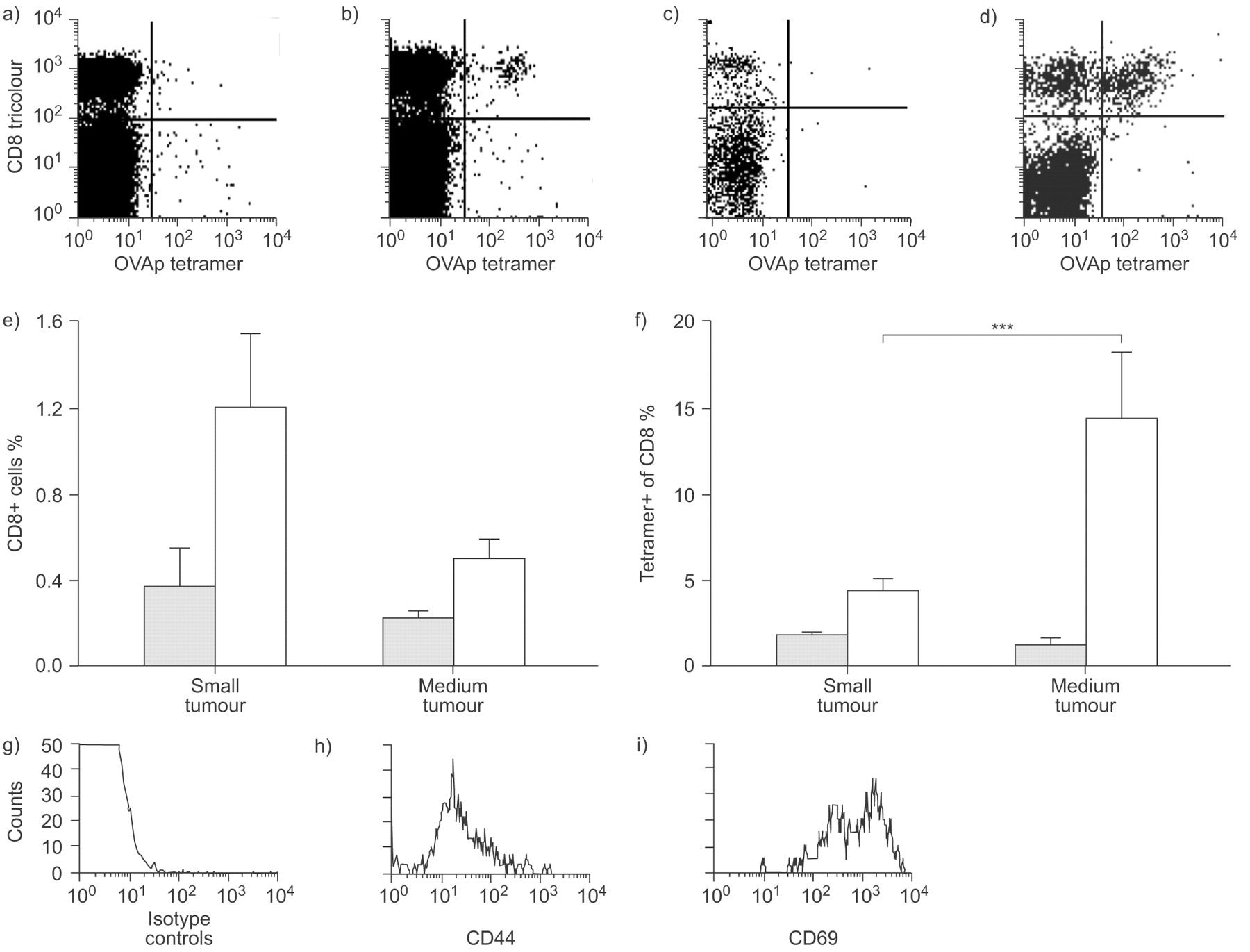
Tumour-specific CD8+ cells in malignant mesothelioma tumours are activated. a and b) Draining lymph node (dLN) and c and d) tumours were harvested from a and c) AE17 and b and d) AE17-secreted ovalbumin (sOVA) tumour-bearing mice and fluorescence-activated cell sorter analysed for co-expression of the OVA257–264 peptide SIINFEKL (OVAp) tetramer and CD8 (a–f), as well as g) an isotype control, h) CD44 or i) CD69. Flow cytometric analysis was performed by gating on CD8+tetramer+ cells. a–d) Representative dot plots and g–i) histograms are shown. Pooled data from two experiments (6 mice·group−1) are shown as mean±sem percent of CD8 cells that are tetramer+ in the e) dLN and f) tumour. ░ AE17; □: AE17-sOVA. ***: p<0.001 comparing small and medium tumours.
- Fig. 5—
Malignant mesothelioma-infiltrating CD4+ T-cells are activated. Tumours and draining lymph nodes (dLN) were harvested from AE17-bearing mice and stained for CD4 expression and fluorescence-activated cell sorter (FACS) analysed. These experiments were repeated twice (6 mice·group−1). a) pooled data are shown as mean±sem percent of CD4+ cells. *: p<0.05 and **: p<0.01 comparing dLN (░) and non-dLN (▒) with normal LN (□). ▪: tumour. Cells were also double stained for CD4 and c and g) isotype controls, or d and h) CD69, e and i) CD44 as well as f and j ) interferon (IFN)-γ. FACS analysis was performed by gating on CD4+ cells and representative FACS histograms are shown (c–j). f and j) ––––: IFN-γ; ······: isotype control. CD4+CD25+ co-expression was also examined in normal LN, dLN and tumours and b) pooled data from two experiments with (6 mice·group−1) are shown as the percent of CD4+ that are CD25+ ±sem.
- Fig. 6—
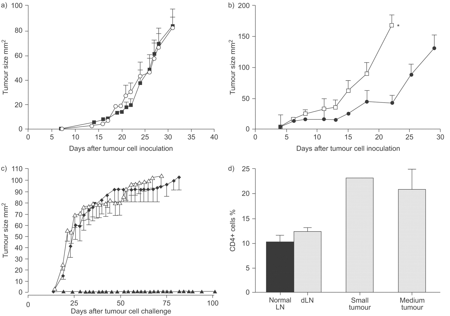
The majority of CD4+ cells are effector cells. The role of CD4+ cells was determined by a) antibody depletion (○: CD4 depletion; ▪: no depletion), and b) by the use of GK mice (mice that never have a mature CD4+ T-cell compartment). Pooled data are shown as mean±sem tumour size from 5 mice·group−1. b) Only GK mice were significantly different to C57BL/6 mice. •: C57BL/6 mice; □: GK CD4-mice. c) To address a possible role for splenic cells, naïve C57BL/6 mice were the recipients of adoptively transferred, unfractionated spleen cells prepared from unmanipulated, healthy mice and from mice with progressing AE17 tumours. All mice, plus tumour growth controls (no transfer), were challenged with AE17 tumour cells 3 weeks later. c) Pooled data from one of two experiments (8 mice·group−1) are shown as mean±sem. ▵: no transfer; ♦: healthy splenocytes; ▴: tumour splenocytes. d) Tumour and draining lymph node (dLN) from AE17 tumour-bearing mice were fluorescence-activated cell sorter analysed for CD4+FoxP3+ co-expression in normal lymph node (▪), tumour dLN and tumours. Pooled data are shown after gating on CD4+ cells from 12 mice·group−1. ░: AE17 mice.
- Fig. 7—
CD25+CD4+ T-regulatory (Treg) cells do not interfere with endogenous anti-malignant mesothelioma immunity. The role of CD4+CD25+ suppressor cells was determined by depletion using the anti-CD25 antibody (PC61). A single intra-tumoural injection of PC61 depleted CD4+CD25+FoxP3+ from the tumour (a, d, g and j), draining lymph node (b, e, h and k), spleen (c, f, i and l) and bone marrow for 10 days (m). Representative dot plots 4 or 6 days after PC61 injection show CD4+CD25+ (a–f) gated on lymphocytes, and FoxP3+CD25+ gated on CD4+CD25+ (g–l). a–c) and g–i) not depleted; d–f) and j–l) PC61 depleted. m) ♦: tumour; ▾: bone marrow; ▵ lymph node; □: spleen. Data are from one of three experiments in which the organs from 5 mice·group−1 were pooled for analysis. n) Mice were given one (▴) or two (▿) PC61 injections (arrows) and tumour growth was monitored (three experiments with 5 mice·group−1); data shown as mean±sem. □: no depletion.
- Fig. 8—
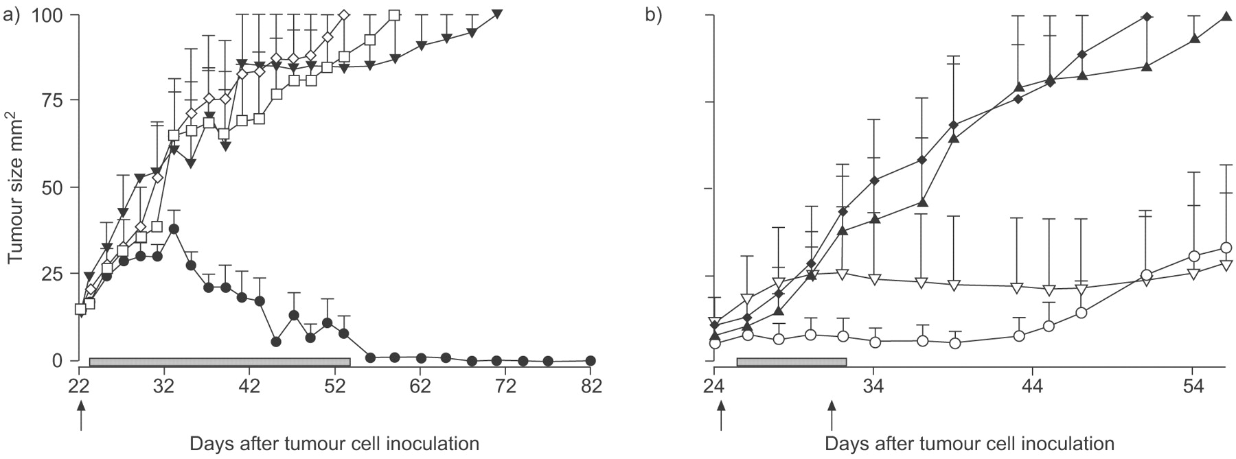
CD25-targeted T-regulator (Treg) cells depletion may confound cytokine-based immunotherapy. a) A single PC61 injection was given 1 day prior to intratumoural interleukin (IL)-2 injections given every 2nd day (two experiments with 5 mice·group−1). b) PC61 injections were given 1 day before each of two hydrodynamic injections of pORF/mIL-21 that were 6 days apart (one experiment with 5 mice·group−1), and tumour growth monitored. ▾: PBS no depletion; ⋄: Treg depletion; •: Il-2 no depletion; □: IL-2 Treg depletion; ▴: saline no depletion; ♦: saline Treg depletion; ○: IL-21 no depletion; ▿: IL-21 Treg depletion; ▓: treatment duration. Arrows: Treg depletion. Data shown as mean±sem.


