
Main menu

  • Home
  • Current issue
  • ERJ Early View
  • Past issues
  • Authors/reviewers
    • Instructions for authors
    • Submit a manuscript
    • Open access
    • COVID-19 submission information
    • Peer reviewer login
  • Alerts
  • Podcasts
  • Subscriptions
  • ERS Publications
    • European Respiratory Journal
    • ERJ Open Research
    • European Respiratory Review
    • Breathe
    • ERS Books
    • ERS publications home

User menu

  • Log in
  • Subscribe
  • Contact Us
  • My Cart

Search

  • Advanced search
  • ERS Publications
    • European Respiratory Journal
    • ERJ Open Research
    • European Respiratory Review
    • Breathe
    • ERS Books
    • ERS publications home

Login

European Respiratory Society

Advanced Search

  • Home
  • Current issue
  • ERJ Early View
  • Past issues
  • Authors/reviewers
    • Instructions for authors
    • Submit a manuscript
    • Open access
    • COVID-19 submission information
    • Peer reviewer login
  • Alerts
  • Podcasts
  • Subscriptions

Diagnostic value of interleukin-12 p40 in tuberculous pleural effusions

L. Valdés, E. San José, J. M. Álvarez Dobaño, A. Golpe, J. M. Valle, P. Penela, F. J. González Barcala
European Respiratory Journal 2009 33: 816-820; DOI: 10.1183/09031936.00085008
L. Valdés
  • Find this author on Google Scholar
  • Find this author on PubMed
  • Search for this author on this site
E. San José
  • Find this author on Google Scholar
  • Find this author on PubMed
  • Search for this author on this site
J. M. Álvarez Dobaño
  • Find this author on Google Scholar
  • Find this author on PubMed
  • Search for this author on this site
A. Golpe
  • Find this author on Google Scholar
  • Find this author on PubMed
  • Search for this author on this site
J. M. Valle
  • Find this author on Google Scholar
  • Find this author on PubMed
  • Search for this author on this site
P. Penela
  • Find this author on Google Scholar
  • Find this author on PubMed
  • Search for this author on this site
F. J. González Barcala
  • Find this author on Google Scholar
  • Find this author on PubMed
  • Search for this author on this site
  • Article
  • Figures & Data
  • Info & Metrics
  • PDF
Loading

Abstract

The diagnosis of tuberculous pleural effusion (TBPE) is frequently problematic. Several markers of TBPE in pleural fluid have been evaluated, with different results.

Pleural effusions from 96 patients were classified on the basis of definitive diagnosis as tuberculous (n = 39), neoplastic (n = 42) or parapneumonic (n = 15). Adenosine deaminase (ADA), ADA isoform ADA-2, interferon (IFN)-γ, CD3+/DR+ T-lymphocytes and interleukin (IL)-12 p40 were determined in all 96 effusions. The efficiency of IL-12 p40 for diagnosis of TBPEs was evaluated, in comparison with those of the other parameters, by comparing the areas under their receiver operating characteristics.

With the threshold value of 550 pg·mL−1, IL-12 p40 had a sensitivity of 92.3% (36 out of 39) and specificity of 70.2% (17 false positives). The misclassification rate of IL-12 p40 was significantly greater than those of ADA-2 and ADA. Among TBPEs, ADA correlated significantly with ADA-2, and IFN-γ with ADA and IL-12 p40.

Although tuberculous pleural effusions show values of interleukin-12 p40 that are significantly higher than neoplastic and parapneumonic fluids, this parameter is less efficient than adenosine deaminase, adenosine deaminase isoform 2 and interferon-γ. Its routine determination is, accordingly, not justified.

  • Interleukin
  • pleural effusion
  • tuberculosis

The diagnosis of tuberculous pleural effusion (TBPE), one of the most frequent causes of pleural effusion 1, is not infrequently problematic due to the unspecificity of the diagnostic tests used 2–5. Some determinations from pleural fluid (mycobacterial DNA detected by PCR 6–12, interferon (IFN)-γ 12–17, lysozyme 15, 18, 19 and adenosine deaminase (ADA) 2, 12, 15, 19–24) can be useful in these diagnostic processes.

It has become clear in recent years that, as part of the immune response to infection by Mycobacterium tuberculosis, interleukin (IL)-12 increases macrophage activation by upregulating the production of IFN-γ by natural killer (NK) cells 25, 26. This raises the possibility that IL-12 or its subunits may also be of diagnostic value. The current authors report the results of a study in which the efficiency of IL-12 p40 in discriminating between TBPE and pleural effusions of neoplastic or parapneumonic origin was evaluated in comparison with the previously characterised parameters.

MATERIAL AND METHODS

The current authors prospectively studied, during a 24-month period, 146 patients who were admitted to the Pulmonology Service of the Complejo Hospitalario Clínico Universitario de Santiago (Santiago de Compostela, Spain) with what proved to be TBPEs (24 males, 15 females), neoplastic pleural effusions (NPPEs; 22 males, 20 females; table 1⇓) or parapneumonic pleural effusions (PPPEs; 12 males, 3 females) according to previous established criteria 15. The Review Board on Human Studies at the Complejo Hospitalario Clínico Universitario de Santiago approved the protocol. Informed consent was obtained from all patients.

View this table:
  • View inline
  • View popup
Table 1—

Types or locations of tumours associated with the 42 neoplastic pleural effusions

Sample collection and analyses

Pleural fluid samples were taken by thoracocentesis at admission before initiation of any treatment. Total cell counts were determined with a Siemens ADVIA 2120 haematology system (Siemens, Madrid, Spain). IL-12 p40 was determined (in pg·mL−1) in 96 effusions (39 tuberculous, 42 neoplastic and 15 parapneumonic) using a sandwich ELISA from AMS Biotechnology (Abingdon, UK). ADA, expressed in U·L−1, was determined by the method of Giusti 27, isoform ADA-2 was determined by inhibition with erythro-9-(2-hydroxy-3-nonyl)adenine 28, IFN-γ was determined (in pg·mL−1) using an ELISA kit (Intertest-γ; Genzyme, Madrid), and CD3+/DR+ T-lymphocyte (CD3+/DR+ T) counts (in cells·mm−3) were determined as previously described 29, 30.

Statistical analysis

Kolmogorov–Smirnov tests were used to check distributional normality; non-normal distributions were subjected to log transforms. Groups were compared using a post hoc multiple comparison test (Bonferroni). The relative abilities of the various diagnostic parameters for pairwise discrimination between TBPEs and non-TBPEs were evaluated by comparing the areas under their receiver operating characteristics (ROCs) 31. The diagnostic performance of the various parameters, when used with the diagnostic thresholds afforded by the ROC analysis (see Results section), was evaluated in terms of sensitivity, specificity and positive and negative likelihood ratios (PLR and NLR, respectively). The PLR indicates how much more frequent positive test results occur in TBPEs, calculated as the proportion of positive test results in patients with TBPEs (sensitivity) divided by the proportion in non-TBPEs (1-specificity). The NLR indicates how much to decrease the probability of disease if the test is negative ((1-sensitivity)/specificity).

RESULTS

The distributions of the biochemical parameters in each group of pleural effusions are summarised in table 2⇓. For all these parameters, the median of the tuberculous group differed significantly from those of the neoplastic and parapneumonic groups in all cases, except comparison with PPPEs with regard to CD3+/DR+ T. The diagnostic thresholds afforded by the ROC analysis for IL-12 p40, ADA, ADA-2, IFN-γ and CD3+/DR+ T were 550 pg·mL−1, 54.3 U·L−1, 44.5 U·L−1, 169 pg·mL−1 and 80.3 cells·mm−3, respectively. However, the ROCs of the diagnostic parameters (fig. 1⇓) show that IL-12 separated TBPEs from NPPEs and PPPEs less well than ADA, ADA-2 and IFN-γ. The area under the IL-12 p40 ROC, 0.837, was significantly smaller than the areas of these other parameters. The area under the IL-12 p40 ROC did not differ significantly from the area under the CD3+/DR+ T ROC. The area under the ADA-2 ROC, 0.996, was significantly higher than the areas of the other parameters.

Fig. 1—
  • Download figure
  • Open in new tab
  • Download powerpoint
Fig. 1—

Receiver operating characteristics of the parameters studied, for diagnosis of tuberculous pleural effusion versus neoplastic or parapneumonic pleural effusion. –––: interleukin-12; ·····: adenosine deaminase (ADA); - - - -: ADA-2; – – – –: interferon-γ; ––··––··––: CD3+/DR+ T-lymphocytes.

View this table:
  • View inline
  • View popup
Table 2—

Descriptive statistics of diagnostic parameters considered, for each type of pleural effusion

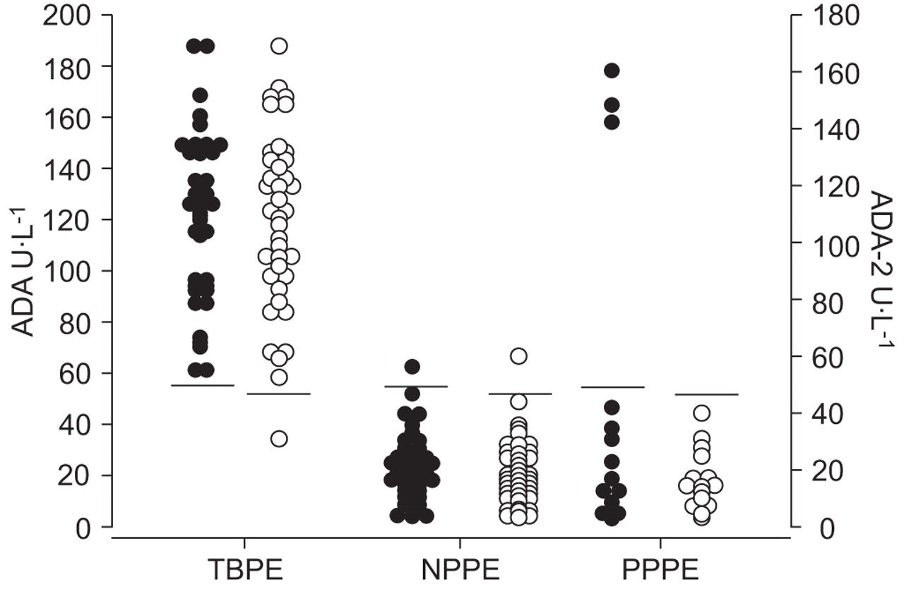
Figure 2⇓ shows the ADA and ADA-2 levels in each group of effusions. All TBPEs except one had values higher than the established diagnostic thresholds (54.3 U·L−1 for ADA and 44.5 U·L−1 for ADA-2). Sub-threshold levels of ADA and ADA-2 were found in 97.6% of NPPEs, while, among PPPEs, 80% had sub-threshold levels of ADA and 100% had sub-threshold levels of ADA-2. Figure 3⇓ shows IL-12 and IFN-γ levels in each group of effusions. IL-12 p40 was above the threshold (550 pg·mL−1) in 92.3% of TBPEs, 35.7% of NPPEs and 13.3% of PPPEs. IFN-γ was above the threshold (169 pg·mL−1) in 82.1% of TBPEs, 7.1% of NPPEs and 6.7%% of PPPEs. CD3+/DR+ T was above the threshold (80.3 cells·mm−3) in 82.1% of TBPEs, 31% of NPPEs and 46.6% of PPPEs.

Fig. 2—
  • Download figure
  • Open in new tab
  • Download powerpoint
Fig. 2—

Adenosine deaminase (ADA; •) and ADA-2 (○) levels in the pleural fluid of the three groups of patients studied (cut-off points of 54.3 and 44.5 U·L−1, respectively). TBPE: tuberculous pleural effusion; NPPE: neoplastic pleural effusion; PPPE: parapneumonic pleural effusion.

Fig. 3—
  • Download figure
  • Open in new tab
  • Download powerpoint
Fig. 3—

Interleukin (IL)-12 p40 (•) and interferon (IFN)-γ (○) levels in the pleural fluid of the three groups of patients studied (cut-off points of 550 and 169 pg·mL−1, respectively). TBPE: tuberculous pleural effusion; NPPE: neoplastic pleural effusion; PPPE: parapneumonic pleural effusion.

Table 3⇓ lists the numbers of misclassifications by each parameter and group. From worst to best, CD3+/DR+ T misclassified 28.1% of effusions, IL-12 20.8%, IFN-γ 11.5%, ADA 5.2% and ADA-2 2.1%. The misclassification rates of ADA and ADA-2 did not differ significantly. The misclassification rate of ADA-2 was significantly lower than those of IFN-γ, IL-12 p40 and CD3+/DR+ T. The misclassification rate of IL-12 p40 did not differ significantly from that of CD3+/DR+ T and IFN-γ, but was significantly greater than those of ADA and ADA-2.

View this table:
  • View inline
  • View popup
Table 3—

Numbers of misclassified effusions of each group, for each diagnostic parameter studied

Table 4⇓ lists other performance parameters, emphasising that IL-12 p40 had a sensitivity of 92.3%, a specificity of 70.2%, a PLR of 3.10 and an NLR of 0.11. Between the studied parameters, no significant differences were found with respect to the sensitivity. The specificity of IL-12 p40 was significantly lower than ADA, ADA-2 and IFN-γ, while ADA-2 showed no significant differences with respect to ADA and IFN-γ. Table 5⇓ lists correlations among the various parameters in the TBPE group.

View this table:
  • View inline
  • View popup
Table 4—

Performance measures for diagnosis of tuberculous pleural effusions by each diagnostic parameter with the stated thresholds

View this table:
  • View inline
  • View popup
Table 5—

Coefficients of correlation between the parameters studied in the tuberculous pleural effusion group

DISCUSSION

The results of the present study appear to confirm that IL-12 p40 levels in the TBPEs are significantly higher than in the NPPEs and the PPPEs, but are less efficient than the established markers ADA, ADA-2 and IFN-γ.

IL-12 is a cytokine composed of two polypeptide subunits, p40 and p35, that are encoded by distinct genes and are linked covalently by disulphide bridges in the active heterodimer, p70. IL-12 stimulates the production of IFN-γ by NK cells, and thereby indirectly promotes the activation of macrophages. It is, therefore, believed to play an important role in the immune response to intracellular pathogens such as M. tuberculosis. Previous studies have in fact found, like the present study, that IL-12 p40 levels are significantly higher in TBPEs than in NPPEs and other pleural effusions 32–36, and have reported areas under the IL-12 p40 ROC similar to the 0.837 observed in the current study 33; but this value is significantly less than the areas under the ROCs of IFN-γ, ADA and ADA-2.

The current results confirm that most TBPEs are identifiable by their high IL-12 p40 levels (sensitivity 92.3%), but their specificity is poor (70.2%). In a previous study using a diagnostic threshold of 560 pg·mL−1, a similar sensitivity but a higher specificity (85.4%) was found 34, the difference probably being mainly due to this previous study having included transudates, which only rarely have IL-12 p40 levels above the threshold. The current authors believe that the specificity obtained when excluding transudates from the study is the more relevant result for clinical practice, because the aetiological diagnosis of transudates is generally unproblematic; it is to distinguish among TBPEs, NPPEs and PPPEs that tests are needed. The overall misclassification rate of IL-12 p40 in the present study was 20.8%, a figure that is significantly larger than the misclassification rates of ADA (5.2%) and ADA-2 (2.1%).

Other studies have reported IL-12 p40 to have a specificity of 96.9% and a PLR of 17.45 but less sensitivity (54.5%) 35. The immediate cause of the wide discrepancy between these values and those of the current study is the previous study having used a diagnostic threshold more than twice as high (1,296 pg·mL−1) as that of the current study. That this was the optimal threshold for their sample is in turn probably attributable mainly to that sample having included no PPPEs and three times as many NPPEs as TBPEs, an aetiological distribution very different from that of the current study, as well the small size of their sample (43 effusions) and a different method to determine IL-12 p40.

The yield of the rest of the parameters was similar to previous studies 37. Although the overall misclassification rate of ADA-2 (2.1%) did not differ significantly from that of ADA (5.2%), it did have a significantly larger area under its ROC.

IFN-γ was positively correlated with IL-12 p40, in keeping with current accounts of the role of IL-12 in inducing IFN-γ production during infection by M. tuberculosis. As expected, ADA-2 was closely correlated with ADA among TBPEs.

In conclusion, the observed elevation of interleukin-12 p40 levels in tuberculous pleural effusions is consistent with interleukin-12 playing a role in the immune response to infection by Mycobacterium tuberculosis. The correlation between interleukin-12 p40 and interferon-γ in these effusions is in line with current knowledge about the relationship between them. However, interleukin-12 p40 is less efficient in this role than adenosine deaminase, adenosine deaminase isoform 2 or interferon-γ, and its routine determination is, accordingly, not recommended.

Statement of interest

None declared.

  • Received June 5, 2008.
  • Accepted November 10, 2008.
  • © ERS Journals Ltd

References

  1. ↵
    Valdés L, Álvarez D, Valle JM, Pose A, San José E. The etiology of pleural effusions in an area with high incidence of tuberculosis. Chest 1996;109:158–162.
    OpenUrlCrossRefPubMedWeb of Science
  2. ↵
    Valdés L, Álvarez D, San José E, et al. Tuberculous pleurisy: a study of 254 cases. Arch Intern Med 1998;158:2017–2021.
    OpenUrlCrossRefPubMedWeb of Science
  3. Conde MB, Loivos AC, Rezende VM, et al. Yield of sputum induction in the diagnosis of pleural tuberculosis. Am J Respir Crit Care Med 2003;167:723–725.
    OpenUrlCrossRefPubMedWeb of Science
  4. Berger HW, Mejia E. Tuberculous pleurisy. Chest 1973;63:88–92.
    OpenUrlCrossRefPubMedWeb of Science
  5. ↵
    Valdés L, Pose A, San José E, Martínez Vázquez JM. Tuberculous pleural effusions. Eur J Intern Med 2003;14:77–88.
    OpenUrlCrossRefPubMed
  6. ↵
    de Wit D, Maartens G, Steyn L. A comparative study of the polymerase chain reaction and conventional procedures for the diagnosis of tuberculous pleural effusion. Tuber Lung Dis 1992;73:262–267.
    OpenUrlCrossRefPubMedWeb of Science
  7. de Lassence A, Lecossier D, Pierre C, Cadranel J, Stern M, Hance AJ. Detection of mycobacterial DNA in pleural fluid from patients with tuberculous pleurisy by means of the polymerase chain reaction: comparison of two protocols. Thorax 1992;47:265–269.
    OpenUrlAbstract/FREE Full Text
  8. Querol JM, Mínguez J, García-Sánchez E, Farga MA, Gimeno C, García-de-Lomas J. Rapid diagnosis of pleural tuberculosis by polymerase chain reaction. Am J Respir Crit Care Med 1995;152:1977–1981.
    OpenUrlPubMedWeb of Science
  9. Villena V, Rebollo MJ, Aguado JM, Galán A, López Encuentra A, Palenque E. Polymerase chain reaction for the diagnosis of pleural tuberculosis in immunocompromised and immunocompetent patients. Clin Infect Dis 1998;26:212–214.
    OpenUrlFREE Full Text
  10. Nagesh BS, Sehgal S, Jindal SK, Arora SK. Evaluation of polymerase chain reaction for detection of Mycobacterium tuberculosis in pleural fluid. Chest 2001;119:1737–1741.
    OpenUrlCrossRefPubMedWeb of Science
  11. Kuwano K, Minamide W, Kusunoki S, et al. Evaluation of nested polymerase chain reaction for detecting mycobacterial DNA in pleural fluid. Kansenshogaku Zasshi 1995;69:175–180.
    OpenUrlPubMed
  12. ↵
    Villegas MV, Labrada LA, Saravia NG. Evaluation of polymerase chain reaction, adenosine deaminase, and interferon-γ in pleural fluid for the differential diagnosis of pleural tuberculosis. Chest 2000;118:1355–1364.
    OpenUrlCrossRefPubMedWeb of Science
  13. Ribera E, Ocaña I, Martínez-Vázquez JM, Rossell M, Español T, Ruibal A. High level of interferon-γ in tuberculous pleural effusion. Chest 1988;93:308–311.
    OpenUrlCrossRefPubMedWeb of Science
  14. Shimokata K, Saka H, Murate T, Hasegawa Y, Hasegawa T. Cytokine content in pleural effusion. Comparison between tuberculous and carcinomatous pleurisy. Chest 1991;99:1103–1107.
    OpenUrlCrossRefPubMedWeb of Science
  15. ↵
    Valdés L, San José E, Álvarez D, et al. Diagnosis of tuberculous pleurisy using the biologic parameters adenosine deaminase, lysozyme, and interferon-γ. Chest 1993;103:458–465.
    OpenUrlCrossRefPubMedWeb of Science
  16. Villena V, López-Encuentra A, Echave-Sustaeta J, Martín-Escribano P, Ortuño-de-Solo B, Estenoz-Alfaro J. Interferon-γ in 388 immunocompromised and immunocompetent patients for diagnosing pleural tuberculosis. Eur Respir J 1996;9:2635–2639.
    OpenUrlAbstract
  17. ↵
    Wongtim S, Silachamroon U, Ruxrungtham K, et al. Interferon-γ for diagnosing tuberculous pleural effusions. Thorax 1999;54:921–924.
    OpenUrlAbstract/FREE Full Text
  18. ↵
    Verea Hernando HR, Masa Jimenez JF, Domínguez Juncal L, Garcia-Buela JP, Martin Egaña MT, Fontan Bueso J. Meaning and diagnostic value of determining the lysozyme level of pleural fluid. Chest 1987;91:342–345.
    OpenUrlCrossRefPubMed
  19. ↵
    Fontán Bueso J, Verea Hernando H, García-Buela JP, Domínguez Juncal L, Martin Egaña MT, Montero Martinez MC. Diagnostic value of simultaneous determination of pleural adenosine deaminase and pleural lysozyme/serum lysozyme ratio in pleural effusions. Chest 1988;93:303–307.
    OpenUrlCrossRefPubMedWeb of Science
  20. Ocaña I, Martínez-Vázquez JM, Segura RM, Fernandez-De-Sevilla T, Capdevila JA. Adenosine deaminase in pleural fluids. Test for diagnosis of tuberculous pleural effusion. Chest 1983;84:51–53.
    OpenUrlCrossRefPubMedWeb of Science
  21. Ena J, Valls V, Pérez de Oteyza C, Enríquez de Salamanca R. Utilidad y limitaciones de la adenosina desaminasa en el diagnóstico de la pleuresía tuberculosa. Estudio metaanalítico [The usefulness and limitations of adenosine deaminase in the diagnosis of tubercular pleurisy. A meta-analytical study]. Med Clin (Barc) 1990;95:333–335.
    OpenUrlPubMed
  22. Burgess LJ, Maritz FJ, Le Roux I, Taljaard JJ. Combined use of pleural adenosine deaminase with lymphocyte/neutrophil ratio. Increased specificity for the diagnosis of tuberculosis pleuritis. Chest 1996;109:414–419.
    OpenUrlCrossRefPubMedWeb of Science
  23. Valdés L, San José E, Álvarez D, Valle JM. Adenosine deaminase (ADA) isoenzyme analysis in pleural effusions: diagnostic role, and relevance to the origin of increased ADA in tuberculous pleurisy. Eur Respir J 1996;9:747–751.
    OpenUrlAbstract
  24. ↵
    Pérez-Rodríguez E, Pérez Walton IJ, Sánchez Hernández JJ, et al. ADA1/ADAp ratio in pleural tuberculosis: an excellent diagnostic parameter in pleural fluid. Respir Med 1999;93:816–821.
    OpenUrlCrossRefPubMedWeb of Science
  25. ↵
    Greinert U, Ernst M, Schlaak M, Entzian P. Interleukin-12 as successful adjuvant in tuberculosis treatment. Eur Respir J 2001;17:1049–1051.
    OpenUrlAbstract/FREE Full Text
  26. ↵
    Silva RA, Flórido M, Appelberg R. Interleukin-12 primes CD4+ T cells for interferon-γ production and protective immunity during Mycobacterium avium infection. Immunology 2001;103:368–374.
    OpenUrlCrossRefPubMedWeb of Science
  27. ↵
    Giusti G. Adenosine deaminase. In: Bergmeyer HU, ed. Methods of Enzymatic Analysis. New York, Academic Press, 1974; pp. 1092–1099
  28. ↵
    Giusti G, Gakis C. Temperature conversion factors, activation energy, relative substrate specificity and optimum pH of adenosine deaminase from human serum and tissues. Enzyme 1971;12:417–425.
    OpenUrlPubMedWeb of Science
  29. ↵
    Shapiro HM. Practical Flow Cytometry. 4th Edn. Hoboken, John Wiley & Sons, Inc., 2003
  30. ↵
    San José ME, Valdés L, Saavedra MJ, et al. Lymphocyte populations in tuberculous pleural effusions. Ann Clin Biochem 1999;36:492–500.
    OpenUrlPubMedWeb of Science
  31. ↵
    Zweig MH, Campbell G. Receiver-operating characteristic (ROC) plots: a fundamental evaluation tool in clinical medicine. Clin Chem 1993;39:561–577.
    OpenUrlAbstract/FREE Full Text
  32. ↵
    Zhang M, Gately MK, Wang E, et al. Interleukin 12 at the site of disease in tuberculosis. J Clin Invest 1994;93:1733–1739.
    OpenUrlCrossRefPubMedWeb of Science
  33. ↵
    Hiraki A, Aoe K, Eda R, et al. Comparison of six biological markers for the diagnosis of tuberculous pleuritis. Chest 2004;125:987–989.
    OpenUrlCrossRefPubMedWeb of Science
  34. ↵
    San José-Capilla ME, Valdés-Cuadrado L, Álvarez-García D, Segade-Diéguez A. Interleukina-12 en el derrame pleural tuberculoso [Interleukin-12 in tuberculous pleural effusion]. Quim Clin 2002;21:400
    OpenUrl
  35. ↵
    Okamoto M, Kawabe T, Iwasaki Y, et al. Evaluation of interferon-γ, interferon-γ-inducing cytokines, and interferon-γ-inducible chemokines in tuberculous pleural effusions. J Lab Clin Med 2005;145:88–93.
    OpenUrlCrossRefPubMedWeb of Science
  36. ↵
    Okamoto M, Hasegawa Y, Hara T, et al. T-Helper type 1/T-helper type 2 balance in malignant pleural effusions compared to tuberculous pleural effusions. Chest 2005;128:4030–4035.
    OpenUrlCrossRefPubMedWeb of Science
  37. ↵
    Gopi A, Madhavan SM, Sharma SK, Sahn SA. Diagnosis and treatment of tuberculous pleural effusion in 2006. Chest 2007;131:880–889.
    OpenUrlCrossRefPubMedWeb of Science
View Abstract
PreviousNext
Back to top
View this article with LENS
Vol 33 Issue 4 Table of Contents
European Respiratory Journal: 33 (4)
  • Table of Contents
  • Index by author
Email

Thank you for your interest in spreading the word on European Respiratory Society .

NOTE: We only request your email address so that the person you are recommending the page to knows that you wanted them to see it, and that it is not junk mail. We do not capture any email address.

Enter multiple addresses on separate lines or separate them with commas.
Diagnostic value of interleukin-12 p40 in tuberculous pleural effusions
(Your Name) has sent you a message from European Respiratory Society
(Your Name) thought you would like to see the European Respiratory Society web site.
CAPTCHA
This question is for testing whether or not you are a human visitor and to prevent automated spam submissions.
Print
Citation Tools
Diagnostic value of interleukin-12 p40 in tuberculous pleural effusions
L. Valdés, E. San José, J. M. Álvarez Dobaño, A. Golpe, J. M. Valle, P. Penela, F. J. González Barcala
European Respiratory Journal Apr 2009, 33 (4) 816-820; DOI: 10.1183/09031936.00085008

Citation Manager Formats

  • BibTeX
  • Bookends
  • EasyBib
  • EndNote (tagged)
  • EndNote 8 (xml)
  • Medlars
  • Mendeley
  • Papers
  • RefWorks Tagged
  • Ref Manager
  • RIS
  • Zotero

Share
Diagnostic value of interleukin-12 p40 in tuberculous pleural effusions
L. Valdés, E. San José, J. M. Álvarez Dobaño, A. Golpe, J. M. Valle, P. Penela, F. J. González Barcala
European Respiratory Journal Apr 2009, 33 (4) 816-820; DOI: 10.1183/09031936.00085008
del.icio.us logo Digg logo Reddit logo Technorati logo Twitter logo CiteULike logo Connotea logo Facebook logo Google logo Mendeley logo
Full Text (PDF)

Jump To

  • Article
    • Abstract
    • MATERIAL AND METHODS
    • RESULTS
    • DISCUSSION
    • Statement of interest
    • References
  • Figures & Data
  • Info & Metrics
  • PDF
  • Tweet Widget
  • Facebook Like
  • Google Plus One

More in this TOC Section

  • Predictive value for progression to tuberculosis by IGRA and TST in immigrant contacts
  • Active screening at entry for tuberculosis among new immigrants: a systematic review and meta-analysis
  • Association between Gc genotype and susceptibility to TB is dependent on vitamin D status
Show more Original Articles: Tuberculosis

Related Articles

Navigate

  • Home
  • Current issue
  • Archive

About the ERJ

  • Journal information
  • Editorial board
  • Reviewers
  • Press
  • Permissions and reprints
  • Advertising

The European Respiratory Society

  • Society home
  • myERS
  • Privacy policy
  • Accessibility

ERS publications

  • European Respiratory Journal
  • ERJ Open Research
  • European Respiratory Review
  • Breathe
  • ERS books online
  • ERS Bookshop

Help

  • Feedback

For authors

  • Instructions for authors
  • Publication ethics and malpractice
  • Submit a manuscript

For readers

  • Alerts
  • Subjects
  • Podcasts
  • RSS

Subscriptions

  • Accessing the ERS publications

Contact us

European Respiratory Society
442 Glossop Road
Sheffield S10 2PX
United Kingdom
Tel: +44 114 2672860
Email: journals@ersnet.org

ISSN

Print ISSN:  0903-1936
Online ISSN: 1399-3003

Copyright © 2023 by the European Respiratory Society