


Abstract
There is an intimate relationship between the extracellular matrix (ECM) and smooth muscle cells within the airways. Few studies have comprehensively assessed the composition of different ECM components and its regulators within the airway smooth muscle (ASM) in asthma.
With the aid of image analysis, the fractional areas of total collagen and elastic fibres were quantified within the ASM of 35 subjects with fatal asthma (FA) and compared with 10 nonfatal asthma (NFA) patients and 22 nonasthmatic control cases. Expression of collagen I and III, fibronectin, versican, matrix metalloproteinase (MMP)-1, -2, -9 and -12 and tissue inhibitor of metalloproteinase-1 and -2 was quantified within the ASM in 22 FA and 10 control cases.
In the large airways of FA cases, the fractional area of elastic fibres within the ASM was increased compared with NFA and controls. Similarly, fibronectin, MMP-9 and MMP-12 were increased within the ASM in large airways of FA cases compared with controls. Elastic fibres were increased in small airways in FA only in comparison with NFA cases.
There is altered extracellular matrix composition and a degradative environment within the airway smooth muscle in fatal asthma patients, which may have important consequences for the mechanical and synthetic functions of airway smooth muscle.
- Airway smooth muscle
- extracellular matrix
- fatal asthma
- metalloproteinases
- remodelling
- tissue inhibitor of metalloproteinase
The airway smooth muscle (ASM) is the major effector controlling airway calibre, and structural alterations in the ASM may be the basis of the airway hyperresponsiveness that is characteristic of asthma 1. Pathological studies have described an increased ASM mass along the bronchial tree, with evidence of cell hypertrophy 2 and hyperplasia 2, 3. ASM cells are also recognised as immunomodulators in asthma and secrete several inflammatory mediators, such as cytokines, chemokines and growth factors 4.
The thickened ASM layer in asthma patients also includes extracellular matrix (ECM) components that have important roles in determining the mechanical properties of the ASM, as well as transferring force between ASM cells and from the ASM cells to the surrounding tissues 1. The ECM is composed of collagenous structures as well as noncollagenous structures, such as elastin, proteoglycans and glycoproteins. It influences vital processes in ASM cells, such as proliferation, migration, survival and secretion of mediators 5.
It has previously been demonstrated that ASM cells exposed to serum from patients with asthma produce increased levels of ECM proteins 6. In turn, ECM proteins are able to affect the proliferative and secretory state of the ASM 7. ASM cells secrete matrix metalloproteinases (MMPs) and tissue inhibitors of metalloproteinases (TIMPs) 8, which have a role in the immunomodulatory mechanisms regulating ECM composition in asthma patients.
In asthma patients, abnormal deposition of ECM elements has been described in the submucosal and adventitial areas of large and small airways 9–12. The fractional area of the ECM has also been shown to be increased within the ASM layer in fatal asthma (FA) cases 13, but there are important reasons for better understanding of this subject. First, changes in the ECM composition within the ASM may affect its contractile properties. Digestion with collagenase of the ECM associated with ASM results in increased force generation and shortening in strips of ASM 14. Conversely, a given ECM composition within the ASM layer could constrain shortening and prevent excessive airway narrowing 15. Secondly, describing the expression of MMPs and TIMPs within the ASM could enhance understanding of its involvement in asthma remodelling. Thirdly, an altered ECM composition can influence the phenotype of ASM cells 16.
There are scarcely any data about the in vivo composition of the different ECM components and their regulators within the ASM in asthma. Therefore, in the present study, the expression of major lung ECM elements (collagens, elastic fibres, fibronectin and versican), members of the MMP family (MMP-1, -2, -9 and -12) and tissue inhibitors (TIMP-1 and -2) were determined within the ASM layer in the lung tissue of patients with asthma.
METHODS
Part of the present study population has been previously described 9, 10, 12, 17–19. The present study was approved by the review board for human studies of the São Paulo University Medical School (CAPPesq-FMUSP; São Paulo, Brazil), and by the Sir Charles Gairdner Hospital Ethics Committee (Nedlands, Australia).
Study population
São Paulo cases
Tissue was obtained from subjects who had died of FA and undergone autopsy at the Dept of Pathology, São Paulo University, between 1996 and 2003. All had a previous history of asthma, documented by interviews with the families, and had no other lung disease. Clinical data, including treatment, smoking habits, duration of disease, previous hospitalisations and duration of the final attack, were obtained by administering a questionnaire to the next of kin. Nonasthma control (NAC) subjects were defined as those that had no history of asthma, wheeze, use of asthma medications or other lung disease, and no gross or microscopic lung pathology at autopsy.
Perth cases
The left or right lung was obtained from individuals coming to coroner’s autopsy, if the cause of death was asthma or if death occurred suddenly without chest trauma or illness (other than asthma). Medical histories were obtained retrospectively from family members, usual medical practitioners and hospital files. Cases were subsequently categorised as follows: FA, if the events surrounding death were consistent with a fatal attack of asthma and there were no other significant contributing causes to death; nonfatal asthma (NFA), if death (usually sudden and unexpected) was from nonrespiratory causes and there was a definite history of doctor-diagnosed asthma; or NAC, as defined above for the São Paulo cases.
Tissue processing
Samples from central and peripheral areas of the lung were randomly collected from all patients. Tissue was fixed in 10% buffered formalin, routinely processed and embedded in paraffin wax. Sections 5 µm thick were cut and stained with haematoxylin and eosin for initial analysis.
Histochemistry
For identification of elastic fibres, Weigert’s Resorcin–Fuchsin technique with oxidation was used as described previously 9. For total collagen estimation, Sirius Red was used 20. The pattern of staining was similar for both São Paulo and Perth cases.
Immunohistochemistry
Antigen retrieval and primary antibodies used to label ECM components (type-I and -III collagens, versican and fibronectin), MMPs (MMP-1, -2, -9 and -12) and TIMPs (TIMP-1 and -2) are shown in table 1⇓. Briefly, sections were dewaxed and hydrated. A 3% H2O2 solution was applied for 40 min to inhibit endogenous peroxidase activity, followed by overnight incubation with the primary antibody. The streptavidin–biotin complex (LSAB; DAKO, Glostrup, Denmark) was used after secondary antibodies. All sections were stained within one staining session using antibodies from one batch. For negative controls, the primary antibody was replaced with PBS during the staining process.
Antibodies and processing used in immunohistochemical analyses
Immunohistochemical analysis was performed only in FA and NAC cases collected in São Paulo. During optimisation of the different antibodies, it was observed that staining intensities varied significantly between São Paulo and Perth cases, with a much weaker pattern of staining in the former, almost certainly due to the longer fixation periods in the Perth cases (>1 week), which were initially collected for studies of airway structure and dimensions. Therefore, to assure quality of data, immunohistochemistry was performed only in São Paulo cases.
Morphometry
Two large (basement membrane perimeter (Pbm) >6 mm) and three small (Pbm ≤6 mm) airways cut in transverse section were analysed from each case. Morphometric data were obtained by image analysis using the Image-Pro® Plus 4.1 for Windows® software (Media Cybernetics, Silver Spring, MD, USA) on a compatible microcomputer connected to a digital camera and coupled to a light microscope (Leica DMR; Leica Microsystems GmbH, Wetzlar, Germany). The measurements were performed by two observers who were blinded to the study group. Measurements of ASM around the entire airway circumference were performed at 400× magnification. The area of ASM was measured only in nonoverlapping fields where it was clearly defined; areas of adjacent connective tissue were excluded. The area of positive staining for each antibody within the marked region of ASM was determined by colour threshold (fig. 1⇓) 9–12, 21–23. For this purpose, different sections stained with each antibody (six to eight cases per group), as well as negative controls, were used to achieve the best range of positivity in the cases, which was always checked by two experienced pathologists (M. Dolhnikoff and T. Mauad). These procedures generated a file containing all colour selection data, which were afterwards applied to all cases stained with the same antibody. The fractional area of specific antibody staining was expressed as a percentage of the total ASM area.

Method for measurement of fractional areas within the airway smooth muscle (ASM) layer. After manually delineating ASM at 400× magnification (green line) and determining the area, the positive staining for a given protein within the ASM bundle was determined by colour threshold (red). This example shows elastic fibre staining by Weigert’s Resorcin–Fuchsin staining. Results were expressed as percentages. Ep: epithelial layer; LP: lamina propria. Scale bar = 20 μm.
Data analysis
Demographic data are presented as median (interquartile range). Numerical data are presented as mean±sd, unless otherwise specified. Data were log transformed before analysis, where necessary. If normality was achieved, ANOVA or unpaired t-tests were used for comparison of means. If log-transformed data had a nonparametric distribution, Mann–Whitney U-tests were used. The Bonferroni post hoc test was used to discriminate differences among groups. The fractional areas of ECM components within the ASM were compared in large and small airways using paired t-tests and Wilcoxon tests. Correlations were performed using Pearson’s or Spearman’s coefficient tests. A p-value of <0.05 was considered significant.
RESULTS
Subjects
Subject characteristics are shown in table 2⇓. In the FA group, all subjects had macroscopic and histological changes compatible with FA 17. In general, their clinical characteristics suggested clinically severe asthma, with frequent hospital admissions, use of oral corticosteroids or time away from usual activities owing to asthma. In general, NFA subjects had histories suggesting asthma of mild clinical severity, with few symptoms, only occasional use of β-agonist therapy, low-dose or no inhaled corticosteroids, and no history of hospitalisation or interference with usual activities owing to their asthma. The NAC subjects had no history of asthma or other lung disease, were all nonsmokers and had normal lung histology.
Clinical data of fatal asthma(FA), nonfatal asthma (NFA) and nonasthma control (NAC) subjects
Morphometry
Figure 2⇓ shows lung histology in large airways from NAC and FA cases. There was a more prominent ASM layer in FA compared with NAC. Other asthmatic histological features were mucus plugging, airway inflammation and increase in number of submucosal glands.
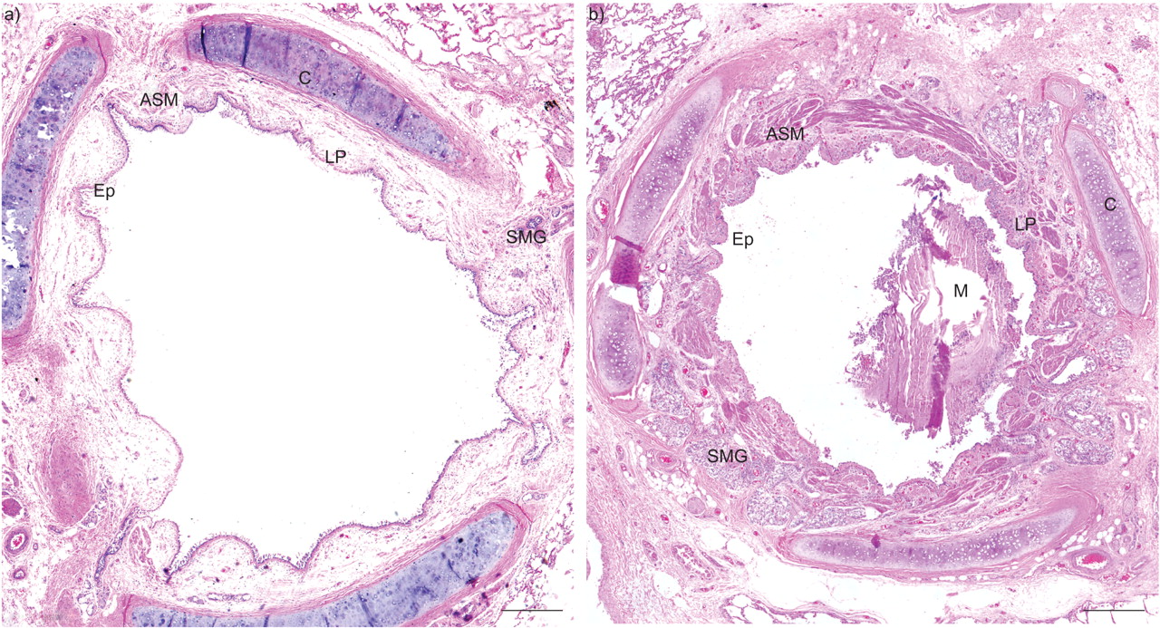
Large airways from a) a nonasthma control and b) a fatal asthma (FA) patient, stained with haematoxylin and eosin. The FA cases showed a very prominent airway smooth muscle (ASM) layer, an increased number of submucosal glands (SMG) and mucus (M) plugging in the airway lumen. Ep: epithelial layer; LP: lamina propria; C: cartilage. Scale bars = 200 μm.
In large airways, the mean±sd Pbm was 10.6±3.4 mm in FA, 12.6±3.1 mm in NFA and 10.3±3.8 mm in NAC (p = 0.23). In small airways, the Pbm was 2.7±1.0 mm in FA, 2.6±0.9 mm in NFA and 2.4±0.5 mm in NAC (p = 0.58). The mean±sd ASM area per field in large airways was 15.4±3.7×104 μm2 in FA, 12.2±4.1×104 μm2 in NFA and 9.1±3.6×104 μm2 in NAC (p<0.001 for FA versus NAC; p = 0.085 for FA versus NFA; p = 0.16 for NFA versus NAC). The ASM area per field in small airways was 5.4±3.5×104 μm2 in FA, 2.5±1.9×104 μm2 in NFA and 2.9±2.1×104 μm2 in NAC (p = 0.03 for FA versus NAC; p = 0.02 for FA versus NFA; p = 0.99 for NFA versus NAC).
Collagen and elastic fibre histochemical analysis
Figure 3⇓ (a and b) shows examples of NAC and FA cases stained for elastic fibres. In NFA and FA patients, there were no differences in total collagen or elastic fibres between large and small airways. In NAC, the fractional area of elastic fibres within the ASM was higher in the small airways than in the larger airways (p = 0.035; table 3⇓).

Fractional areas of elastic fibres (a–c) and fibronectin (d–f) within the airway smooth muscle (ASM) layer in nonasthma controls (NAC; a and d) and fatal asthma (FA) patients (b and e) measured using Weigert’s Resorcin–Fuchsin staining (a and b) and immunohistochemistry (d and e). There was an increased fractional area of these proteins within the ASM layer in FA (c and f). Data are presented as mean±sd. Ep: epithelial layer; LP: lamina propria. ▓: FA; ▪: nonfatal asthma; □: NAC. #: p = 0.007; ¶: p = 0.002; +: p = 0.019; §: p = 0.04. Scale bars = 50 μm.
Fractional areas of extracellular matrix components within the airway smooth muscle in fatal asthma(FA), nonfatal asthma (NFA) and nonasthma control (NAC) cases
The fractional area of elastic fibres in ASM was increased in the large airways in FA compared with NFA (p = 0.002) and NAC (p = 0.007). There were no significant differences between NFA and NAC. In the small airways, the fractional area of elastic fibres was significantly increased in FA compared with NFA (p = 0.019) but not in comparison with NAC. There were no differences in the fractional area of elastic fibres within the ASM of small airways between NFA and NAC. There were no significant differences between groups for the percentage of total collagen within the ASM (table 3⇑).
Immunohistochemical analysis
The results of immunohistochemical analysis are summarised in tables 3⇑ and 4⇓. Among the ECM components, both in FA and NAC patients, the fractional areas of versican, collagen I and collagen III were higher in the small airways than in the larger airways (for FA, p = 0.005, p = 0.0001 and p = 0.001, respectively; for NAC, p = 0.033, p = 0.018 and p = 0.011, respectively; table 3⇑).
Fractional areas of matrix metalloproteinases(MMPs) and tissue inhibitors of metalloproteinases (TIMPs) within the airway smooth muscle in fatal asthma (FA) and nonasthma control (NAC) cases
There was a significant increase in the fractional area of fibronectin in the large airways in FA cases compared with NAC (p = 0.04; fig. 3d–f⇑). Fibronectin expression correlated positively with age (r = 0.48, p = 0.03) in large airways of FA cases. There were no differences between patient groups for versican, collagen I or collagen III fractional areas in large or small airways.
Among the MMPs, FA patients presented higher fractional areas of MMP-12 in large airways than in small airways (p = 0.001). Among NAC patients, the ASM fractional areas of MMP-9 and TIMP-2 were higher in the small airways than in the large airways (p = 0.043 and p = 0.025, respectively; table 4⇑).
The ASM area fractions of MMP-9 (fig. 4a–c⇓) and MMP-12 (fig. 4d–f⇓) were increased in large airways, but not in small airways, in cases of FA compared with NAC. There was a positive correlation between elastic fibres and MMP-12 in large airways in FA cases (r = 0.71, p = 0.003). Both MMP-9 (r = 0.51, p = 0.01) and MMP-12 (r = 0.52, p = 0.02) expression in large airways of FA cases correlated positively with age at onset of asthma. There were no statistically significant differences between the case groups for MMP-1, MMP-2, TIMP-1 or TIMP-2.

Matrix metalloproteinase (MMP)-9 (a–c) and -12 (d–f) expression within the airway smooth muscle (ASM) cells in large airways of nonasthma controls (NAC; a and d) and fatal asthma (FA) patients (b and e) measured using immunohistochemistry. There was increased expression of MMP-9 and MMP-12 within the ASM layer of large airways in FA patients (c and f). MMP-9 and -12 were also expressed in the bronchial epithelium (Ep) and in inflammatory cells in the lamina propria (LP). Data are presented as median and ranges. ▓: FA; □: NAC. #: p = 0.01. Scale bars = 50 μm.
Smokers versus nonsmokers
There were 11 FA subjects who were smokers or ex-smokers. Smokers had a larger fractional area of MMP-9 in large and small airways than nonsmokers (large airways 8.6±10.6 versus 2.8±4.4%, p = 0.024; small airways 6.5±6.0 versus 1.2±1.0%, p = 0.001; analysis performed with São Paulo patients, n = 8). There were no significant differences in elastic fibre or collagen area fractions between smokers and nonsmokers in the NFA cases.
Corticosteroid use
The 13 FA subjects who received steroids regularly had smaller area fractions of elastic fibres in large and small airways compared with those who did not receive any corticosteroids (large airways 8.8±13.7 versus 28.8±9.5%, p = 0.003; small airways 10.0±12.8 versus 20.4±9.5%, p = 0.01). There were no differences in the other parameters analysed.
DISCUSSION
In the present study, the ASM content of different ECM components, MMPs and TIMPs was analysed in lung tissue of patients with and without asthma. Increased area fractions of elastic fibres were found in FA compared with NFA and NAC. Furthermore, there was increased expression of fibronectin, MMP-9 and MMP-12 in the large airways in FA compared with NAC. To the current authors’ knowledge, this is the first in vivo human tissue study to comprehensively analyse several ECM components and their regulators, both at the large and small airway level in the ASM in asthma.
Elastic fibres are an integral component of the ECM of the lung parenchyma and airways. They have a major role in regulating airway patency and lung elastic recoil. It has previously been shown that, in FA, elastic fibres are damaged in the large airways and that elastic fibre content is decreased at the subepithelial and alveolar attachment levels 9, 10. Elastic fibres are abundant within the ASM and, although their physiological role is unclear, they are presumably related to the necessary airway alterations in calibre and length during normal tidal breathing, deep breaths and coughing. Elastic fibres contribute to loads promoting bronchodilation and opposing bronchoconstriction during inspiration 24, 25.
The present study shows an increase in the area fraction of elastic fibres in the ASM in large and small airways in patients with FA, but not in NFA, compared with NAC cases. This contrasts with previous findings of a reduced area fraction of elastic fibres beneath the epithelial basement membrane and in the airway periphery in cases of FA 9, 10. Possible explanations for this varying fraction of elastic fibres may be related to the potential local secretory function of the smooth muscle that promotes elastin formation, and to the elastolytic effects of inflammation, which is prominent in the lamina propria and adventitial layers in FA 17.
There is scarce information about the role of corticosteroids on elastin content in obstructive lung diseases. The present data suggest that asthma severity could be related to increased fractional areas of elastic fibres within the ASM and that this change may be altered by treatment.
Proteoglycans comprise a family of proteins that have major roles in lung biology 26. One of these proteins, the large proteoglycan versican, has the ability to regulate water content in tissues, thereby affecting resiliency 27. Previous studies have demonstrated an increase in versican in the inner wall of small and large airways in cases of asthma, but no differences were detected at the ASM level 12, 21, 27. Pini et al. 21 recently compared the fractional area of versican within the ASM in a small population of moderate and severe asthma cases and control cases and also found no significant differences between asthmatic and nonasthmatic subjects. It is possible that the increased versican in the inner wall is related to accumulation of oedema fluid.
Taken together, these findings indicate that ECM changes, in a fashion similar to inflammatory changes, vary between the different airway compartments in asthma 28, and that trying to understand the relationship between airway structure and function in asthma is not trivial. Functional changes in the airways will ultimately depend on the relative contribution of each protein in each airway compartment, and the interactions among the proteins and with the surrounding parenchyma. It would have been extremely interesting to couple all these data to functional data in the present study patients, but lung function data were unfortunately not available in the large majority. Further studies correlating airway structure and function in asthma are certainly needed.
Fibronectin is also abundant in the ASM and has previously been shown to be increased in the airway wall in asthma 29. The current authors found an increase in its area fraction in large airways in cases of FA. Although the present study is the first to show that there is increased fibronectin within the ASM in vivo, there is a large body of in vitro evidence to show that fibronectin strongly influences ASM biology in asthma. Fibronectin may enhance ASM proliferation 30, migration 31 and cell survival 32. Chan et al. 33 have demonstrated that ASM cells from asthmatics secrete more fibronectin in vitro, and that this autocrine secretion contributes to increased levels of interleukin (IL)-13-dependent eotaxin expression.
Type-I and -III collagens are the major fibrillar collagens in muscle tissues, whereas the network-forming type-IV collagen is the major basement membrane collagen 34. In vitro studies have shown that ASM from asthmatics secretes significantly more collagen I 7. In vivo, some previous studies have found increased collagens at the bronchial submucosal level in asthma 29, 35, 36, whereas others have not 37. In the present study, there was no increase in collagen content within the ASM in FA as detected by Sirius Red staining and immunohistochemistry. Increases in fibrillar collagens would certainly act to limit the force transference among ASM cells and thereby decrease bronchoconstriction, a hypothesis not supported by the present data.
Differential expression of the MMPs has been associated with asthma pathogenesis. Besides their roles in degrading ECM components, MMPs are also involved in inflammatory cell trafficking, host defences and tissue repair 38. In the current study, increases in MMP-9 and -12 were detected, but not in MMP-1 or -2, or in TIMP-1 or -2 in the ASM bundles in cases of FA. The present data favour a degradative environment within the ASM, especially of the MMPs with an elastolytic activity.
There is strong evidence of MMP-9 involvement in asthma, particularly in patients with severe asthma 39 and in acute, severe exacerbations 40. MMP-9 is expressed by inflammatory cells and structural cells of the lungs, including the ASM cells. Interestingly, the present study also showed that smokers have an increased fractional area of MMP-9 in the ASM in comparison with nonsmoking subjects. MMP-12 cleaves elastin and also type-IV collagen, fibronectin, laminin and gelatin 41. It has recently been demonstrated that ASM cells express MMP-12, which is highly inducible by IL-1β 41. The current data showed an increased expression of MMP-12 in the ASM cells of subjects who died of asthma. Additionally, in cases of FA, there was a positive correlation between elastic fibres and MMP-12, consistent with the idea that a dynamic turnover of elastic fibres occurs in asthma. In adult tissues, functional repair of elastic fibres is difficult because it requires the coordinated re-expression of all the molecules that make up the microfibril as well as the enzymes critical for cross-linking elastin 42. It is possible to speculate that, although the fractional expression of elastic fibres is increased within the ASM in cases of FA, elastic fibre structure is abnormal, with consequent impaired function. This fact may contribute to altered mechanisms of ASM contraction and relaxation in asthma.
The present study has some important limitations, such as the limited clinical and functional data available for the asthmatic subjects. In addition, despite having normal lung histology, the controls were not devoid of other diseases. Although it was not possible to stain the Perth cases immunohistochemically, providing information in FA, NFA and NAC for “total” collagen and elastic fibres was very important, since these two major lung proteins are likely to have major roles in ASM mechanics. Furthermore, histochemical data on “total” collagen was confirmed by immunohistochemistry data in the São Paulo subset of patients.
There were statistically significant correlations between some proteins and age and asthma onset and, although the relevance of these findings is unclear, Bai et al. 13 have previously shown a relationship between age and the area fraction of ECM within the smooth muscle layer in asthma. The current data are based on histo- and immunohistochemistry techniques. It would have been useful to confirm the findings at RNA level. Unfortunately, attempts to extract RNA from microdissected ASM of the autopsy material were not successful, due to extensive post mortem RNA degradation.
Despite the fact that the fractional areas of some of the studied proteins were higher in the small airways, most of the changes between FA and NAC were found within the large airways. Recently, Brown et al. 43 and Permutt 44 have emphasised the role of airway structure at large airway level as a major determinant of airway hyperresponsiveness in asthma. It is possible that the observed ECM changes within the ASM contribute more significantly to the intrinsic airway alterations in asthma in large airways than in small airways.
In conclusion, elastic fibres and fibronectin are increased within the airway smooth muscle in fatal asthma, with increased matrix metalloproteinase-9 and -12 expression. The present in vivo data confirm that changes in extracellular matrix and its regulators occur within the airway smooth muscle in asthma, but do not necessarily reflect those elsewhere within the airway wall. They may, however, have important consequences for airway smooth muscle function and excessive airway narrowing in asthma.
Support statement
This study received support from the Coordenação de Aperfeiçoamento de Pessoal de Nível Superior (Brasilia, Brazil), the Conselho Nacional de Desenvolvimento Científico e Tecnológico (Brasilia) and the Australian National Health and Medical Research Council (Canberra, Australia), grant ID 343601.
Statement of interest
None declared.
Acknowledgments
The authors wish to thank the Serviço de Verificação de Óbitos da Capital (SVOC), São Paulo University (São Paulo, Brazil), for permission to collect the fatal asthma cases. They also thank all members of the Pulmonary Pathology group of the Dept of Pathology, São Paulo University Medical School, for organising the tissue collection.
- Received November 6, 2007.
- Accepted February 26, 2008.
- © ERS Journals Ltd
References


