
Main menu

  • Home
  • Current issue
  • ERJ Early View
  • Authors/reviewers
    • Instructions for authors
    • Submit a manuscript
    • Open access
    • COVID-19 submission information
    • Peer reviewer login
  • Alerts
  • Podcasts
  • Subscriptions
  • ERS Publications
    • European Respiratory Journal
    • ERJ Open Research
    • European Respiratory Review
    • Breathe
    • ERS Books
    • ERS publications home

User menu

  • Log in
  • Subscribe
  • Contact Us
  • My Cart

Search

  • Advanced search
  • ERS Publications
    • European Respiratory Journal
    • ERJ Open Research
    • European Respiratory Review
    • Breathe
    • ERS Books
    • ERS publications home

Login

European Respiratory Society

Advanced Search

  • Home
  • Current issue
  • ERJ Early View
  • Authors/reviewers
    • Instructions for authors
    • Submit a manuscript
    • Open access
    • COVID-19 submission information
    • Peer reviewer login
  • Alerts
  • Podcasts
  • Subscriptions

Necrotising sarcoid granulomatosis: clinical, functional, endoscopical and radiographical evaluations

C. Quaden, I. Tillie-Leblond, A. Delobbe, L. Delaunois, A. Verstraeten, M. Demedts, B. Wallaert
European Respiratory Journal 2005 26: 778-785; DOI: 10.1183/09031936.05.00024205
C. Quaden
  • Find this author on Google Scholar
  • Find this author on PubMed
  • Search for this author on this site
I. Tillie-Leblond
  • Find this author on Google Scholar
  • Find this author on PubMed
  • Search for this author on this site
A. Delobbe
  • Find this author on Google Scholar
  • Find this author on PubMed
  • Search for this author on this site
L. Delaunois
  • Find this author on Google Scholar
  • Find this author on PubMed
  • Search for this author on this site
A. Verstraeten
  • Find this author on Google Scholar
  • Find this author on PubMed
  • Search for this author on this site
M. Demedts
  • Find this author on Google Scholar
  • Find this author on PubMed
  • Search for this author on this site
B. Wallaert
  • Find this author on Google Scholar
  • Find this author on PubMed
  • Search for this author on this site
  • Article
  • Figures & Data
  • Info & Metrics
  • PDF
Loading

Figures

  • Tables
  • Fig. 1—
    • Download figure
    • Open in new tab
    • Download powerpoint
    Fig. 1—

    Computed tomography scan of the lower lung zones showing bilateral airspace consolidation.

  • Fig. 2—
    • Download figure
    • Open in new tab
    • Download powerpoint
    Fig. 2—

    Computed tomography scan at the level of the right bronchus intermedius showing small airway disease in the right upper lobe with areas of airspace consolidation in the lingula and the apical segment of the left lower lobe. The peripheral location of the axillary mass with irregular borders should be noted.

  • Fig. 3—
    • Download figure
    • Open in new tab
    • Download powerpoint
    Fig. 3—

    Computed tomography scan at the level of the right bronchus intermedius, showing three nodular opacities with irregular borders, one located in the lingula and one located in the left lower lobe. The presence of areas of ground-glass attenuation in the posterior parts of both lower lobes corresponds to hypoventilated lung.

  • Fig. 4—
    • Download figure
    • Open in new tab
    • Download powerpoint
    Fig. 4—

    Computed tomography scan at the level of the right bronchus intermedius, showing large areas of airspace consolidation in the right middle lobe, lingula and the apical segments of both lower lobes. The presence of cavitation within the right and left lower lobe masses should be noted.

  • Fig. 5—
    • Download figure
    • Open in new tab
    • Download powerpoint
    Fig. 5—

    Histological stain showing coagulative necrosis and individual granulomas.
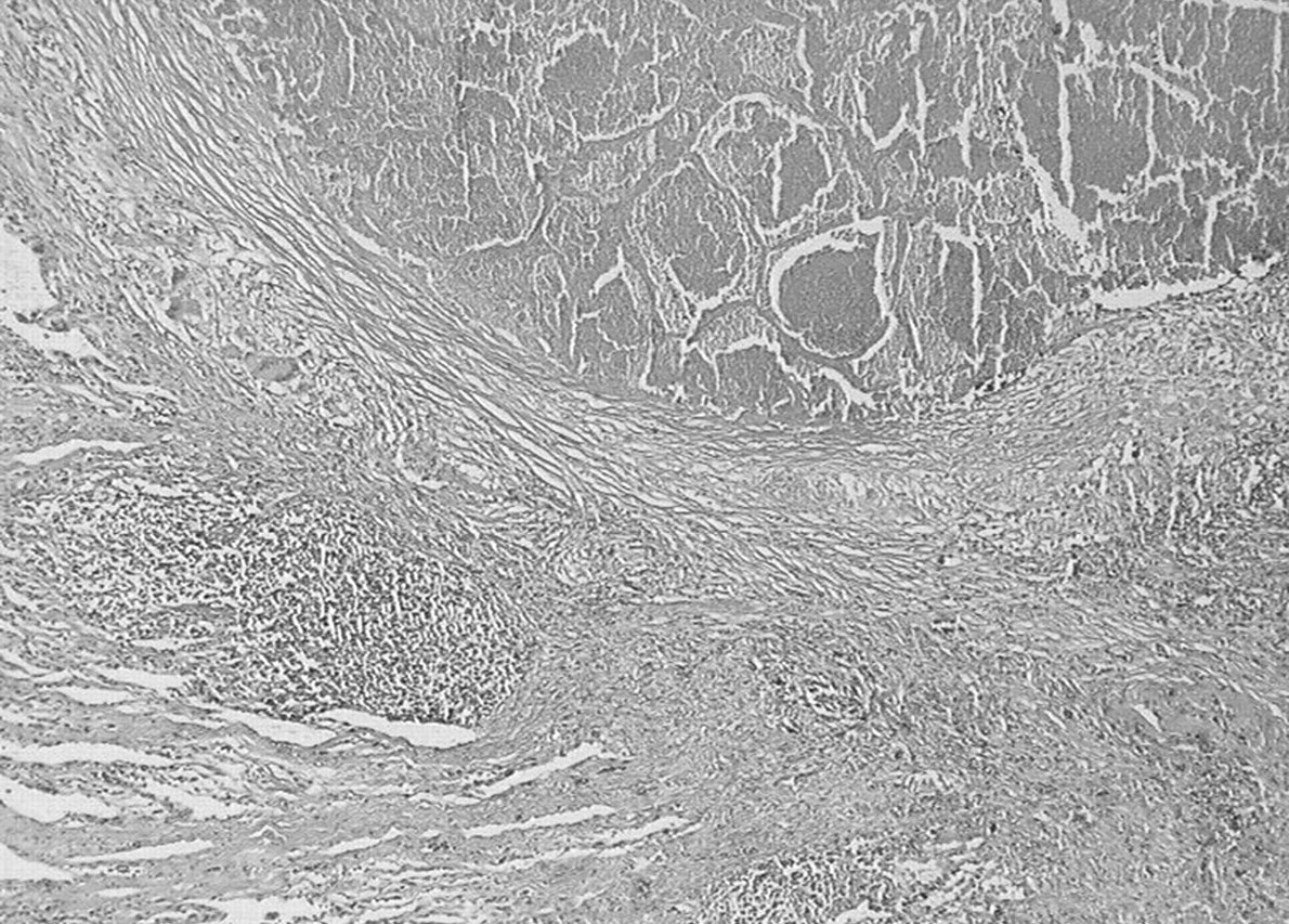
  • Fig. 6—
    • Download figure
    • Open in new tab
    • Download powerpoint
    Fig. 6—

    Histological stain showing vascular involvement with extensive granulomatous infiltration of the media.

Tables

  • Figures
  • Table. 1—

    Clinical features at time of diagnosis in 14 necrotising sarcoid granulomatosis patients

    FeaturesPatients
    Smoking habits
     Nonsmokers9 (64)
     Ex-smokers1 (7)
     Smokers4 (29)
    Respiratory manifestations8 (57)
     Cough5 (36)
     Dyspnoea4 (29)
      Exertional4 (29)
      At rest0 (0)
     Chest pain4 (29)
    Extrarespiratory manifestations12 (86)
     Systemic symptoms8 (57)
      Easily tired7 (50)
      Fever5 (36)
      Weight loss4 (29)
      Night sweats2 (14)
     Arthralgia5 (36)
     Ocular manifestations3 (21)
     Sicca syndrome2 (14)
     Neurological symptoms1 (7)
     Nasal symptoms1 (7)
     Skin involvement1 (7)
    Physical findings
     Crackles1 (7)
     Joint swelling1 (7)
     Splenomegaly1 (7)
    • Data are presented as n (%).

  • Table. 2—

    Results of pulmonary function tests in 14 patients with necrotising sarcoid granulomatosis

    Patient No.FEV1 L (% pred)FVC L (% pred)FEV1/FVC % predTLC L (% pred)RV L (% pred)RV/TLC % predDL,CO/VA % pred
    13.1 (118)3.9 (125)805.2 (110)1.3 (85)2574
    22.6 (80)3.0 (80)825.6 (104)2.6 (160)46105
    32.8 (82)3.1 (92)864.3 (82)1.1 (82)26109
    43.8 (102)4.7 (101)826.3 (88)1.1 (52)1874
    52.6 (111)3.0 (109)865.0 (99)2.1 (110)4274
    62.3 (78)3.0 (80)765.4 (83)1.7 (95)3178
    73.1 (88)3.2 (89)954.3 (91)1.1 (109)2583
    83.3 (116)3.4 (95)966.2 (98)2.8 (116)4560
    92.3 (80)2.7 (83)844.1 (85)1.3 (87)3274
    102.2 (91)2.6 (101)764.3 (103)1.4 (90)32NA
    112.1 (91)2.6 (95)815.5 (106)2.1 (101)38NA
    121.8 (52)2.7 (62)695.2 (79)2.7 (134)5274
    133.0 (110)3.5 (113)866.1 (123)2.6 (150)4371
    143.5 (84)3.9 (74)84NANANANA
    • FEV1: forced expiratory volume in one second; % pred: percentage of predicted; FVC: forced vital capacity; TLC: total lung capacity; RV: residual volume; DL,CO: carbon monoxide diffusing capacity of the lung; VA: alveolar volume; NA: not available.

  • Table. 3—

    Computed tomography findings in 14 patients with necrotising sarcoid granulomatosis

    FindingPatients
    Infiltrates7 (50)
    Solitary nodule4 (29)
    Bilateral/multiple nodules3 (21)
    Cavitation2 (14)
    Hilar or mediastinal adenopathy5 (36)#
    Pleural thickening1 (7)
    • Data are presented as n (%). #: bilateral in all cases.

  • Table. 4—

    Examination of bronchoalveolar lavage samples in eight patients with necrotising sarcoid granulomatosis

    Patient No.103 cells·mL−1Macrophage %Lymphocyte %Neutrophil %Eosinophil %
    119290820
    218093610
    43892.552.50
    5150826120
    637590280
    7240603730
    8229821413
    1088643240
  • Table. 5—

    Treatment and clinical course in 14 patients with necrotising sarcoid granulomatosis

    Patient No.TreatmentIndicationRespiratory courseExtrarespiratory courseFollow-up months
    1SteroidSystemic symptomsResolution relapseResolution60
    2SteroidArthralgiaResolution relapse#Persistent arthralgia102
    3NoneResolutionResolution32
    4SurgeryDiagnostic procedureResolutionResolution21
    5SteroidSystemic symptomsResolution relapseResolution101
    6S, C#, A, EICerebral involvementResolutionDeath (neurological)114
    7SteroidSystemic symptomsResolutionResolution28
    8SurgeryDiagnostic procedureRelapse of noduleNone42
    9NoneResolutionResolution18
    10SurgeryDiagnostic procedureRelapse of nodulesPericarditis C-myopathy36
    11SurgeryDiagnostic procedureResolutionNone24
    12NoneSequelar cavities¶Resolution56
    13NoneResolutionResolution52
    14NoneResolutionPersistence of skin lesions79
    • S: steroid; C: cyclophosphamide; A: azathioprine; EI: encephalic irradiation. #: neoplasy occurrence; ¶: aspergilloma and lung cancer.

PreviousNext
Back to top
View this article with LENS
Vol 26 Issue 5 Table of Contents
  • Table of Contents
  • Index by author
Email

Thank you for your interest in spreading the word on European Respiratory Society .

NOTE: We only request your email address so that the person you are recommending the page to knows that you wanted them to see it, and that it is not junk mail. We do not capture any email address.

Enter multiple addresses on separate lines or separate them with commas.
Necrotising sarcoid granulomatosis: clinical, functional, endoscopical and radiographical evaluations
(Your Name) has sent you a message from European Respiratory Society
(Your Name) thought you would like to see the European Respiratory Society web site.
CAPTCHA
This question is for testing whether or not you are a human visitor and to prevent automated spam submissions.
Print
Citation Tools
Necrotising sarcoid granulomatosis: clinical, functional, endoscopical and radiographical evaluations
C. Quaden, I. Tillie-Leblond, A. Delobbe, L. Delaunois, A. Verstraeten, M. Demedts, B. Wallaert
European Respiratory Journal Nov 2005, 26 (5) 778-785; DOI: 10.1183/09031936.05.00024205

Citation Manager Formats

  • BibTeX
  • Bookends
  • EasyBib
  • EndNote (tagged)
  • EndNote 8 (xml)
  • Medlars
  • Mendeley
  • Papers
  • RefWorks Tagged
  • Ref Manager
  • RIS
  • Zotero

Share
Necrotising sarcoid granulomatosis: clinical, functional, endoscopical and radiographical evaluations
C. Quaden, I. Tillie-Leblond, A. Delobbe, L. Delaunois, A. Verstraeten, M. Demedts, B. Wallaert
European Respiratory Journal Nov 2005, 26 (5) 778-785; DOI: 10.1183/09031936.05.00024205
del.icio.us logo Digg logo Reddit logo Technorati logo Twitter logo CiteULike logo Connotea logo Facebook logo Google logo Mendeley logo
Full Text (PDF)

Jump To

  • Article
    • Abstract
    • MATERIALS AND METHODS
    • RESULTS
    • DISCUSSION
    • Acknowledgments
    • References
  • Figures & Data
  • Info & Metrics
  • PDF
  • Tweet Widget
  • Facebook Like
  • Google Plus One

More in this TOC Section

  • Clinical predictors of pulmonary hypertension in sarcoidosis
  • Fluid-containing emphysematous bullae: a spectrum of illness
  • Left ventricular diastolic dysfunction in idiopathic pulmonary fibrosis: a tissue Doppler echocardiographic study
Show more Original Articles: Clinical forum

Related Articles

Navigate

  • Home
  • Current issue

About the ERJ

  • Journal information
  • Editorial board
  • Reviewers
  • Press
  • Permissions and reprints
  • Advertising

The European Respiratory Society

  • Society home
  • myERS
  • Privacy policy
  • Accessibility

ERS publications

  • European Respiratory Journal
  • ERJ Open Research
  • European Respiratory Review
  • Breathe
  • ERS books online
  • ERS Bookshop

Help

  • Feedback

For authors

  • Instructions for authors
  • Publication ethics and malpractice
  • Submit a manuscript

For readers

  • Alerts
  • Subjects
  • Podcasts
  • RSS

Subscriptions

  • Accessing the ERS publications

Contact us

European Respiratory Society
442 Glossop Road
Sheffield S10 2PX
United Kingdom
Tel: +44 114 2672860
Email: journals@ersnet.org

ISSN

Print ISSN:  0903-1936
Online ISSN: 1399-3003

Copyright © 2022 by the European Respiratory Society