


Figures
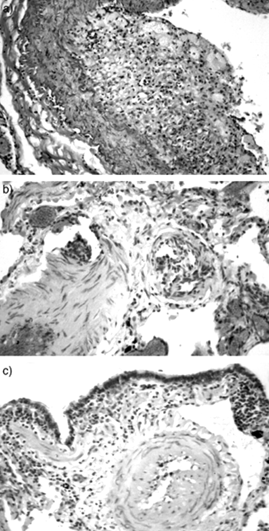
- Fig. 1.—
Histology. a) In the large pulmonary arteries foam cell aggregates and a few lymphocytes were dominant (Elastica van Gieson stained, original magnification ×100). b) Media hypertrophy, intima fibrosis, and rarely plexiform lesions were found (haematoxylin and eosin stained, original magnification ×100). c) Sometimes slight chronic inflammation was seen in the respiratory bronchioles (haematoxylin and eosin stained, original magnification ×100).
- Fig. 2.—
Immunofluorescence and electron microscopy. a) Confocal laser scanning microscopy with antiserum against Chlamydia spp. shows bright spots within the hypertrophic media of small arteries demonstrating the positive immunofluorescence reaction to Chlamydia spp. (double staining with actin (pale staining), objective magnification ×50). b) Spherical bodies with a diameter of 0.61 µm were seen in lytic areas of the thickened intima, macrophages were found nearby (scanning electron microscopy, original magnification ×10,000). c) These had a double membrane, were lying in small groups, and were sometimes detected within myofibroblasts (transmission electron microscopy, original magnification ×20,000).


