


Figures
- FIGURE 1
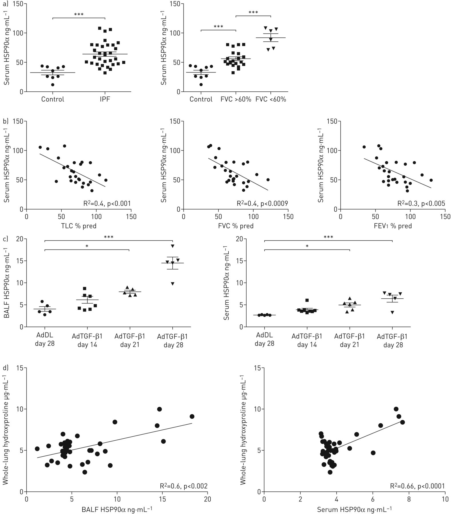
Circulating heat shock protein (HSP) isoform HSP90α is upregulated in idiopathic pulmonary fibrosis (IPF) patients and in adenovector-mediated transforming growth factor-β1 (AdTGF-β1)-induced lung fibrosis. a) Serum level of HSP90α measured by ELISA on IPF patients and respective aged-matched healthy volunteers. Data presented as mean±sem; n=9 and n=31 for controls and IPF patients, respectively. IPF patients were clustered in two groups: moderate IPF (FVC % pred >60%) and severe IPF (FVC % pred <60%). Data presented as mean±sem; n=9, n=25 and n=6 for controls, moderate IPF and severe IPF patients, respectively. b) Correlation curves between the serum level of HSP90α in IPF patients and clinical lung function parameters: total lung capacity (TLC), forced vital capacity (FVC) and forced expiratory volume in 1 s (FEV1). c) Bronchoalveolar lavage fluid (BALF) and serum levels of HSP90α of rats treated with AdTGF-β1 (or AdDL as control). Data presented as mean±sem; AdDL: n=5; AdTGF-β1 day 14: n=8; AdTGF-β1 day 21: n=5; AdTGF-β1 day 28: n=5. d) Correlation curves between BALF and serum levels of HSP90α and hydroxyproline lung content in rats treated with AdTGF-β1. *: p<0.05; ***: p<0.001.
- FIGURE 2
Mechanical stretch and tissue stiffness induce heat shock protein (HSP) isoform HSP90α secretion. a) Level of HSP90α and HSP90β in supernatant of untreated control/primary idiopathic pulmonary fibrosis (IPF) lung fibroblasts and A549 cells treated or not with recombinant transforming growth factor (rTGF)-β1. HSP90α and HSP90β were measured by ELISA after cells were placed in serum-free medium for 24 h. Data presented as mean±sem; n=3. b) Western blot analysis of HSP90α and HSP90β expression in supernatant (with corresponding densitometry analysis for HSP90α). Cells were cultured either on hydrogels of stiffness 1 kPa (soft) or 50 kPa (stiff) or on regular tissue culture plates (1 GPa) and placed in serum-free medium for 24 h before supernatant collection. Ponceau staining served as loading control. Data presented as mean±sem; n=4. c) Western blot analysis of HSP90α and HSP90β expression release by nonfibrotic and fibrotic lung slices (from adenovector-mediated transforming growth factor-β1 (AdTGF-β1)-treated rats) before and after stretch. Western blots are representative of three independent experiments. Western blots of fibrotic and nonfibrotic lung slices were performed on separated gels but run simultaneously. Ponceau staining served as loading control. d) Western blot analysis of HSP90α and HSP90β expression release by fibrotic lung slices (from AdTGF-β1-treated rats) before and after stretch with or without protein transport inhibitor (PTI) treatment (with corresponding densitometry analysis for HSP90α). S: stimulated; US: unstimulated. Ponceau staining served as loading control. Data presented as mean±sem; n=6. *: p<0.05; **: p<0.01; ***: p<0.001.
- FIGURE 3
Extracellular heat shock protein (HSP) isoform HSP90α induces myofibroblast differentiation. a) Western blot analysis (and corresponding densitometry) of E-cadherin, collagen 1A, phosphorylated extracellular signal-regulated kinase (p-ERK), ERK, α-smooth muscle actin (α-SMA), phosphorylated Smad2 (p-Smad2) and Smad2 on A549 cells treated with recombinant (r) HSP90α or vehicle (control) for 48 h at 10 µM. Glyceraldehyde 3-phosphate dehydrogenase (GAPDH) staining served as loading control. Data presented as mean±sem; n=4. b) Western blot analysis (and corresponding densitometry) of collagen 1A, p-ERK, ERK, α-SMA, p-Smad2 and Smad2 on human primary control fibroblasts treated with rHSP90α or vehicle (control) for 48 h at 10 µM. GAPDH staining served as loading control. Data presented as mean±sem; n=4. c) Western blot analysis (and corresponding densitometry) of collagen 1A, p-ERK, ERK and α-SMA on human primary idiopathic pulmonary fibrosis (IPF) fibroblasts treated with a blocking HSP90α antibody (AbHSP90α) or vehicle (control) for 48 h at 40 µM. GAPDH staining served as loading control. Data presented as mean±sem; n=4. d) Representative images of the wound closure assay performed on A549 cells cultured with conditioned medium (CM) from control or IPF primary fibroblasts and treated with AbHSP90α or vehicle (control) at 40 µM. Images are representative of three independent experiments. The graph represents the percentage of wound closure measured at 36 h after the wound was treated. Corresponding proliferating cell nuclear antigen (PCNA) expression of A549 cells showed no increase in proliferation during the wound closure assay. Data presented as mean±sem; n=3. *: p<0.05; **: p<0.01; ***: p<0.001.
- FIGURE 4
Heat shock protein (HSP) isoform HSP90α signals through low-density lipoprotein receptor-related protein 1 (LRP1). a) Immunoprecipitation (IP) of HSP90α and HSP90β performed on plasma membrane extracted proteins followed by immunodetection of LRP1 and HSP90α. Control: control human primary fibroblasts; IPF: idiopathic pulmonary fibrosis human primary fibroblasts; IgG: nonrelevant antibody; input: nonimmunoprecipitated extracts. Na,K-ATPase served as loading control of the plasma membrane fraction. b) Immunoprecipitation of LRP1 performed on plasma membrane extracted proteins followed by immunodetection of HSP90α, HSP90β and LRP1. Immunoprecipitation was performed with or without prior cross-linking of extracellular/membrane proteins: control human primary fibroblasts and human primary IPF fibroblasts. NC: non-cross-linked; C: cross-linked. c) Western blot analysis of LRP1, HSP90α and HSP90β expression on IPF human primary fibroblasts treated with small interfering (si) RNA for HSP90α or HSP90β (Scramble siRNA used as control) and treated with or without MG132 for 6 h at 50 µM. Glyceraldehyde 3-phosphate dehydrogenase (GAPDH) staining served as loading control. The Western blot is representative of three independent experiments. d) Western blot analysis of phosphorylated extracellular signal-regulated kinase (p-ERK), ERK, α-smooth muscle actin (α-SMA), phosphorylated Smad2 (p-Smad2), Smad2, LRP1 and collagen 1A expression on human primary IPF fibroblasts, A549 cells and control fibroblasts treated with siRNA for LRP1 (Scramble siRNA used as control) and treated with recombinant (r) HSP90α for 48 h at 10 µM. GAPDH staining served as loading control. Data presented as mean±sem; n=3. *: p<0.05; **: p<0.01; ***: p<0.001.
- FIGURE 5
HS-30, an inhibitor of extracellular heat shock protein HSP90, prevents HSP90α/low-density lipoprotein receptor-related protein 1 (LRP1) signalling. a) Western blot analysis of collagen 1A, α-smooth muscle actin (α-SMA), phosphorylated extracellular signal-regulated kinase (p-ERK), ERK, HSP90α and LRP1 expression on human primary control fibroblasts treated with recombinant (r) HSP90α for 48 h at 10 µM with or without HS-30 at 1 mM. Glyceraldehyde 3-phosphate dehydrogenase (GAPDH) staining served as loading control. The Western blot is representative of three independent experiments. b) Western blot analysis of α-SMA, p-ERK, ERK, phosphorylated Smad2 (p-Smad2), Smad2, HSP90α and LRP1 expression on human primary idiopathic pulmonary fibrosis (IPF) fibroblasts treated with rHSP90α for 48 h at 10 µM with or without HS-30 at 1 mM. GAPDH staining served as loading control. The Western blot is representative of three independent experiments. c) Immunoprecipitation (IP) of LRP1 performed on plasma membrane extracted proteins followed by immunodetection of HSP90α and LRP1. C: cross-linked; input: nonimmunoprecipitated extracts; IgG: nonrelevant antibody. Immunoprecipitation was performed with prior cross-linking of extracellular/membrane proteins. Immunoprecipitation performed on IPF fibroblasts treated with or without HS-30 at 1 mM. d) Immunofluorescence of LRP1 (red) and HSP90α (green) on control fibroblasts, IPF fibroblasts and IPF fibroblasts treated with HS-30 at 1 mM. Nuclear staining: 4′,6-diamidino-2-phenylindole (blue). Images are representative of three independent experiments. Scale bar: 50 μm. Pearson's coefficient for each condition presented as mean±sem; n=3. **: p<0.01; ***: p<0.001.
- FIGURE 6
Heat shock protein (HSP) isoform HSP90α/low-density lipoprotein receptor-related protein 1 (LRP1) colocalise in idiopathic pulmonary fibrosis (IPF) patients. Immunofluorescence of LRP1 (red) and HSP90α (green) on human lung tissue from controls, moderate IPF patients and severe IPF patients. IgG as inserts. Nuclear staining: 4′,6-diamidino-2-phenylindole (blue). Images presented are representative of three independent experiments. Scale bar: 50 μm. Pearson's coefficient for each condition is presented as mean±sem; n=3. ***: p<0.001.
- FIGURE 7
HS-30 has antifibrotic properties on fibrotic lung tissue ex vivo. a) Quantitative reverse transcription PCR analysis of α-smooth muscle actin (α-SMA) and collagen 1A expression on lung slices from rats receiving adenovector-mediated transforming growth factor-β1 (AdTGF-β1). Lung slices were collected at day 21 after AdTGF-β1 administration and then cultured ex vivo with HS-30 or vehicle for 72 h. Data presented as mean±sem; n=4. b) Western blot analysis (and corresponding densitometry) of α-SMA and TGF-β1 expression on lung slices from rats receiving AdTGF-β1. Lung slices were collected at day 21 after AdTGF-β1 administration and then cultured ex vivo with HS-30 or vehicle for 72 h. Glyceraldehyde 3-phosphate dehydrogenase (GAPDH) staining served as loading control. Data presented as mean±sem; n=4. *: p<0.05; **: p<0.01.


Supplementary Materials
Supplementary Material
Please note: supplementary material is not edited by the Editorial Office, and is uploaded as it has been supplied by the author.
Supplementary material ERJ-00386-2017_Supplement
Figure S1. Mechanical stretch machine. a) Top-down schematic representation of the ex vivo model to apply mechanical stretch to lung strips. b) Photograph of the ex vivo model to apply mechanical stretch. ERJ-00386-2017_Figure_S1
Figure S2. Circulating HSP90α is upregulated in IPF patients and in AdTGF-β1 induced lung fibrosis. a) Serum level of HSP90β measured by ELISA on IPF patients and respective aged-matched healthy volunteers (Left panel). Results are presented as mean±SEM, n=9 and n=31 for controls and IPF patients respectively. IPF patients have been clustered in 2 groups (right panel): moderate IPF (FVC % predicted >60%) and severe IPF (FVC % predicted <60%). Results are presented as mean±SEM. ***p<0.001, n=9, n=25 and n=6 for controls, moderate IPF and severe IPF patients respectively. b) Western blot analysis of HSP90 expression in BALF of rats treated with AdTGF-β1 (AdDL=control). WB presented is representative of 3 independent experiments. c) Correlation curves between the serum and BALF level of HSP90α in rats treated with AdTGF-β1 and Ashcroft scores of corresponding lungs. ERJ-00386-2017_Figure_S2
Figure S3. Mechanical stretch and tissue stiffness induce HSP90α secretion. A. Western blot analysis of HSP90α and HSP90β expression in cells’ supernatant of untreated control/IPF primary lung fibroblasts and A549 treated or not with rTGF-β1. WB presented are representative of 3 independent experiments. Ponceau staining served as loading control. b) Correlation curves between the levels of HSP90α released by fibrotic lung strips after stretch and Youngâs modulus (stiffness) of corresponding lung strips. ERJ-00386-2017_Figure_S3
Figure S4. HSP90α does not induce pro-fibrotic markers in IPF fibroblasts Western blot analysis (and corresponding densitometry) of collagen 1A, p-ERK, ERK, p-Smad2, Smad2 and α-SMA on human primary IPF fibroblasts treated with rHSP90α or vehicle (ctrl) for 48 h at 10 μM. GAPDH staining served as loading control. Results are presented as mean±SEM, n=4. ERJ-00386-2017_Figure_S4
Figure S5. rHSP90α does not interact with TGFβRI. a) Immunoprecipitation (IP) of HSP90α performed on plasma membrane extracted proteins followed by immuno-detection of TGFβRI and HSP90α. IP was performed with prior crosslinking of extracellular/membrane proteins. IP performed on IPF fibroblasts treated with or without rHSP90α for 48 h at 10 μM. IgG: non-relevant antibody; input: non-immunoprecipitated extracts. ERJ-00386-2017_Figure_S5
Figure S6. HSP90α signaling in IPF fibroblasts is independent of TGF-β1 pathway. a) Western blot analysis of TGF-β1, α-SMA, p-ERK and ERK expression on human primary control fibroblasts treated with rHSP90α for 48 h at 10 μM and with or without SD-208, an ALK5 inhibitor at 30 μM. GAPDH staining served as loading control. WB presented is representative of 3 independent experiments. b) Total and active TGF-β1 levels measured by ELISA on control fibroblasts treated with rHSP90α for 48 h at 10 μM and with or without SD-208, an ALK5 inhibitor at 30 μM. Results are presented as mean±SEM, **p<0.01, ***p<0.001, n = 5. c) Total and active TGF-β1 levels measured by ELISA on control fibroblasts treated with rHSP90α for 48 h at 10 μM and with or without HS-30 at 1 mM. Results are presented as mean±SEM, **p<0.01, ***p<0.001, n = 5. ERJ-00386-2017_Figure_S6
Supplementary Material
T. Haystead ERJ-00386-2017_Haystead
P. Hughes ERJ-00386-2017_Hughes
M. Kolb ERJ-00386-2017_Kolb


