


Figures
- FIGURE 1
Interferon (IFN) production in co-infected human lung tissue. Lung explants were ex vivo mock infected or challenged with influenza A virus Pan/99(H3N2) (1×10⁶ PFU·mL−1). After 24 h, dedicated explants were either infected with Streptococcus pneumoniae D39 (1×10⁶ CFU·mL−1) or mock infected. Supernatants were collected 16 h after pneumococcal infection and assayed for a) type I IFN-α2; b) IFN-β; c) type II IFN-γ; and d) type III IFN-λ1 release. Data are presented as mean±sem of six donors within independent experiments. ns: nonsignificant. **: p≤0.01.
- FIGURE 2
Co-infection with influenza A virus Pan/99(H3N2) (IAV) and treatment with interferons (IFN) inhibits the Streptococcus pneumoniae D39-induced interleukin (IL)-1β–granulocyte-macrophage colony-stimulating factor (GM-CSF) axis in human lungs. Lung explants were ex vivo mock infected or challenged with (a and b) IAV (1×10⁶ PFU·mL−1) for 24 h or (c–e) a combination of IFN-β and IFN-γ (100 U·mL−1 each) for 16 h. Afterwards, dedicated specimen were infected with the S. pneumoniae (1×10⁶ CFU·mL−1). Supernatants were collected 16 h after pneumococcal infection and assayed for release of a) IL-1β; b) GM-CSF; c) IL-1β; d) GM-CSF. e) Lung tissue was treated with the IL-1β receptor antagonist anakinra (1 ng·mL−1) or control medium. After 4 h, indicated lung specimen were either mock infected, challenged with S. pneumoniae (1×10⁶ CFU·mL−1) or stimulated with recombinant IL-1β (10 ng·mL−1) for 16 h, and subsequently assayed for GM-CSF release. f) Lung tissue was either mock stimulated or treated with tumour necrosis factor (TNF)-α (100 ng·mL−1) for 16 h in the presence or absence of anakinra (1 ng·mL−1). After 4 h, lung specimens were either mock infected or challenged with S. pneumoniae (1×10⁶ CFU·mL−1) for 16 h, and subsequently assayed for GM-CSF release. Data are presented as mean±sem of at least four donors within independent experiments. *: p≤0.05; **: p≤0.01.
- FIGURE 3
Alveolar epithelial type II cells (AEC II) and alveolar macrophages (AM) produce type I, II, and III interferons (IFN) upon influenza A virus Pan/99(H3N2) (IAV) infection, which suppresses AM-induced interleukin (IL)-1β leading to suppression of AEC-produced granulocyte-macrophage colony-stimulating factor (GM-CSF). AEC II and AM, both isolated from fresh human lung tissue, were cultured for either 3–4 days (AEC II) or 2 days (AM). To identify the cellular source of IFN, the release of a) IFN-α; b) IFN-β; c) IFN-γ; and d) IFN-λ1 was determined in supernatants of IAV (1×10⁶ PFU·mL−1; 24 h) infected cells compared to mock-infected cells. e) Measurement of IL-1β and f) GM-CSF in AEC II challenged with Streptococcus pneumoniae (S.p.) D39 (1 multiplicity of infection (MOI)) for 16 h. f) Additional stimulation of AEC II with IL-1β (5 ng·mL−1) showed induction of GM-CSF in AEC II. g) Measurement of IL-1β and h) GM-CSF in AM challenged with S. pneumoniae (MOI 1) for 16 h. i) AM were mock challenged or stimulated with a combination of IFN-β and IFN-γ (100 U·mL−1 each) 16 h before pneumococcal infection and supernatants were assayed for release of IL-1β. j) AEC II were treated with anakinra (1 ng·mL−1) or control medium for 4 h. Afterwards cells were incubated for 20 h with supernatants (sn) obtained from AM challenged with control (C) medium or S. pneumoniae (MOI 1 for 16 h). Supernatants of AEC II were assayed for release of GM-CSF. Data are presented as mean±sem of at least three donors within independent experiments. ns: nonsignificant. *: p≤0.05; **: p≤0.01.
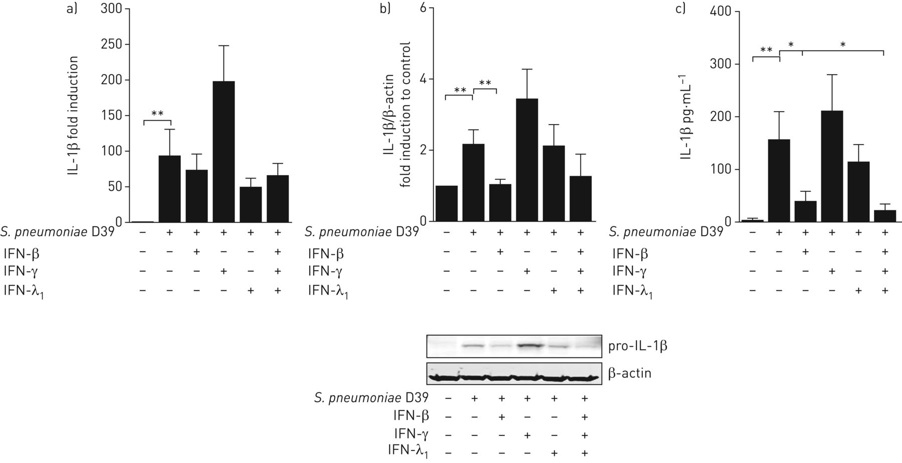
- FIGURE 4
Type I interferon (IFN)-β is the main suppressor of alveolar macrophage (AM)-induced interleukin (IL)-1β protein expression and secretion. AMs, isolated from fresh human lung tissue, were cultured for 2 days and challenged with type I IFN-β, type II IFN-γ, type III IFN-λ1 or a combination of all three (100 U·mL−1 each) for 16 h. Afterwards, cells were infected with Streptococcus pneumoniae (1 multiplicity of infection). RNA, protein and supernatants were collected 16 h after pneumococcal infection and assayed for release of a) IL-1β mRNA expression; b) pro-IL-1β protein expression; and c) IL-1β secretion. Data are presented as mean±sem of at least six donors within independent experiments. *: p≤0.05; **: p≤0.01.
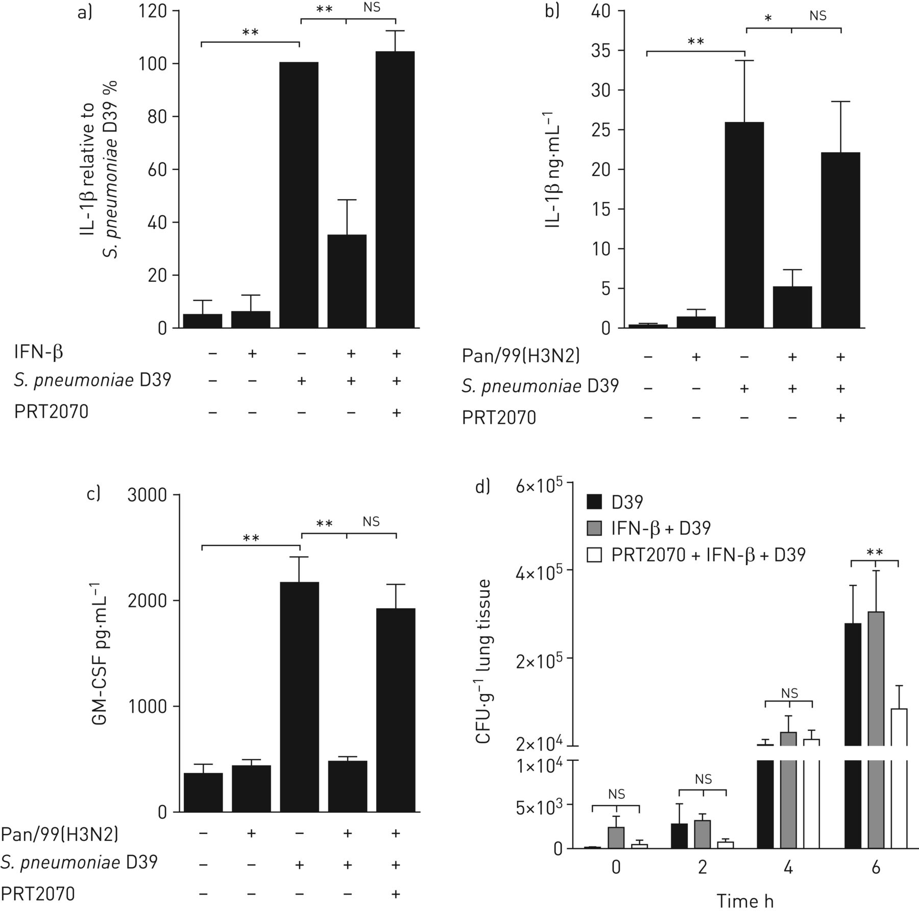
- FIGURE 5
Block of type I and III interferon (IFN) signalling with the tyrosine kinase 2 inhibitor PRT2070 rescues Streptococcus pneumoniae D39-induced interleukin (IL)-1β and granulocyte-macrophage colony-stimulating factor (GM-CSF) in alveolar macrophages (AM) and human lung tissue and decreases bacterial growth. a) AMs isolated from fresh human lung tissue were cultured for 2 days and treated with the inhibitor PRT2070 (1 µM). Afterwards, cells were challenged with IFN-β (100 U·mL−1) for 16 h and infected with S. pneumoniae (1 multiplicity of infection; 16 h) and supernatants were assayed for release of IL-1β. b) and c) Lungs were pretreated with PRT2070 (1 µM) for 1 h. Indicated lung specimens were challenged with influenza A virus Pan/99(H3N2) (IAV) (1×10⁶ PFU·mL−1) for 24 h and with S. pneumoniae (1×10⁶ CFU·mL−1) for a further 16 h. Release of b) IL-1β and c) GM-CSF was measured. Data in a) are presented as percentage of S. pneumoniae-treated cells, because of high variations between AM donors for secreted IL-1β. Amounts of IL-1β secretion in pg·mL−1 are documented in online supplementary table S1. d) Bacterial growth was measured in human lung tissue after S. pneumoniae infection, IFN-β treatment followed by S. pneumoniae infection and pretreatment with PRT2070 (1 µM) followed by IFN-β and S. pneumoniae. Data are presented as mean±sem of at least five donors within independent experiments. ns: nonsignificant. *: p≤0.05; **: p≤0.01.
- FIGURE 6
a) Proposed model for the cellular interplay controlled by the early cytokine response in the human alveolus under i) single bacterial or ii) viral and bacterial co-infection. In i) Streptococcus pneumoniae is recognised by alveolar macrophages (AM) releasing inflammasome-dependent interleukin (IL)-1β, which serves as driving factor for granulocyte-macrophage colony-stimulating factor (GM-CSF) production in alveolar epithelial type II cells (AECII). GM-CSF probably supports the inflammatory reaction by contribution to immune cell recruitment, phagocytic function and surfactant homeostasis, as well as the initiation of proliferation and repair processes. In ii), the pre-infection with influenza A virus initiates an antiviral interferon (IFN) response, which subsequently blocks IL-1β and GM-CSF expression, thereby reducing protective GM-CSF effects and promoting alveolar damage. b) Inhibition of tyrosine kinase 2 (Tyk2) with PRT2070 (cerdulatinib) restores the influenza A virus Pan/99(H3N2) (IAV)-induced and IFN type I- and III-mediated impairment of the antibacterial immune response. Jak: Janus kinases; AC: alveolar capillary; IFNR: IFN receptor.


Supplementary Materials
Supplementary Material
Please note: supplementary material is not edited by the Editorial Office, and is uploaded as it has been supplied by the author.
Supplementary material ERJ-01953-2016_Supplement
Figure S1. Localisation of influenza A virus Pan/99(H3N2) (IAV) and Streptococcus pneumoniae D39 (S. pneumoniae) in single and co-infected human lung tissue and IAV induced interferon (IFN) expression in single and co-infected human lung tissue. (A) Human lung tissue was either mock infected, (B) challenged with the seasonal IAV for 24 h, (C) S. pneumoniae for 16 h, or (D) co-infected with the IAV subsequently followed by S. pneumoniae. IAV (green, white arrowheads) typically replicated in alveolar epithelial type II cells indicated by pro-SP-C (blue, asterisk, B, D). S. pneumoniae closely attached to alveolar epithelial cells (open arrowheads, C, D) and alveolar macrophages (white arrow, D). Lung structure was visualised by differential interference contrast (grey) and nuclei were counterstained using DAPI (orange). Scale bar 10 µm. ERJ-01953-2016_Supplementary_Figure_S1
Figure S2. Co-infection with influenza A virus Pan/99(H3N2) (IAV) and Streptococcus pneumoniae (S. pneumoniae) shows differential cytokine regulation in human lungs. Lung explants were ex vivo mock infected or challenged with IAV (1 × 10⁶ PFU/ml) for 24 h. (A-F) Afterwards, dedicated specimen were either infected with the S. pneumoniae (1 × 10⁶ CFU/ml) (A - D) or (E, F) the clinical isolate of serotype 3 S. pneumoniae (1 × 10⁶ CFU/ml), respectively. Supernatants were collected 16 h after pneumococcal infection and assayed for release of (A) IL-6, (B) IL-8, (C) IL-10, (D) TNFα, (E) IL-1β, (F) GM-CSF as indicated. Data are presented as mean ± SEM of six donors within independent experiments. **p≤0.01. ERJ-01953-2016_Supplementary_Figure_S2
Figure S3. Influenza A virus Pan/99(H3N2) (IAV) infection or interferon (IFN) treatment of human lung tissue fails to suppress Streptococcus pneumoniae D39 (S. pneumoniae) induced tumour necrosis factor α (TNFα) release. Lung explants were ex vivo mock infected or challenged for 24 h with influenza A virus Pan/99(H3N2) (IAV) (1 × 10⁶ PFU/ml) or 16 h pre-treated with a combination of IFNβ and IFNγ (100 U/ml each). Mock or S. pneumoniae (1 × 10⁶ CFU/ml) infection followed and supernatants of lung specimen were analysed after additional 16 h for release of TNFα. Data are presented as mean ± SEM of six donors within independent experiments. **p≤0.01. ERJ-01953-2016_Supplementary_Figure_S3
Figure S4. TNFα stimulation induces cyclooxygenase-2 (COX-2) expression and granulocyte macrophage - colony stimulating factor (GM-CSF) expression is time shifted to IL-1β in human lungs. (A) Stimulation with TNFα (100 ng/ml) of human lung specimen for 16 h induces pro-inflammatory COX-2. (B) Human lung explants were either mock infected or challenged with S. pneumoniae (1 × 10⁶ CFU/ml) for 2, 4, 6, 8, and 16 h to demonstrate time shifted release of GM-CSF in relation to earlier IL-1β liberation. Data are presented as fold of control and mean ± SEM of three/four donors within independent experiments. ERJ-01953-2016_Supplementary_Figure_S4
Figure S5. Alveolar macrophages (AM) and alveolar epithelial type II cells (AEC II) were isolated from fresh human lung tissue and seeded on glass coverslips, fixed and phenotyped with different cell markers by immunofluorescence and confocal microscopy. (A) AM, cultured for 2 days, were positive for CD-68 (red). AEC II were cultured for 4 days after isolation. (B) Cells were positive for pan-Cytokeratin (red) and pro-SP-C (green). For nuclear counterstaining DAPI (blue) was used. Microscope: Carl Zeiss LSM780, Plan Apo-Chromat 63× oil/NA 1.4. Scale bar 5 µm ERJ-01953-2016_Supplementary_Figure_S5
Figure S6. Tumour necrosis factor α (TNFα) expression was not suppressed by interferons (IFN) in isolated human alveolar macrophages (AM). Isolated AM were cultured for 2 days and either mock challenged or stimulated with a combination of interferon β and γ (100 U/ml each) for 16 h before pneumococcal infection. Afterwards, supernatants were assayed for release of TNFα. Data are represented as mean ± SEM of four donors within independent experiments. *p≤0.05. ERJ-01953-2016_Supplementary_Figure_S6
Figure S7. IL-1β induced granulocyte macrophage - colony stimulating factor (GM-CSF) release in human lung tissue is suppressed by interferons (IFN). Human lungs were pre-treated with a combination of IFNβ and IFNγ (100 U/ml each) for 16 h and subsequent IL-1β stimulation (10 ng/ml) for 16 h. Supernatants of lung specimen were analysed for release of GM-CSF. Data are presented as mean ± SEM of three donors within independent experiments. *p≤0.05. ERJ-01953-2016_Supplementary_Figure_S7
Supplementary Material
J. Berg ERJ-01953-2016_Berg
D. Fatykhova ERJ-01953-2016_Fatykhova
A.C. Hocke ERJ-01953-2016_Hocke
K. Zscheppang ERJ-01953-2016_Zscheppang


