


Figures
- Figure 1–
Tumour growth in both groups of mice. At day 14, tumour volume in the animals subjected to intermittent hypoxia (n=7) was significantly greater (p<0.001 by two-way ANOVA with Holm-Sidak post-test) than that of the normoxia group (n=8). Tumour weight at day 14 was significantly greater (p=0.012 by unpaired t-test) in the intermittent hypoxia group than in the normoxia group. #: p=0.012; ***: p<0.001.
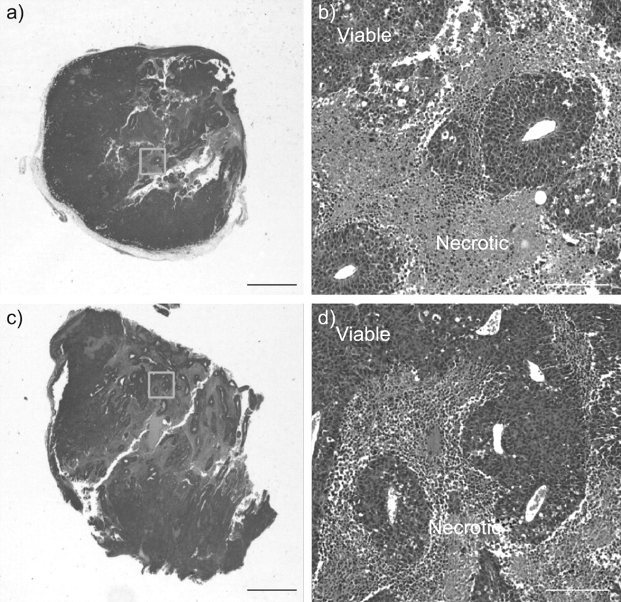
- Figure 2–
Representative tumour tissue preparations stained with haematoxylin–eosin corresponding to a, b) the normoxic group and c, d) animals subjected to intermittent hypoxia. a, c) whole section of the tumour, where viable tumour areas appear darker than necrotic areas. Areas marked within a square are enlarged in b) and d), respectively. a, c) Scale bars=2 mm; b, d) scale bars=200 μm.


