


Abstract
Staphylococcus aureus has been recognised as a cause of community-acquired pneumonia, albeit uncommon, and an important cause of healthcare-associated (HA) pneumonia, including ventilator-associated pneumonia. Resistance of S. aureus to methicillin developed shortly after its introduction into clinical practice. Since then, methicillin-resistant S. aureus (MRSA) has predominantly been a feature of hospital-acquired, or latterly HA, infections as the boundaries became more blurred between the community and hospital environments.
However, more recently true community-acquired (CA)-MRSA infections have been detected and are becoming increasingly common, especially in the USA. Europe has not been immune to the development of MRSA in healthcare settings and although the prevalence of CA-MRSA is currently relatively low, there is the risk of wider spread. These new CA-MRSA strains appear to behave differently to HA-MRSA strains. Although predominantly causing skin and soft tissue infections, mainly as boils and abscesses requiring drainage, life threatening invasive infections including necrotising pneumonia can also occur. This article summarises the pathogenesis and clinical presentations of MRSA-related lung infections.
- Community-acquired pneumonia
- healthcare associated
- hospital-acquired pneumonia
- lung infections
- methicillin-resistant Staphylococcus aureus
- pulmonology
SERIES “MRSA AND THE PULMONOLOGIST”
Edited by M. Woodhead and A. Torres
Number 2 in this Series
Staphylococci are Gram-positive spherical bacteria that occur in microscopic clusters resembling grapes. Staphylococcus aureus mainly colonises the nasal passages, but it may be found regularly in most other anatomical sites. Carrier rates in adults vary from 20–50% with people being persistent carriers, intermittent carriers or noncarriers. A large study found that 24% of people persistently carry S. aureus and 57% are intermittent carriers whilst 20% were never colonised 1.
S. aureus causes a number of important infections both in the community and in the healthcare setting. These include skin and soft tissue infections, bone and joint infections, bacteraemia, endocarditis and pneumonia. It is also important in a variety of toxinoses, for example food poisoning by the superantigen enterotoxin (SAE), scalded skin syndrome in neonates by the exfoliative toxin, and toxic shock syndrome by the toxic shock syndrome toxin (TSST).
S. aureus has a large repertoire of virulence factors, including structural and secreted products that play a role in pathogenesis (fig. 1⇓).
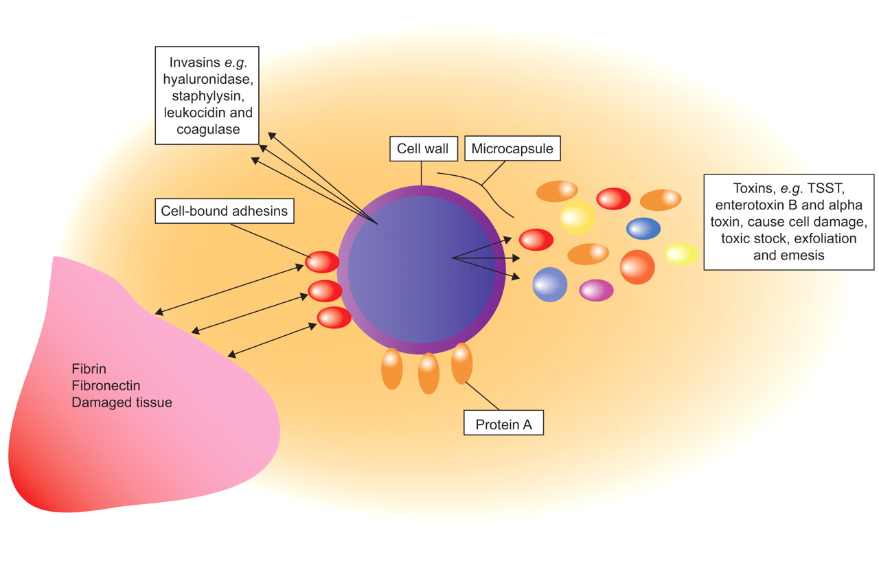
Some of the virulence determinants of Staphylococcus aureus. TSST: toxic shock syndrome toxin.
Some examples of S. aureus virulence factors include the following. 1) Surface proteins, for example protein A, that promote adherence and hence colonisation of host tissues. Different S. aureus strains may have different groups of these proteins predisposing them to different kinds of infections. 2) Invasions that promote bacterial spread in tissues (leukocidin, kinases and hyaluronidase). 3) Membrane damaging toxins that lyse eukaryotic cell membranes (haemolysins, leukotoxin and leukocidin). 4) Exotoxins that damage host tissues or otherwise provoke symptoms of disease (SAE-G, TSST-1, exfoliatin toxin and Panton–Valentine leukocidin (PVL)). 5) Inherent and acquired resistance to antimicrobial therapeutic agents.
The expression of these virulence factors is influenced by different environmental signals, and there are a number of regulatory genes directly involved in their control, such as agr, spa, sar, and sigB 2, 3. The accessory gene regulatory locus, agr, regulates an array of genes including the haemolysins α, β, γ and δ as well as leukocidin and PVL 3. The spa regulatory gene controls for protein A synthesis 3. Therefore, different S. aureus strains may contain different adhesins or toxins, or differ in their ability to form biofilm. In lung infections, various mouse pneumonia models have shown the importance of protein A, α-haemolysin and PVL in the pathogenesis of pneumonia. The mouse pneumonia model by Labandeira-Rey et al. 4 compared PVL-positive strains of methicillin-resistant Staphylococcus aureus (MRSA) to PVL-negative strains showing that the former caused necrotising pneumonia similar to that seen in humans, whereas the PVL-negative strains did not. However, the precise role of PVL in pulmonary infections has not been fully explained. In a different pneumonia mouse model, Bubeck Wardenburg et al. 5 found that agr and spa mutants were unable to cause lethal lung infections raising the possibility that α-haemolysin and protein A are important in lung parenchymal damage. Indeed, immunisation against α-haemolysin appears to protect mice against lethal pneumonia and may offer an early insight into potential future vaccine strategies 6. It is likely that a combination of many factors cause the enhanced virulence of certain strains of S. aureus.
Staphylococci have developed resistance to methicillin by various mechanisms. The mechanism characteristic of MRSA is the acquisition of the mecA gene 7, probably via a mobile genetic element, known as the staphylococcal cassette chromosome mec (SCCmec), from coagulase-negative staphylococci. At least five types of the SCCmec (SCCmec I–V) genetic element have been identified 8, 9. The mecA gene codes for a variant penicillin binding protein with a low affinity for the β-lactam antibiotics, thus, effectively reducing the activity of these drugs.
Initially, MRSA was exclusively associated with acquisition from hospitals and other healthcare settings 10, 11. Apparently, community-acquired (CA)-MRSA, described in the 1980s and1990s, could generally be linked to healthcare settings 12. This led to a new terminology of “healthcare-associated” (HA)-MRSA and “community-associated” MRSA. The strains of HA-MRSA and community-associated MRSA were very similar in terms of molecular typing and antimicrobial susceptibility, with both being associated with SCCmec I–III resistance elements against many classes of antibiotic 8–10.
However, in the late 1990s true CA-MRSA was identified as the cause of severe and fatal infections occurring in clusters of previously healthy children in North America, who had no identifiable associations with healthcare settings 9, 13, 14. Cases of CA-MRSA causing skin and soft tissue infections and necrotising pneumonia have since been widely reported in otherwise healthy individuals 8, 9. CA-MRSA, as distinct from HA-MRSA, is associated with SCCmec IV and V which, in addition to the mecA gene, can contain genes encoding for toxin production 8–10. One such toxin, the PVL toxin, destroys leukocytes and causes extensive tissue necrosis, thus having a clear role in the pathogenesis of necrotising pneumonia 4, 15, 16. As stated previously it is unlikely that PVL alone accounts for the increased invasiveness of CA-MRSA, instead a combination of virulence factors including protein A and the haemolysins α and γ are likely to play a role in the invasiveness of CA-MRSA lung infections. The principle differences between CA-MRSA and HA-MRSA are summarised in table 1⇓ 17. The epidemiology and clinical burden of MRSA infections in Europe will be discussed later.
Typical differences between HA-MRSA and CA-MRSA
MRSA AND THE LUNG
The effect of S. aureus on the airways, from asymptomatic colonisation to severe pneumonia, depends on the interplay of patient, environmental and bacterial factors.
Colonisation of the lower respiratory tract by S. aureus and, therefore, MRSA can occur in the setting of chronic pulmonary disease, such as chronic obstructive pulmonary disease and suppurative lung disease, or due to breaches in natural defences, such as endotracheal intubation. Although this colonisation may be asymptomatic, it paves the way for overt infection, i.e. pneumonia, if the balance between host defence and bacterial virulence is shifted in the favour of bacteria. MRSA pneumonia can also occur in previously healthy patients with no risk factors for colonisation.
Hospital-acquired pneumonia (HAP) or nosocomial pneumonia is usually defined as pneumonia developing ≥48 h after admission to hospital that was not incubating at the time of admission. Ventilator-associated pneumonia (VAP) is usually defined as pneumonia developing ≥48 h after implementation of endotracheal intubation and/or mechanical ventilation and which was not present prior to intubation 18, 19. VAP can be divided into early and late onset. Early-onset disease occurs within 4–5 days of admission and tends to be caused by antibiotic-susceptible community-type pathogens, whereas late-onset disease tends to be caused by antibiotic-resistant pathogens. However, some studies have found an increasing frequency of early-onset HAP caused by pathogens more commonly associated with nosocomial disease. This has contributed to the concept of healthcare-associated pneumonia (HCAP), involving pathogens associated with recent prior hospitalisation and/or antimicrobial therapy. HCAP has been defined as pneumonia occurring in any patient who had: been admitted to an acute care hospital for ≥2 days within 90 days of the infection; been a resident in a nursing home or long-term care facility (LTCF); attended a hospital or haemodialysis clinic; or received recent intravenous antibiotic therapy, chemotherapy or wound care within the 30 days prior to the current infection. A sub-population of these patients is those with nursing home-acquired pneumonia. While MRSA is considered by many in North America and elsewhere 20, 21 as an important pathogen in the nursing home/LTCF setting, the data from Europe is not consistent with this and is in need of further evaluation 22. In this context, the pneumonia presentation is clinically similar to that caused by Gram-negative organisms, and has an associated all-cause mortality of 55.5% regardless of early appropriate therapy 23.
In the European setting, S. aureus remains an unusual primary cause of community-acquired pneumonia (CAP) 24, although it is an important cause of pneumonia and death following influenza 25. The role of CA-MRSA is even more poorly defined, although emergent in the UK and Europe 17. Infections due to CA-MRSA have symptom onset before or within 48 h of admission to hospital and patients have no significant previous healthcare contact. CAP, which is due to CA-MRSA, classically presents in a young, previously healthy, individual with rapidly progressive, severe respiratory disease. The aggressive nature of CA-MRSA, due to toxin production, causes massive destruction in previously normal lungs.
PNEUMONIA: CLINICAL FEATURES
CAP
Staphylococcal pneumonia has been a changing clinical entity since being initially reported in the late 19th and early 20th Centuries. It was recognised in young healthy military personnel during World War 1 as a post-influenza pneumonia with a rapid onset of symptoms. Descriptions of cases highlighted the presence of isolated tachypnoea without other signs of severe illness and normal chest radiography (CXR) appearance in the first 24–48 h. Death ensued rapidly and occurred in ∼80–90% of cases in this pre-antibiotic era. Post mortem findings revealed pulmonary haemorrhage and microabscess formation 26, 27. If the patients did survive until day 4–5, clinical signs of bronchopneumonia then developed and CXR revealed cavities, empyema and pyopneumothoraces 27, 28.
In the 1950s, cases of Staphylococcal pneumonia began to be reported in people without any prior influenza infection. Usually they had some predisposing risk factors, such as cardiopulmonary disease, alcoholism or diabetes mellitus, or had acquired an infection in hospital. These strains included methicillin-sensitive strains, as well as MRSA. The clinical presentation was usually less explosive than had previously been described, and the mortality in the antibiotic era ranged from 20% in young adults to 30–50% in post-influenza cases through to 83% in patients with bacteraemic primary pulmonary pneumonia 29–33.
In addition to primary pneumonia resulting from direct inoculation of the lungs, Staphylococcal pneumonia can also develop by haematogenous spread to the lungs from another primary infective source, such as endocarditis or bone and joint infection.
HAP and VAP
Until recently S. aureus accounted for ∼1–5% of CAP cases and ∼10–15% of HAP cases 34, but over the past 10–20 yrs there has been important changes in the epidemiology of Staphylococcal pneumonia. First, there has been a dramatic increase in the proportion of S. aureus infections due to MRSA, which is now responsible for >50% of all S. aureus infections in some intensive care units (ICUs). At the same time, there has been an increase in ventilatory support of an ageing population who often have significant comorbidity. Together, this has fuelled an increase in MRSA pneumonia, with 20–40% of all HAP cases in the USA, including VAP, being due to MRSA 23. VAP due to MRSA is associated with a worse outcome and increased resource utilisation than VAP due to other organisms 35.
HAP is estimated to occur at a rate of 4–50 cases per 1,000 admissions in community hospitals and general medical wards, and 120–220 cases per 1,000 ICU admissions 36. It accounts for >50% of the antibiotics prescribed in ICUs, and has an associated attributable mortality of 33–50% 18.
HAP requires the entry of microbial pathogens into the lower respiratory tract followed by colonisation which, if the body's defences are overwhelmed, leads to overt infection. Factors such as the severity of the patient's underlying disease, prior surgery, exposure to antibiotics, other medications, and exposure to invasive respiratory devices and equipment are important in the pathogenesis of HAP and VAP 18.
The time of onset, relative to hospital admission, of HAP and VAP is an important risk factor for patient outcome and for specific pathogens. Early-onset disease, defined as within 4 days of hospitalisation, has a better prognosis and is more likely to be caused by antibiotic-sensitive bacteria. Exceptions to this are patients with recent previous hospitalisation and/or antibiotic use and elderly people residing in LTCFs. These HCAP patients have a spectrum of pathogens that more closely resemble late-onset HAP and VAP with up to 33% being due to MRSA 18, 23.
VAP due to MRSA appears to have significant excess morbidity and mortality regardless of appropriate antimicrobial treatment and patient characteristics. Compared to matched ICU controls, an estimated excess mortality of 22.7% was observed in Spanish ICU patients with MRSA VAP 37; this was despite appropriate glycopeptide therapy 37. In the same study, cases with MRSA VAP also had an increased length of ICU stay compared with controls (median 33 and 21 days, respectively) 23, 37.
A prospective study comparing VAP outcome by causative pathogen demonstrated that, in patients who received appropriate initial antimicrobial therapy, cases due to MRSA still had a significantly slower clinical resolution than those due to other pathogens. Resolution of fever and hypoxia within 72 h occurred in only 30% of MRSA VAP cases, compared to 93.3% of methicilin-sensitive S. aureus (MSSA) VAP cases, 100% due to H. influenzae and 73% due to Pseudomonas aeruginosa 35. Clinical resolution and ventilator dependence in patients with MRSA VAP treated appropriately was similar to that in patients with VAP due to P. aeruginosa who received inappropriate antibiotic therapy 35.
A new form of CAP
There has been an emergence of CA-MRSA CAP being reported on both sides of the Atlantic. Although CA-MRSA is primarily a cause of skin and soft tissue infections, it can also cause severe necrotising pneumonia 16, 38–42. Some of these respiratory infections have been associated with septic shock, haemoptysis, respiratory failure and intensive care admission for ventilatory or circulatory support. These infections in young previously healthy patients resemble those reported in the early part of the 20th Century.
Characteristics of CA-MRSA CAP often frequently occur in young previously healthy adults, up to 75% of cases, with a preceding flu-like illness 16, 43. Sufferers rapidly develop severe respiratory symptoms, often including haemoptysis, hypotension and a high fever. Characteristically, leukopenia occurs and C-reactive protein is elevated (>350 g·L−1). CXR findings of multilobar cavitating alveolar infiltration are also consistent with CA-MRSA 16, 40, 43, 44. These features are not specific to CA-MRSA but rather are consistent with PVL producing S. aureus and, therefore, can be applicable to some strains of MSSA. However, young age in CA-MRSA CAP has been a consistent finding in studies from Europe and the USA 16, 43, 45. Table 2⇓ summarises when CA-MRSA in CAP should be suspected and table 3⇓ summarises the risk groups with increased rates of CA-MRSA colonisation.
Summary of when to suspect CA-MRSA in community-acquired pneumonia
Risk groups with increased rates of CA-MRSA colonisation
INVESTIGATIONS FOR STAPHYLOCOCCAL PNEUMONIA
Obtaining isolates of the organism
Endotracheal cultures should not be used to diagnose VAP as bronchoalveolar lavage specimens are preferred. For HAP, the least expensive, least invasive and most rapid sampling technique requiring minimal expertise should be used for establishing the microbiological diagnosis, e.g. nonbronchoscopy-directed (blind) bronchoalveolar lavage 19, 23.
Blood cultures are more likely to be positive in secondary pneumonia where the primary source is elsewhere, such as infective endocarditis or discitis, rather than in primary pneumonia (90% versus 20%) 38. Blood cultures are also more likely to be positive in VAP than HAP, 24–36% compared with 5–15%, respectively 46, 47. Therefore, because blood cultures are frequently negative it is important to obtain an adequate respiratory tract specimen prior to initiating therapy. Appropriate specimens include endotracheal sampling or pleural fluid but not sputum, as S. aureus is frequently present in the upper respiratory secretions of healthy individuals.
The antibiogram: antibiotic sensitivities
After isolating S. aureus, sensitivity testing for various antibiotics will determine whether it is MRSA or not, and which antibiotics may be clinically effective. Until the emergence of CA-MRSA, isolates of MRSA (HA-MRSA) were not only methicillin resistant and therefore resistant to all β-lactams, but were often multiresistant and resistant to a range of other groups of antibiotics. However, the antibiogram of CA-MRSA is commonly only resistant to the β-lactams and susceptible to most other antibiotic classes. This difference in the laboratory findings may provide a clue that the patient has a CA-MRSA isolate as opposed to an HA-MRSA isolate. However, with time, CA-MRSA is likely to acquire the resistance genes that will make it more difficult to differentiate from HA-MRSA by routine antimicrobial susceptibility testing.
Molecular techniques
Novel laboratory techniques, including microarrays to detect PVL and possibly other staphylococcal toxins or superantigens, may aid in the diagnosis of CA-MRSA pneumonia. These microarrays can reveal if the isolates are harbouring the genes for several toxins including PVL and leukocidin, which have been linked with pulmonary disease and have been associated with CA-MRSA isolates. Also under investigation and development are molecular-based rapid tests to detect PVL, mecA and SCCmec type IV 23, 48.
Radiological investigations
No radiological features are highly specific for Staphylococcal pneumonia. Early in the disease progression of CAP with S. aureus there may be minimal infiltrates but they rapidly progress, even within hours. There may be a unilateral consolidation or bilateral infiltrates, especially in PVL producing CA-MRSA. Compared with HA-MRSA pneumonia these infiltrates are more likely to cavitate, which may be seen on serial CXR and best confirmed by a computed tomography scan 49, 50. Pleural effusions, pneumatoceles and pneumothoraces are also common findings. However, for HAP and VAP there are no radiological features that distinguish MRSA from any other causative organism and, in addition, VAP may be missed on CXR in up to 26% of cases 23. Clinically however, there may be a suspicion of MRSA as the causative organism in VAP as patients tend to have more severe disease and respond more slowly to appropriate antimicrobial therapy 35. Accordingly, radiological progression may be faster than with other organisms (fig. 2⇓).

Necrotising pneumonia on a) a chest radiograph and b) a computed tomography (CT) scan obtained on day 3. The CT scan shows multiple bilateral nodular and cavity lesions. Reproduced from 51 with permission from the publisher.
CONCLUSIONS
The frequency of pneumonia caused by HA-MRSA and CA-MRSA is increasing. HA-MRSA traditionally occurs in the hospital setting, although it has been suggest that many elderly patients, who often have comorbidities and who reside in LTCFs in the community, may acquire respiratory infections caused by pathogens more traditionally found in the hospital or healthcare setting. The evidence that this is the case in Europe is equivocal and requires further investigation. A new form of MRSA, more virulent and frequently more toxin producing (including PVL toxin), is emerging from the community. It is primarily affecting young healthy individuals and has a high mortality rate. In light of this, a strong clinical suspicion is needed to instigate adequate therapy. Vancomycin has been disappointing in treating MRSA pneumonia and although linezolid may be a better choice, more data is required to support this as a standard of care. Combination therapy with clindamycin and immunoglobulin may be helpful in cases of CA-MRSA where there is PVL production causing haemorrhagic and necrotising pneumonia.
Statement of interest
A statement of interest for D. Nathwani can be found at www.erj.ersjournasl.com/misc/statements.dtl
Footnotes
-
Previous articles in this series: No. 1: Pantosti A, Venditti M. What is MRSA? Eur Respir J 2009; 34: 1190–1196.
- Received July 31, 2009.
- Accepted August 27, 2009.
- © ERS Journals Ltd
References


