


Figures
- Fig. 1—
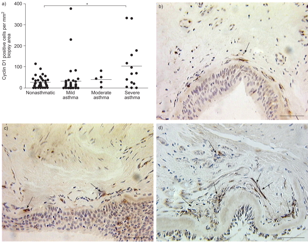
a) Quantitation of cyclin D1-positive cell numbers in biopsies from subjects with increasing asthma severity. Individual and median values from nonasthmatic (n = 34), mild asthmatic (n = 38), moderate asthmatic (n = 5) and severe asthmatic (n = 14) subjects are shown. Immunohistochemistry for cyclin D1 in endobronchial biopsies from b) a nonasthmatic, c) a mild asthmatic and d) a severe asthmatic subject. Arrows indicate cyclin D1-positive cells. *: p<0.05, Kruskal–Wallis test. Scale bars = 50 μm.
- Fig. 2—
Comparative immunohistochemistry for human airway myofibroblast (a, b, e, f, i and j) and smooth muscle (c, d, g, h, k and l) cultures, derived from an endobronchial biopsy and a lung resection specimen, respectively. Immunostaining was performed for α-smooth muscle actin (a and c), proline-4-hydroxylase clone 5B5 (b and d), vimentin (e and g), smooth muscle myosin (f and h) and pan cytokeratin (i and k). j and l) Immunoglobulin G1 as negative controls. The patterns shown are representative of three experiments. Scale bars = 100 μm.
- Fig. 3—
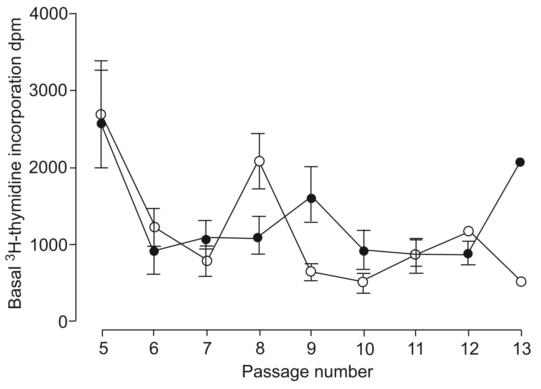
Effect of passage number on basal 3H-thymidine uptake in myofibroblasts from nonasthmatic (○) and asthmatic (•) subjects. Results are presented as the mean±sem 3H-thymidine uptake over a 4-h incubation period for cells plated at the same density. For nonasthmatics and asthmatics, there were ≤19 and ≤25 cultures at each passage, respectively; p>0.05.
- Fig. 4—
Effects of a) fibroblast growth factor (FGF)-2 (300 pM), b) thrombin (0.3 U·mL−1) and c) foetal calf serum (FCS; 5% volume (v)/v) on proliferation of myofibroblasts from nonasthmatic and asthmatic subjects. Confluent cells were serum-deprived for 24 h then stimulated with mitogen for 48 h in the presence of essential progression factors. Cell numbers were determined by haemocytometry. Individual data points represent the average response from between one and six replicates within a given culture, expressed as a percentage of the unstimulated cell number. The mean responses are also shown. Numbers of cultures: a) seven nonasthmatic, 16 asthmatic; b) 18 nonasthmatic, 21 asthmatic; c) seven nonasthmatic, 14 asthmatic. *: p<0.05, unpaired t-test.
- Fig. 5—
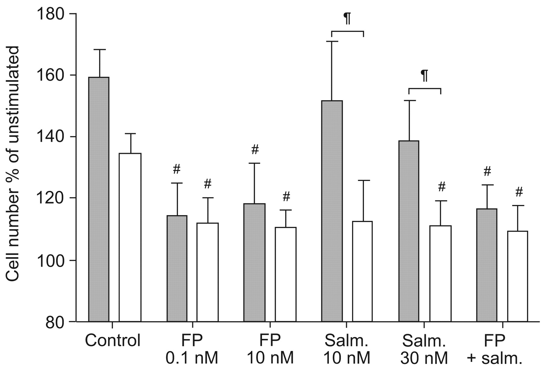
Effect of fluticasone propionate (FP) and salmeterol (salm.) on thrombin (0.3 U·mL−1)-induced proliferation of myofibroblasts from nonasthmatic (▓) and asthmatic (□) subjects. FP (0.1 or 1 nM), salm. (10 or 30 nM) or both (FP 0.1 nM, salm. 30 nM) were added to serum-deprived cells 30 min prior to thrombin addition. The control cells received thrombin alone. Cell numbers were determined by haemocytometry after 48 h. Data are presented as mean±sem of the response, expressed as a percentage of the unstimulated cell number (in the absence of thrombin). One to four replicates were performed for each culture (nine nonasthmatic, seven asthmatic). #: p<0.05 for comparison with control within asthma status group; ¶: p<0.05 for comparison with nonasthmatic under same conditions.
- Fig. 6—
Effect of interleukin (IL)-1α (1 ng·mL−1) on supernatant levels of inflammatory mediators for myofibroblasts from nonasthmatic and asthmatic subjects. Confluent cells were serum-deprived for 24 h then incubated with IL-1α for 48 h. Supernatant levels of granulocyte-macrophage colony-stimulating factor (GM-CSF) and IL-8 were measured using ELISA. Individual data points represent the average increase in response to IL-1α above unstimulated levels from between one and 11 replicates within a given culture. The median responses are also shown. Numbers of cultures: a) 12 nonasthmatic, 14 asthmatic; b) 11 nonasthmatic, 12 asthmatic. *: p<0.05, unpaired t-test on log-transformed data.


Tables
- Table 1—
Demographic data for subjects from the Melbourne Epidemiological Study of Childhood Asthma cohort from whom biopsies were obtained for immunohistochemistry and cell culture
Immunohistochemistry Cell culture Nonasthmatic Mild asthma# Moderate asthma# Severe asthma# Nonasthmatic Asthmatic¶ Subjects (M/F) n 34 (21/13) 38 (24/14) 5 (4/1) 14 (6/8) 22 (15/7) 25+ (16/9) FEV1 % pred 104 (96–115) 102 (93–108) 67 (66–71)** 60 (57–74)*** 101 (98–110) 97 (85–115) Atopic 65 85 100 71 68 77 Current smokers 71 49§ 67 0ƒ 82 52 Current treatment β2-adrenoceptor agonists 0 74 100 100 0 76 Inhaled steroids 0 37 80 100 0 40 Oral steroids 0 0 0 64 0 0 Data presented as median (interquartile range) or %, unless otherwise stated. M: male; F: female; FEV1: forced expiratory volume in one second; % pred: % predicted. #: classified using Global Initiative for Asthma guidelines; ¶: all asthmatics were combined for analysis since few biopsies from moderate or severe asthmatics were available for culture; +: 20 mild, four moderate and one severe asthmatic; §: smoking status unknown in 3%; ƒ: smoking status unknown in 14%. **: p<0.01; ***: p<0.001 for Kruskal–Wallis test followed by Dunn’s post hoc test.
- Table 2—
Total area and smooth muscle area of biopsies from nonasthmatic and asthmatic subjects used for cyclin D1 immunohistochemistry
Nonasthmatic Mild asthma Moderate asthma Severe asthma Subjects n 34 38 5 14 Total biopsy area mm2 0.22 (0.17–0.34) 0.28 (0.15–0.36) 0.33 (0.16–0.33) 0.26 (0.17–0.50) Smooth muscle area % 0.7 (0–5.7) 3.0 (0–7.8) 1.1 (0–20.5) 19.9 (3.8–34.9)** Data are presented as median (interquartile range), unless otherwise stated. **: p<0.01 compared with nonasthmatic (Kruskal–Wallis test followed by Dunn’s post hoc test).
- Table 3—
Effects of various stimuli on3H-thymidine uptake in myofibroblasts from nonasthmatic and asthmatic subjects
Stimulus Nonasthmatic Asthmatic p-value Uptake# Cultures Uptake# Cultures Thrombin 0.3 U·mL−1 155±9 20 182±12 25 0.08 FGF-2 300 pM 147±14 17 177±20 21 0.27 FCS 5% v/v 211±22 14 183±15 18 0.27 Data are presented as mean±sem or n, unless otherwise stated. 3H-Thymidine uptake was measured over a 4-h incubation period, 24–28 h after the addition of stimulus. FGF: fibroblast growth factor; FCS: foetal calf serum; v: volume. #: percentage of uptake in unstimulated cells (2,023±634 dpm, n = 18, and 1,721±451 dpm, n = 26, for nonasthmatic and asthmatic, respectively; p>0.05).
Supplementary methods
Files in this Data Supplement:


