


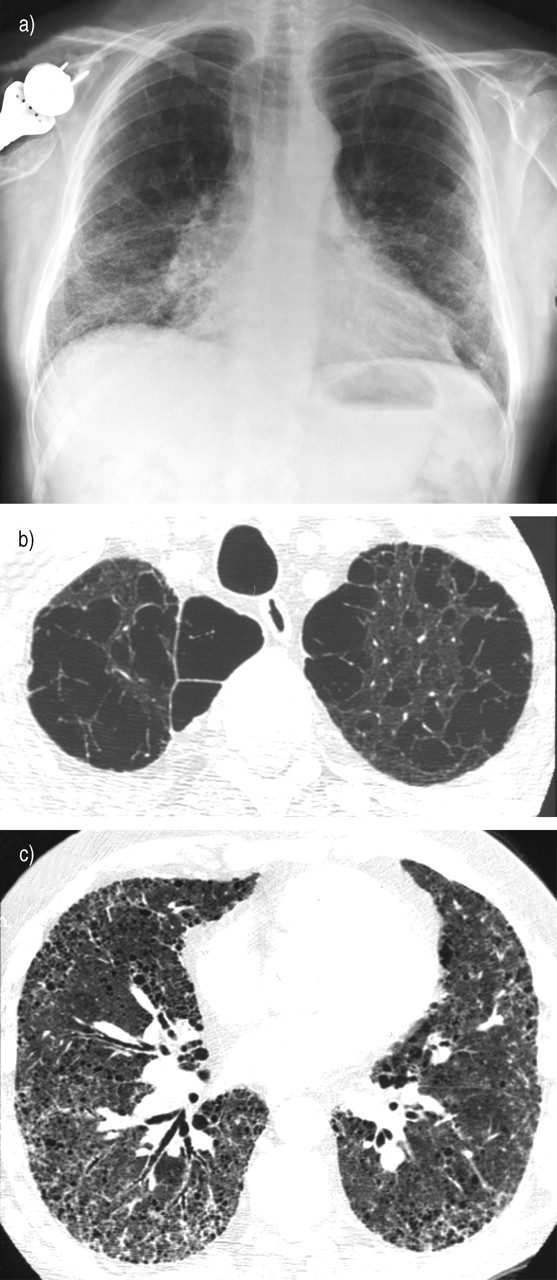
Fig. 1—
Imaging of a typical case of combined pulmonary fibrosis and emphysema. a) Chest radiograph showing bilateral infiltrative opacities of the lower lobes and hyperlucent upper zones. b) Chest computed tomography (CT) of the upper zones of the lungs showing predominant centrilobular and paraseptal emphysema. c) Chest CT of the lower zones of the lungs showing reticular opacities, honeycombing, and traction bronchiectasis. Lung biopsy performed in this patient showed centrilobular emphysema of the upper lobes and usual interstitial pneumonia of the lower lobes.


