


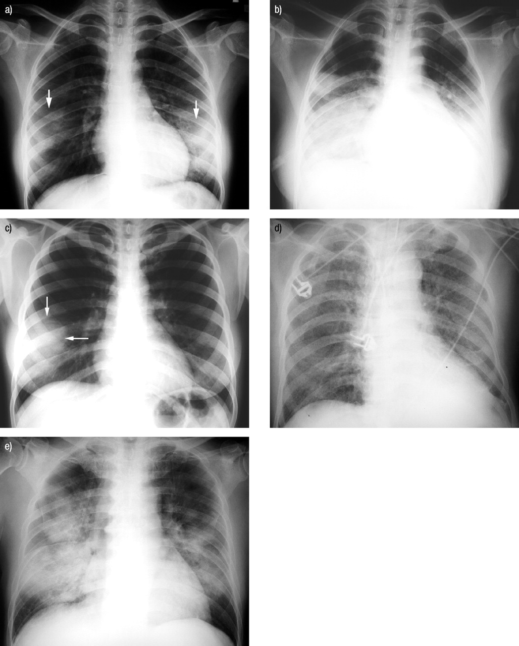
Fig. 1.—
Five different radiographical patterns of severe acute respiratory syndrome (SARS) are shown. Chest radiographs at a) presentation and b) day 4 after admission of a 24-yr-old female with SARS show rapid progression of patchy consolidation (arrows) in both lower zones to diffuse consolidation affecting both mid and lower zones. c) Chest radiograph of another female with SARS showing confluent consolidation (arrows) in the right lower zone. d) Chest radiograph of a 54-yr-old male with SARS showing small diffuse nodular opacities. e) Chest radiograph of a 38-yr-old male showing diffuse acute respiratory distress syndrome-type airspace opacities.


