


Abstract
Loss of skeletal muscle mass is now recognised as an important feature of chronic obstructive pulmonary disease (COPD) which contributes to symptoms and influences prognosis. The changes in skeletal muscle remain poorly understood, largely because only a few studies have been performed to define the adaptations in whole body and muscle protein metabolism in COPD.
The first sections of this review summarise background information about skeletal muscle wasting in COPD, and focuses on the studies concerned with amino acid profiles and protein synthesis and degradation rates. To aid interpretation some discussion of the techniques commonly used is included.
A variety of different catabolic factors may determine whether chronic obstructive pulmonary disease patients become cachectic. The precise role for each one of these factors as well as the intracellular pathways activated in muscle as a result of chronic obstructive pulmonary disease are unknown and remain to be defined. Details of the actions of a range of different catabolic factors and potential mechanisms will be discussed.
A significant proportion of patients with chronic obstructive pulmonary disease (COPD) develop peripheral skeletal muscle wasting and weakness 1, 2. Loss of skeletal muscle mass is reflected by the reduction in fat-free mass (FFM) observed in ∼20–40% of patients with COPD 3, 4, and this has a number of consequences: 1) many studies have now shown that lower FFM is associated with reduced exercise tolerance 3, 5–7. 2) Because muscle power is directly proportional to cross-sectional area, loss of muscle mass is also associated with reduced peripheral muscle strength in COPD 5, 8, 9. 3) Many patients with COPD suffer debilitating symptoms and there is now evidence that those with lower FFM also have worse symptom scores in standard COPD‐related quality of life assessment tools, even when compared with COPD patients who have lost fat mass but not FFM 5. 4) Finally, low body weight has been identified previously as a poor prognostic indicator of survival in COPD 10, 11, but a recent retrospective analysis of 142 COPD patients showed that mid-thigh muscle cross-sectional area was a better indicator of prognosis than body mass index 12. This latter finding reinforces the concept that loss of FFM and in particular muscle mass is a common feature of COPD which has important implications for physical function, health status and survival.
Despite increasing interest in skeletal muscle wasting in COPD, the underlying mechanisms remain unclear. Exercise-based pulmonary rehabilitation programmes have been shown to improve muscle function and exercise tolerance for many affected individuals. The success of exercise therapy suggests that deconditioning due to reduced physical activity plays a major role in COPD‐related skeletal muscle dysfunction, but it also seems likely that other catabolic factors are involved. Furthermore initial studies using anabolic steroids 13–15 or growth hormone supplements 16, in addition to pulmonary rehabilitation, have been successful in increasing FFM, but improvements in most measures of muscle function or exercise capacity have been disappointing. Thus, much remains to be learned about the pathophysiological adaptations in muscle protein metabolism in COPD. This review includes discussion of recent studies that have begun to explore the alterations in protein synthesis and degradation rates, amino acid levels in plasma and muscle, and the intracellular molecular mechanisms relevant to the changes in muscle protein metabolism in COPD.
Indicators of skeletal muscle dysfunction in chronic obstructive pulmonary disease
Reduction of lean body mass and skeletal muscle mass in chronic obstructive pulmonary disease
Muscle usually forms between 60–80% of FFM or lean body mass, and so FFM can be used as a surrogate marker for muscle mass. In 255 consecutive COPD patients admitted for pulmonary rehabilitation, 35% had reduced FFM by bioelectrical impedance analysis (BIA) and, importantly, about one quarter of these had normal body weight 3. In addition, in the same study the creatinine-height index was used to confirm that reduced FFM correlated with reduced muscle mass in these patients 3. Using the same criteria 18–20% of 72 COPD outpatients with moderate airflow obstruction had reduced FFM 4. More recent direct comparisons between groups of COPD patients and healthy controls have also confirmed lower FFM or body cell mass in subjects with COPD 17, 18. Furthermore those with COPD and reduced whole body FFM, also have less lean tissue in their limbs, compared to controls 9. Other studies in COPD patients, showed a reduced cross-sectional area of calf (13% by magnetic resonance imaging) 19 and quadriceps (24% by computed tomography) 20. Attempts to correlate reductions in FFM with impaired lung function have shown that patients with a low diffusing capacity have the lowest values for FFM 9, and a similar relationship between reduced FFM and severity of airflow obstruction has been found in some studies but not in others 9, 21.
Morphometric and metabolic adaptations in skeletal muscle fibres in chronic obstructive pulmonary disease
Several studies have measured the proportions and sizes of the different skeletal muscle fibre types, or their corresponding myosin isoforms in COPD. Most studies have focused on the vastus lateralis muscle, and show a reduction in the proportion of type I slow-twitch type muscle fibres and a corresponding increase in type II fibres in COPD relative to controls 22–25. Studies have shown that muscle fibres from COPD subjects are often smaller than those in controls 18, 25, and in one study mean fibre cross-sectional area correlated with FFM 18. In addition the capillary-fibre ratio in muscles from subjects with COPD was reduced in one report 23, though this was not reproduced in another larger study 25. The changes in fibre type proportions correlate with the reduced activity of certain oxidative enzymes in affected muscles 22, 26. However adaptations in muscles other than the vastus lateralis may differ, and in deltoid biopsies of COPD patients, no changes in fibre type proportions were observed, and the activity of citrate synthase was preserved or even increased 27.
The adaptations in respiratory muscles in COPD appear to be the reverse of those detected in some peripheral muscles. Diaphragm muscle biopsies from subjects with severe COPD showed increased slow myosin heavy chain and reduced fast myosin heavy chain protein levels, with corresponding immunohistochemical changes, compared to controls 28. Furthermore increased oxidative enzyme capacity in all diaphragm muscle fibre types in COPD patients, compared with controls, have been confirmed 29. Despite the differences in the adaptations in diaphragm muscle compared to those in peripheral skeletal muscle in COPD, reductions in FFM are associated with impaired respiratory muscle strength measurements 13, 30. However, a proportion of the apparent weakness of these muscles is undoubtedly due to mechanical disadvantage due to changes in chest wall shape and hyperinflation 31.
Assessing amino acid status in plasma and skeletal muscle
Amino acids play a pivotal role in intermediary metabolism both as the building blocks of proteins, and as precursors for other functionally important compounds such as nucleotides and neurotransmitters. Skeletal muscle is the body's major protein store and under certain conditions e.g. fasting, muscle supplies amino acids to other tissues. However muscle also has a characteristic pattern of amino acid metabolism: the branched-chain amino acids leucine, isoleucine and valine are rapidly degraded as are other nonessential amino acids, including alanine, glutamate and aspartate. However unlike other tissues, muscle does not degrade the carbon skeletons of several other amino acids such as phenylalanine and tyrosine. Leucine is an important energy source for muscle tissue particularly during fasting, and under these conditions leucine inhibits oxidation of glucose. In muscle the breakdown of branched-chain amino acids is accelerated after injury or during fasting, and yields amino groups, which are used for synthesis of alanine (from pyruvate) and glutamine (from glutamate). The de novo synthesis of alanine and glutamine is coupled to degradation of the branched-chain amino acids, and alanine and glutamine are normally exported from muscle in much larger amounts than predicted simply from their occurrence in muscle protein. Alanine is avidly taken up by the liver and used for gluconeogenesis, but glutamine is an important energy source for cells such as leucocytes and fibroblasts and is also metabolised extensively by both the gut and kidney. Glutamate is central to all transamination reactions in muscle, and is one of the most abundant amino acids in proteins and in the free amino acid pool in skeletal muscle, but is present at a low concentration in plasma.
Alterations in amino acid profiles in plasma and skeletal muscle in chronic obstructive pulmonary disease
Studies of the alterations in amino acid concentrations in muscle and plasma may give important information about changes in amino acid metabolism in COPD.
Plasma glutamate, glutamine and alanine
In one study of eight underweight patients with emphysema and muscle wasting, reduced plasma concentrations of glutamine, glutamate and alanine were reported 32. Other comparative studies of COPD patients with moderate-to-severe airflow obstruction and reduced body weight and FFM, also found lower levels for these same amino acids compared to controls 33, 34. In contrast, other studies found elevated plasma levels of glutamine and glutamate in normal weight ambulatory COPD patients 35, and increased glutamine levels in underweight patients with advanced emphysema 36. Interestingly, the studies reporting reduced levels of glutamine in COPD also found an inverse relationship between plasma glutamine concentration and resting energy expenditure 33, 34. These data imply a relationship between plasma glutamine depletion and hypermetabolism in COPD possibly resulting from presence of systemic inflammation 37.
Plasma phenylalanine and tyrosine
The results obtained for the changes in plasma levels of the aromatic amino acids phenylalanine and tyrosine, also vary considerably in different studies. Increased, decreased and unaltered plasma levels of these amino acids have been reported in COPD 33, 34, 36, 38, 39.
Plasma branched chain amino acids: leucine, isoleucine, valine
Many studies have confirmed that plasma levels of branched-chain amino acids (BCAAs), particularly leucine, are reduced in patients with COPD 36, 40. The total BCAA concentration was lower in underweight (<90% ideal body weight) than in normal weight COPD patients 35, and a significant association was found between low levels of BCAAs and depletion of FFM 34. In one study muscle-to-plasma leucine gradient was greater in COPD patients compared with controls, and this was associated with correspondingly higher plasma insulin concentrations (which would favour retention of amino acids within insulin-sensitive tissues like muscle) 40. However the exact reasons for the raised insulin levels observed in this group of patients with COPD are unclear.
Muscle amino acid levels
Stable COPD patients had similar levels of muscle BCAAs but lower concentrations of glutamate compared with healthy age-matched controls 33, 40. The reduction in muscle glutamate concentration observed in COPD is independent of the severity of airflow obstruction but those with emphysema have particularly low glutamate levels 33, 38, 40. Furthermore, the reduction in glutamate status was associated with reduced muscle glutathione levels. After stratification into COPD subtypes, those with emphysema were found to have much lower concentrations of nearly all amino acids 40.
The changes observed in alanine, glutamine, glutamate and BCAA levels in plasma and muscle in patients with COPD, are certainly consistent with the disturbance in skeletal muscle metabolism. However further information is needed about the regulation of inter-organ transport of amino acids to allow confidence in interpreting the significance of the changes in these amino acids levels. Thus further mechanistic studies are needed using techniques to determine the fate of amino acids at both the whole-body level, and in the skeletal muscle compartment (see below).
Measuring muscle protein synthesis and degradation
In an average 70 kg male there is ∼7 kg of skeletal muscle protein of which the majority is formed by contractile proteins including actin and myosin. In addition ∼60% of the free amino acids in the body are thought to derive from muscle. All muscle proteins are continually being synthesised and degraded, and the combined effect of changes in synthesis and degradation alter the total protein turnover. Several factors influence muscle protein turnover including contractile activity, changes in nutritional intake and the circulating levels of a variety of hormones. Clearly measurement of muscle or lean body mass may fail to reveal important imbalances in the rates of muscle protein synthesis or degradation. Furthermore determining whether a reduction in muscle mass is due to a reduction in protein synthesis, or an acceleration in proteolysis, or both, is important to identify underlying mechanisms, and, possibly, to direct treatments.
In humans, the techniques commonly used to estimate total protein turnover, or synthesis or degradation rates, include 3‐methylhistidine (3‐MH) excretion, amino acid balance studies and tracer studies employing infusion of stable isotopes of amino acids (13C, 2H or 15N). 3‐MH, acquires its methyl group after translation, is not re-utilised for protein synthesis or metabolised after release during proteolysis, and is excreted in the urine. As >90% of 3‐MH residues are in actin and myosin in skeletal muscle, timed urinary excretion can be used to estimate whole body skeletal muscle proteolysis. 3‐MH can also be measured in blood draining from a limb to give an indication of proteolysis in the limb muscle. Several factors can affect the accuracy of urinary excretion measurements of 3‐MH, including renal function, dietary creatinine or meat intake and incomplete 24 h urine collections. In addition it has been proposed that a significant proportion of urinary 3‐MH is derived from nonskeletal muscle tissue e.g. smooth muscle in the gut, where protein turnover rates are high.
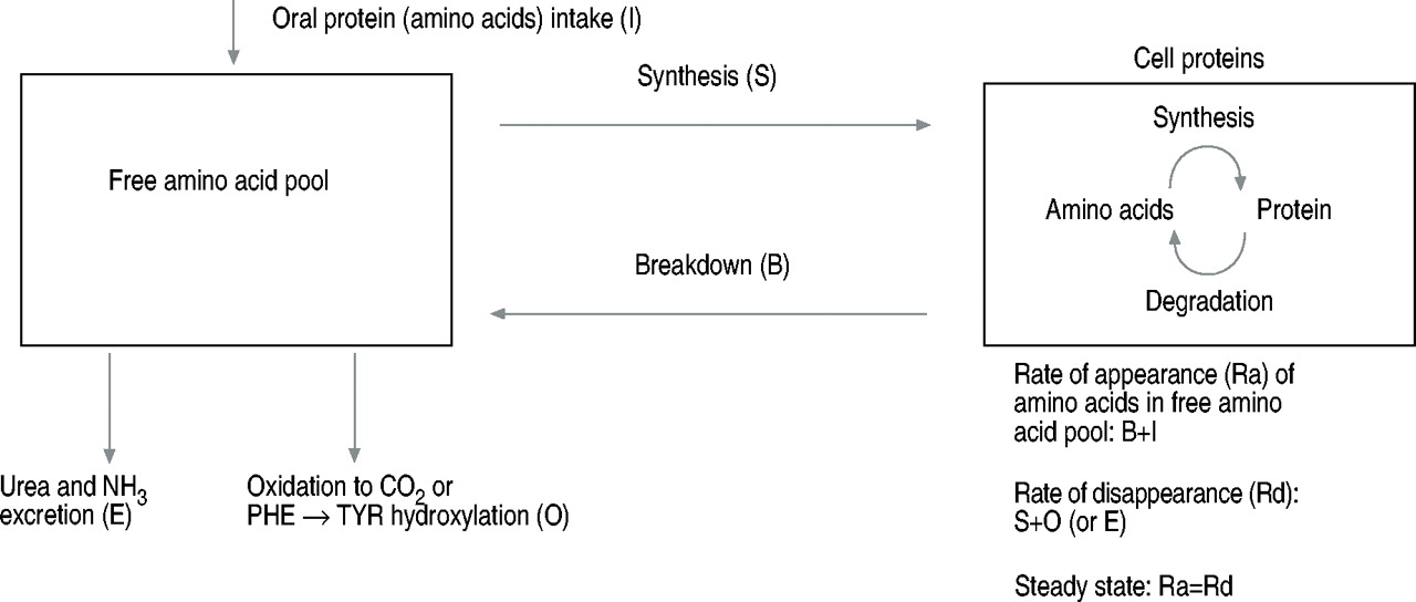
With the use of stable isotope (tracer) methodology rates of whole body protein synthesis and breakdown can be estimated. Metabolic studies using isotopically labelled amino acids have been used extensively for investigation in human subjects, though only a limited number of studies have used this technique in the context of COPD. Primed, constant infusion techniques are commonly used to determine the fate of the labelled amino acid. Under steady state conditions whole body protein synthesis and degradation rates can be derived (fig. 1⇓), and the use of labelled essential amino acids, which are, by definition, not synthesised in the body, makes these calculations easier. Selective cannulation techniques are more invasive but can be used to measure the arterio-venous gradient of tracer across muscle groups, such as the lower limb or forearm, and to calculate protein turnover in that muscle. When whole-body synthesis and degradation rates and muscle balance studies are combined, measurements of muscle protein synthesis and breakdown can be obtained. The calculations of protein synthesis and degradation rates rely on a number of assumptions about the achievement of steady state, and the uniform dispersal of the tracer in the tissue of interest. In addition accurate measurement of isotope enrichment (the amount the tracer isotope which is in excess of that naturally occurring in that subject) requires a mass spectrometer, and accurate measurements of blood flow are needed for protein turnover studies in the lower limb or forearm.

A simple model for calculation of whole body protein synthesis and breakdown rates in tracer experiments.
Studies of protein turnover rates in chronic obstructive pulmonary disease
In 1988 a study using 1‐13C‐leucine tracer in underweight emphysema patients with evidence of muscle wasting and hypoxia, showed that whole body protein turnover was reduced. This reduction was principally due to significantly lower whole body protein synthesis rates 32. Tyrosine can be used to calculate net protein breakdown in muscle as it is incorporated into proteins during protein synthesis and released after proteolysis but it is not degraded in skeletal muscle. Measurements of arterio-venous difference across leg muscle, showed an increase of 47% in the net release of tyrosine from limb muscle in the COPD group 32. Whether the net protein breakdown was the result of reduced muscle protein synthesis and/or enhanced muscle protein breakdown was not determined.
To investigate the effect of COPD on protein metabolism before clinical signs of muscle wasting are apparent, whole body protein turnover was measured in 14 severe COPD patients without evidence for weight loss of muscle wasting and in 7 healthy control subjects, using combined infusion of the stable isotopes of phenylalanine and tyrosine 41. Significantly higher levels of whole body protein synthesis and breakdown were found in COPD patients compared to healthy age-matched controls after overnight fasting. However, overnight fasting did not lead to more net protein catabolism in COPD. In the same study, no changes in protein turnover were found when assessed using labelled leucine 41. The differences in results with different tracers may suggest some specific defect in the metabolism of leucine, which would be consistent with previous data showing lower plasma BCAAs in COPD (see above).
To study the effects of feeding on protein metabolism in COPD, Aguilaniu et al. 42 studied 8 severely malnourished subjects with COPD. All patients had measurements of whole body, and limb, muscle protein degradation via urinary 3‐MH excretion and leg 3‐MH exchange measurement respectively. Measurements were performed at home, after 3 days in hospital on meat-free diet, and after starting hypercaloric high-lipid total parenteral nutrition (TPN). Immediately prior to starting TPN net release of 3‐MH from the leg was similar to that in healthy controls and even in cachectic cancer patients, indicating that COPD patients did not have higher rates of lower limb muscle proteolysis. However after starting TPN the whole body 3‐MH excretion, but not leg 3‐MH exchange, was reduced and nitrogen balance improved. Thus despite no evidence of increased muscle proteolysis compared with controls, intervention with TPN led to a reduction in whole body, but not muscle, proteolysis which explained about 50% of the improvement in nitrogen balance.
These initial results suggest that in more advanced COPD with nutritional depletion, whole body, and possibly muscle, protein synthesis is reduced, and that the normal feeding-induced inhibition of muscle proteolysis is impaired.
The pathways of protein synthesis and proteolysis in muscle
The molecular mechanisms underlying the changes in synthesis and degradation in muscle are still being elucidated, but it appears likely that in COPD both protein synthesis and degradation rates are altered by a variety of factors. The protein kinases, Akt and mTOR are key components of a signalling pathway which is activated in response to nutrients and growth-promoting hormones in eukaryote cells. This pathway appears to play a central role in promoting protein synthesis particularly via enhanced translation initiation, and signalling via the Akt/mTOR pathway is required for muscle hypertrophy 43. Many studies have suggested that the ubiquitin-proteasome pathway is responsible for the majority of the accelerated degradation of muscle proteins in different wasting conditions but the evidence for this in human disease is still lacking.
In studies, largely performed using rodents, acceleration of proteolysis via the ubiquitin-proteasome pathway is the dominant feature of fasting, cancer cachexia, metabolic acidosis, denervation, disuse, diabetes, sepsis, burns, hyperthyroidism, and excess glucocorticoids 44. However to date there is only a limited amount of data from patients with wasting diseases, and some of this suggests that activation of other proteolytic pathways may precede or accompany activation of the ubiquitin-proteasome pathway. It should also be noted that the majority of studies have only measured messenger ribonucleic acid (mRNA) levels for polyubiquitin and one or two 20S proteasome subunits, and the relationship between changes in mRNA levels and muscle proteolysis rates or nutritional status have frequently not been reported. Two studies from surgical patients with gastrointestinal cancer found increased mRNAs for ubiquitin and some proteasomal subunits in muscle, which were more marked in those with more advanced disease but did not correlate with measures of nutritional status 45, 46. In patients with early lung cancer, however no increase in mRNAs for components of the ubiquitin-proteasome pathway were found in muscle, even in depleted patients, but mRNAs for a lysosomal protease, cathepsin B were increased 47. In trauma patients a correlation was found between ubiquitin mRNA levels and proteolysis rates in leg muscle 48, and increased levels of transcripts for ubiquitin-proteasome pathway components, calcium-dependent and lysosomal proteases have been demonstrated in head trauma patients with negative nitrogen balance and raised whole-body proteolysis rates 49. Cachectic acquired immunodeficiency syndrome (AIDS) patients were found to have raised ubiquitin and proteasome subunit mRNAs in muscle 50, and correcting acidosis in subjects with renal failure led to improved nutritional status and lowered muscle ubiquitin mRNA levels 51. However no increase in mRNAs for genes related to any proteolytic pathways were found in patients with Cushing's syndrome 52. Thus the case for a role for the ubiquitin-proteasome pathway in causing the muscle wasting is stronger in some diseases e.g. trauma and acidotic renal failure, than it is in others e.g. Cushings syndrome, and there is, as yet, no data on the role of this pathway in muscle wasting in COPD.
What are the systemic factors which negatively influence protein metabolism and thus promote muscle loss in chronic obstructive pulmonary disease?
There are still many gaps in current understanding of the mechanisms underlying the adaptations in muscle of patients with COPD. However, it is possible to identify a range of systemic factors which are known to be abnormal in certain COPD patients, and are likely to have an impact on muscle protein metabolism (fig. 2⇓).
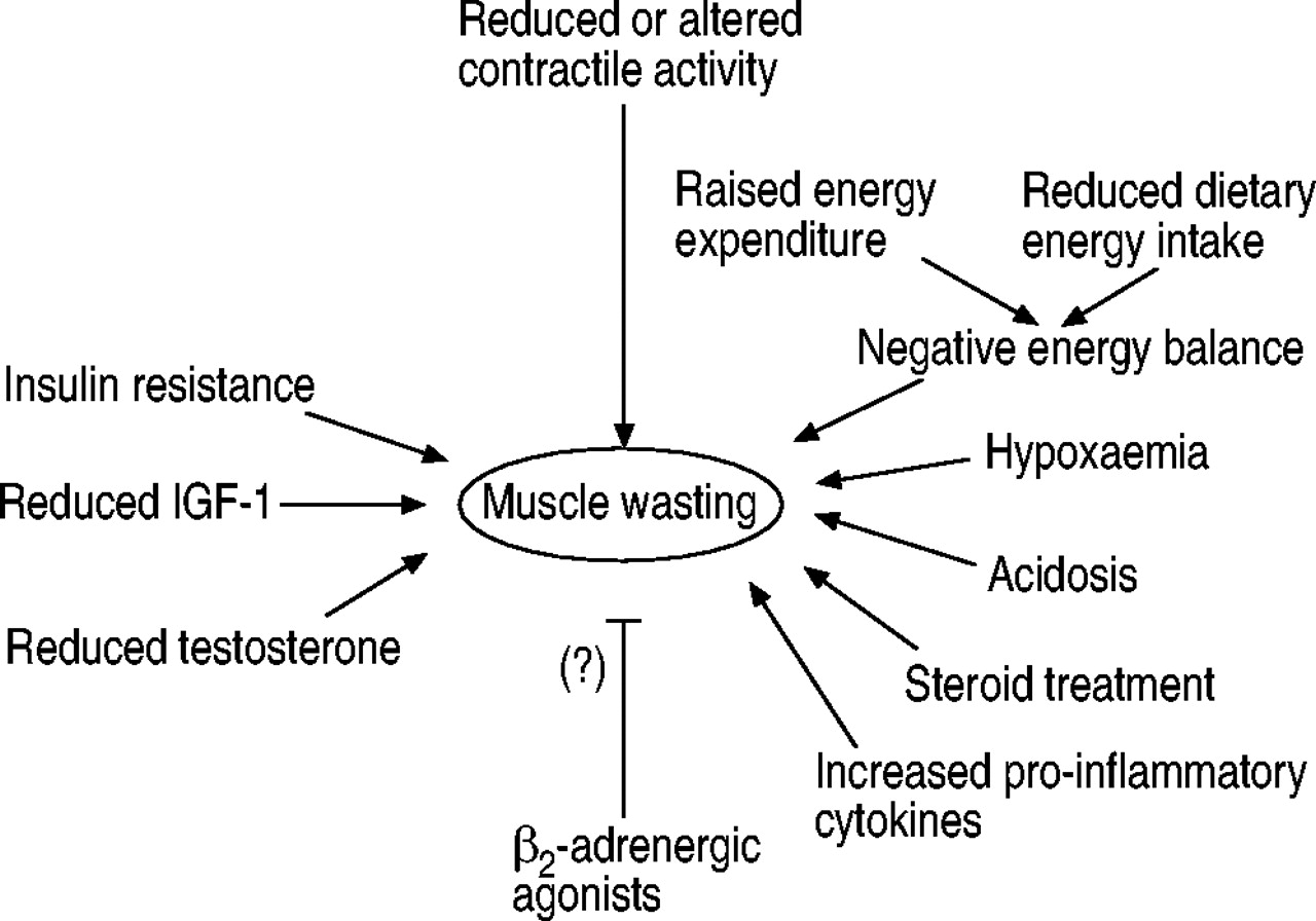
The extracellular factors likely to be involved in muscle wasting in chronic obstructive pulmonary disease. IGF‐1: insulin-like growth factor 1.
Reduced contractile activity
The plasticity of muscle tissue is evident in the way that altering loading has dramatic effects on the size and the metabolic capacity of muscle fibres. Experiments in rats showed unloading leads to muscle wasting, due to reduced protein synthesis and accelerated degradation, in slow-twitch muscles 53. In studies of humans on two weeks' bed rest 54 or after 5 weeks' limb immobilisation in a plaster cast 55, a dramatic reduction in protein synthesis was noted. However, even short-term disuse can result in loss of muscle bulk 56, and in a rodent model of disuse atrophy, a rise in proteolysis was seen within 9 days and was largely due to activation of the ubiquitin-proteasome pathway 57. In addition, recent knockout studies have shown that removing one of the components of the ubiquitin-proteasome pathway in muscle inhibits muscle weight loss due to denervation and disuse by up to 60% 58. With more prolonged immobilisation or disuse a preferential loss of the slow-twitch fibres occurs 55, 59, 60. This pattern of type I fibre atrophy is similar to that described in vastus lateralis muscle in COPD. Furthermore reduced muscle fibre size can be, at least partially, reversed with exercise in these patients 25. Thus reduced muscular activity due to breathlessness, is likely to be a major factor initiating muscle wasting in COPD. However it remains possible that other factors act to exacerbate the muscle loss, by further disturbing the balance of protein synthesis and degradation.
Negative energy balance
Muscle wasting can result from negative energy balance due to inadequate dietary energy intake and/or increased total energy expenditure. In simple short-term fasting there is an initial rise in muscle proteolysis due to activation of the ubiquitin-proteasome pathway 61, and a rapid fall in protein synthesis. However if food is withheld this initial rise in proteolysis is followed by a fall to subnormal levels and coincides with a reduction in basal energy expenditure. When other sources of energy i.e. fat, are exhausted muscle proteolysis rises again and this usually signals the terminal phases of starvation 62.
Reduced dietary energy intake is certainly a problem in COPD and in particular in those with more severe disease with weight loss 63. However, though increased nutritional energy intake alone can increase body weight in COPD 13, this is usually the result of an increase in fat mass rather than lean body mass. In addition, a number of studies have shown increases in resting energy expenditure in COPD patients, and those losing weight are more likely to have higher resting energy expenditure than those who are weight stable 63. Energy requirements are determined by total daily energy expenditure (TDE), rather than resting energy expenditure (REE), and in free-living COPD patients, increased REE does not correlate with an increase in TDE 64. However a study of stable COPD patients undergoing pulmonary rehabilitation showed they had 19% higher TDE than a group of active elderly controls from an earlier study 65. Conversely, when studied in a respiration chamber, COPD subjects and controls had similar TDE 66. In acute exacerbations of COPD any negative energy imbalance is likely to be exacerbated by increases in REE and the often dramatic reductions in dietary intake 67. Thus inadequate energy intake is a factor promoting loss of muscle protein in some individuals. However for most subjects increased energy intake alone does not increase muscle mass, and in some cases has no effect on body weight at all 13.
In addition to the importance of adequate dietary energy content, consideration should be given to the composition of diet, as this may also have a role in modulating muscle protein metabolism. Interestingly, if energy requirements are met by dietary intake, but dietary protein intake is restricted, muscle protein synthesis and proteolysis fall, presumably as part of a protein-sparing mechanism 68. This also coincides with lower muscle oxidation of branched chain amino acids and thus reduced export of alanine and glutamine. Branched chain amino acids and leucine in particular, stimulate protein synthesis at the translational level, even at normal physiological concentrations 69, and have an inhibitory effect on proteolysis in muscle 70. These findings suggest that, in addition to correcting deficiencies in dietary energy intake, separate assessment of the adequacy of protein intake, and possibly supplementing branched chain amino acid intake, might have additional benefits in restricting loss of muscle protein in COPD.
The effects of hypoxia and acidosis on muscle proteolysis
Protein synthesis is an energy consuming process, and hypoxia, which limits energy production, has been shown to depress muscle protein synthesis. After 6 h of hypoxia (inspiratory oxygen fraction: 11%) muscle protein synthesis rate was reduced by 14–17% in rat muscle 71. Proteolysis via the ubiquitin-proteasome pathway also requires hydrolysis of adenosine triphosphate (ATP) 44, thus low tissue levels of oxygen which inhibit oxidative metabolism and ATP‐production, may inhibit rather than stimulate proteolysis via this pathway. However in experiments with isolated rodent muscles, failure to restrict contraction leads to activation of another group of proteases, namely, the Ca2+-dependent proteases 72. It has been proposed that this results from impaired oxygen diffusion into the contracted muscle causing ischaemic membrane damage which then allows calcium release into the cytosol, and activates Ca2+-dependent proteolytic enzymes. These and other studies of post-mortem changes in meat tenderness 73 suggest that Ca2+-dependent proteases may play a major role in muscle proteolysis in ischaemic conditions.
Studies on human subjects in hypoxic conditions simulating high altitude, have shown reduced muscle mass with smaller slow and possibly fast fibres 74. Hypoxia may well have a role in altered muscle metabolism in COPD as hypoxaemia is a hallmark of endstage COPD, and a substantial proportion (mainly emphysema patients) exhibit arterial oxygen desaturation during daily life activities 75. It is difficult to be certain about the degree of tissue hypoxia in these patients but the delivery of oxygen to muscle tissue may be further impaired by reduced capillary to fibre ratios 23. In severe acute exacerbations of COPD, acidosis often accompanies hypoxaemia, and acidotic conditions stimulate muscle proteolysis via the ubiquitin-proteasome pathway 76, and lead to enhanced oxidation of BCAAs 77. Correction of metabolic acidosis due to renal failure in humans reduces whole body proteolysis 78, and correcting acidosis in uraemic rats reduces muscle proteolysis 76.
Thus in patients with COPD, hypoxia alone will inhibit protein synthesis, and may stimulate Ca2+-dependent proteolysis in muscle, but additional acidosis may contribute to a dramatic enhancement of muscle proteolysis via the ubiquitin-proteasome pathway.
Glucocorticoids
It is well known that pharmacological doses of steroids can induce muscle wasting due to an initial increase in proteolysis which wanes after some days, and is accompanied by a more sustained decrease in protein synthesis 79. What is less well known is that, in animal models of muscle wasting, physiological levels of glucocorticoids are required for the accelerated proteolysis via the ubiquitin-proteasome pathway which accompanies fasting 80, metabolic acidosis, diabetes 81 and sepsis 82, and that adrenalectomy or blocking the action of glucocorticoids inhibits muscle wasting in these conditions.
Muscle wasting due to intermittent use of steroids as in COPD appears to be uncommon, and florid steroid myopathy is more usually associated with prolonged treatment with high dose corticosteroids. However there is evidence from clinical studies that cumulative dose of steroid treatment for COPD correlates with impaired muscle function, including respiratory muscle strength, irrespective of the severity of airflow obstruction 83. Thus in most COPD patients it is likely that the beneficial effects of even short courses of oral steroids during exacerbations, must be balanced against the deleterious long-term effects of accelerated loss of muscle protein.
Pro-inflammatory cytokines and muscle wasting
Conditions characterised by high levels of pro-inflammatory mediators e.g. sepsis or certain types of cancer cachexia, often lead to muscle wasting, but the precise role of individual cytokines has been surprisingly difficult to define. Tumour necrosis factor‐α (TNF‐α), interleukin (IL)‐1 and IL‐6, and interferon gamma (IFN‐γ) are the main candidate catabolic inflammatory cytokines, but the importance of each cytokine varies in different catabolic conditions. For example, in animal models it appears that IL‐6 is not involved in the acceleration in proteolysis due to sepsis 84, and in animals made cachectic by tumour, the muscle wasting observed is related to a variety of different soluble catabolic factors, depending on the tumour under consideration 85. Several different experimental designs have been used to study the effects of these molecules including direct injection into experimental animals, addition to the medium bathing isolated muscle preparations or cultured muscle cells. In general, experiments using single or combinations of pro-inflammatory cytokines, have shown that they reduce protein synthesis and enhance proteolysis to a variable extent, but many experiments have produced different results. Potential sources of discrepancies between different in vivo studies include the confounding effects of cytokine-related anorexia, and of circulating levels of amino acids such as leucine, and stress hormones such as glucocorticoids (see above). However, even recent experiments using cultured muscle cells have yielded conflicting results. When TNF‐α was added to C2C12 myotubes and rat primary muscle cultures activation of the ubiquitin-proteasome pathway was observed, with time- and concentration-dependent loss of protein and reduction in heavy chain myosin content 86. Using the same cell line, others showed that though TNF‐α disrupted the normal myocyte differentiation program, it did not cause loss of muscle-specific protein from more mature myotubes 87, and co-administration of IFN‐γ was required before myosin heavy chain protein levels were reduced 88. More recently a bimodal response has been reported: at low doses TNF‐α treatment of C2C12 myotubes in culture caused a reduction in protein content, but at higher doses an anabolic effect was observed with an increase in protein synthesis and reduction in proteolysis 89.
COPD is characterised by the presence of a chronic low-grade systemic inflammatory response which is not significantly related to indices of lung function 90, 91, but circulating markers of systemic inflammation are particularly evident in those with higher REE and lower FFM 37. Furthermore during acute exacerbations of COPD increased levels of acute phase proteins (C‐reactive protein and lipopolysaccharide binding protein) and soluble TNF receptors are found during the first days of treatment 91. Studies in other conditions have shown that during an acute inflammatory response to infection whole body protein synthesis and breakdown rates are increased 92. However it is difficult to extrapolate from whole body studies to estimate muscle protein turnover, as changes in protein metabolism in several different organs are occurring simultaneously. In particular the changes in whole body protein turnover include a substantial increase in synthesis of acute phase proteins by the liver. Increased muscle proteolysis and wasting in conditions characterised by a sustained inflammatory response, are thought to result from the need to supply amino acids for the synthesis of these acute phase proteins. If the inflammatory response is limited skeletal muscle can recover rapidly, but in prolonged systemic inflammation a net drain on the muscle protein stores results in wasting. Thus the presence of a systemic inflammatory response is likely to impact on muscle protein metabolism in two ways: firstly by increasing demand for amino acids to synthesise acute phase proteins in the liver, and secondly by the direct effects of circulating pro-inflammatory cytokines on muscle protein synthesis and degradation. The combination of these direct and indirect effects presumably explains why interventions, such as increased dietary intake and exercise, aimed at reversing muscle wasting in COPD, have had only limited success.
Pro-inflammatory cytokines, such as TNF‐α, can promote deoxyribonucleic acid (DNA) fragmentation in skeletal muscle 93, and this may be relevant to the increase in markers of apoptosis in muscle of patients with COPD 94. Also these effects of TNF‐α and the inhibition of myocyte differentiation noted above, may be particularly relevant to the fate of muscle satellite cells in COPD. These cells are important for the regeneration of muscle fibres after injury when they proliferate and fuse with other muscle cells. In prolonged denervation muscle atrophy satellite cell numbers decline sharply, and this has been linked to an increase in their susceptibility to apoptosis 95. The combination of reduced contractile activity, and raised TNF‐α may induce a significant loss of muscle satellite cells through apoptosis. In addition there may be impairment in the ability of the remaining satellite cells to respond to muscle damage e.g. after exercise or the toxic effects of reactive oxygen species 96.
Beta‐2‐agonists and their effects on muscle proteolysis
Catecholamines released from the adrenal medulla or from adrenergic nerve endings can exert an anabolic effect on muscle principally by reducing the Ca2+-dependent proteolysis 97, 98 but also by increasing protein synthesis. These anabolic effects can also be mimicked by drugs such as the adrenergic β2‐agonist, clenbuterol, which has been used to increase carcass and muscle weights in broiler chickens 99, and to inhibit muscle wasting in a range of conditions including hindlimb suspension, and denervation in rats 100. Furthermore in a hamster model of emphysema, 12 week's treatment with clenbuterol caused increased skeletal muscle including diaphragm weight, and restored the diaphragm twitch pressures to normal 101. Conversely β2‐agonists increase resting energy expenditure in COPD 102 and overactivity of the adrenergic system may contribute to negative energy balance by accelerating metabolic rate and increasing energy demands leading to loss of muscle protein. For this reason β‐adrenergic blockade with propranolol was used to reduce protein loss in children after severe burn injury 103. Whether the doses of β2‐agonists used in COPD have any role in countering or contributing to muscle catabolism remains unclear.
Insulin and insulin-like growth factor‐1
Insulin plays a key role in the regulation of protein metabolism in muscle and other insulin-sensitive tissues. A number of different experiments have shown that insulin increases rates of protein synthesis and inhibits protein breakdown to a variable degree. Insulin treatment stimulated synthesis of muscle proteins such as myosin heavy chain in some studies 104 but in others only muscle mitochondrial protein synthesis was increased 105. Some studies of the lower limb muscles in healthy humans using stable isotopes showed that insulin infusion increased protein synthesis without affecting protein breakdown 106, but others have shown a marked anti-proteolytic effect of insulin on muscle 107. The discrepant effects of insulin in different studies may be due to a number of confounding factors e.g. the independent anabolic effects of nutrients including glucose and amino acids some of which have been mentioned above, the cross-reactivity of insulin with insulin-like growth factor (IGF) receptors at high doses of insulin, and the effects of other hormones in particular glucocorticoids (see above).
Resistance to the actions of insulin, as measured using glucose uptake, is a feature of a number of chronic diseases 108–110, as well as acute sepsis 111, 112 and trauma 113. In addition insulin resistance can be induced after prolonged inactivity or bed rest 114, steroid treatment 115 and by high circulating levels of amino acids 116, and pro-inflammatory cytokines such as TNF‐α 117. Many of these factors are relevant to COPD, particularly during acute exacerbations, but on formal testing no insulin resistance was found in a group of patients with stable severe COPD 118. Nevertheless there is some evidence for impaired glucose tolerance in hypoxaemic COPD patients 119 and for elevated insulin levels in COPD 40. Thus other studies are needed to define the degree of any insulin-resistance and in particular the resistance to protein anabolic actions of insulin in COPD.
IGF‐1 is secreted by the liver in response to the action of growth hormone (GH), and both IGF‐1 and GH promote muscle growth by enhancing protein synthesis and inhibiting proteolysis 120, 121. One report has suggested that there are low levels of circulating IGF‐1 in COPD 122, and if so this may have a profound effect on muscle size, as IGF‐1 has a central role in muscle hypertrophy 123. In a controlled trial of GH supplementation, underweight COPD patients showed an increase in muscle mass but no change in muscle strength or exercise tolerance 16. Other studies have attempted to use IGF‐1 to reduce catabolism. This was successful in animals with burns where muscle proteolysis was reduced and synthesis increased 124. However septic animals were resistant to the antiproteolytic effects of IGF‐1, despite an increase in protein synthesis rates 125. It should be remembered that though circulating IGF‐1 is largely derived from the liver, muscle also produces IGF‐1 including a muscle-specific isoform 126. The relative importance of circulating and locally produced IGF‐1 in muscle anabolism is still unclear. Furthermore the actions of IGFs are modified by a range of extracellular binding proteins which may enhance or inhibit the interaction with the cell surface IGF receptor, depending on their relative local concentrations. The expression of these binding proteins is regulated by muscle cells 127, 128 and may prove to be critical in determining the nature of the response of muscle to IGF‐1.
Anabolic steroids
Low levels of testosterone may also be common in COPD 129 and this may be relevant to loss of muscle mass given the clear anabolic effect this hormone exerts on muscle 130. Interestingly, the anabolic effects of testosterone therapy on muscle, like those of growth hormone, may be indirect and mediated by IGF‐1 131. Nevertheless, testosterone supplements have been used successfully in treating muscle wasting in human immunodeficiency virus (HIV)‐infected males with low testosterone levels 132, and in limited muscle loss after burn injury 133 by improving protein synthetic efficiency and reducing muscle protein degradation rates. In COPD three different studies have used testosterone or other anabolic steroids in conjunction with pulmonary rehabilitation. Each study confirmed that adjunctive treatment with anabolic steroids increased FFM but though one showed an increase in respiratory muscle strength no additional increase in exercise tolerance was detected 13–15. Thus anabolic steroids may aid weight gain including increasing lean body mass, but to date any associated improvements in muscle function have been limited.
A model of muscle wasting in chronic obstructive pulmonary disease
It appears that many different factors combine to determine the net effect on muscle protein metabolism in COPD. However because of the complexity of the interactions between these factors, and the lack of experimental evidence in COPD, an accurate model of the causes of muscle wasting in COPD cannot be drawn with any certainty. Nevertheless the major features of such a model would probably include the following: 1) primary loss of muscle mass in response to reduced physical activity and muscle load, possibly exacerbated by a chronic low-grade inflammatory response; 2) accelerated muscle loss during acute exacerbations due to further restrictions on physical activity, increased circulating pro-inflammatory cytokines, hypoxaemia and acidosis, and treatment with steroids most of which would be expected to accelerate proteolysis particularly via the ubiquitin-proteasome pathway; 3) impaired capacity to recover chronic and acute loss of muscle protein, possibly due to chronically raised levels of proteolysis, but more likely to impaired protein synthesis. This latter effect may be the result of reduced circulating anabolic hormones and possibly other changes in muscle such as reduced secretion of muscle-derived IGF‐1, or IGF‐binding proteins. In addition, pro-inflammatory cytokines, tissue hypoxaemia and disuse may favour selective loss of muscle satellite cells, or impair their ability to differentiate and mature, which would further compromise the muscle's ability to remodel or respond to injury (fig. 3⇓).

A model for the development of muscle wasting in chronic obstructive pulmonary disease.
Conclusions and future research questions
Muscle wasting in chronic obstructive pulmonary disease is associated with impaired skeletal muscle function, worse quality of life and poorer prognosis. Despite widespread acceptance of the importance of muscle wasting in chronic obstructive pulmonary disease, research into the mechanisms underlying these changes in muscle is still in its infancy. Many potential catabolic factors have been identified but the precise contribution of each one remains unclear. At one level the loss of muscle protein is simply due to an imbalance in the rates of muscle protein synthesis and breakdown, and basic descriptive studies are still needed to measure muscle proteolysis and synthesis rates in chronic obstructive pulmonary disease, both in stable patients and during acute exacerbations. Such studies will lay the basis for many further mechanistic or intervention studies. Evidence from other conditions, and animal models suggests that accelerated proteolysis via the ubiquitin-proteasome pathway is likely to have a role in the loss of muscle protein in chronic obstructive pulmonary disease. However, to date, no studies have confirmed that activation of any intracellular proteolytic pathways, including the ubiquitin-proteasome pathway, occurs in muscle in chronic obstructive pulmonary disease. Establishing when such activation occurs during the course of the disease, will also be an important step in designing therapeutic interventions. Many of the catabolic factors discussed above inhibit protein synthesis, and identifying whether there are specific hormonal or biochemical mechanisms which limit muscle hypertrophy in chronic obstructive pulmonary disease should also be a research priority. In addition to loss of muscle mass, other adaptations including changes in muscle fibre type proportions and apoptosis of myonuclei are features of chronic obstructive pulmonary disease. This raises the possibility that interventions, designed to limit loss of type I fibres e.g. by increasing expression of the transcription co-activator peroxisome proliferator-activated receptor-gamma coactivator (PGC)-1 134, or to specifically inhibit apoptosis in muscle, may have long-term benefits.
- Received June 30, 2003.
- Accepted June 30, 2003.
- © ERS Journals Ltd
References


Jump To
- Article
- Abstract
- Indicators of skeletal muscle dysfunction in chronic obstructive pulmonary disease
- The pathways of protein synthesis and proteolysis in muscle
- What are the systemic factors which negatively influence protein metabolism and thus promote muscle loss in chronic obstructive pulmonary disease?
- A model of muscle wasting in chronic obstructive pulmonary disease
- Conclusions and future research questions
- References
- Figures & Data
- Info & Metrics